

- •VOLUME 1
- •CONTRIBUTOR LIST
- •PREFACE
- •LIST OF ARTICLES
- •ABBREVIATIONS AND ACRONYMS
- •CONVERSION FACTORS AND UNIT SYMBOLS
- •ABLATION.
- •ABSORBABLE BIOMATERIALS.
- •ACRYLIC BONE CEMENT.
- •ACTINOTHERAPY.
- •ADOPTIVE IMMUNOTHERAPY.
- •AFFINITY CHROMATOGRAPHY.
- •ALLOYS, SHAPE MEMORY
- •AMBULATORY MONITORING
- •ANALYTICAL METHODS, AUTOMATED
- •ANALYZER, OXYGEN.
- •ANESTHESIA MACHINES
- •ANESTHESIA MONITORING.
- •ANESTHESIA, COMPUTERS IN
- •ANGER CAMERA
- •ANGIOPLASTY.
- •ANORECTAL MANOMETRY
- •ANTIBODIES, MONOCLONAL.
- •APNEA DETECTION.
- •ARRHYTHMIA, TREATMENT.
- •ARRHYTHMIA ANALYSIS, AUTOMATED
- •ARTERIAL TONOMETRY.
- •ARTIFICIAL BLOOD.
- •ARTIFICIAL HEART.
- •ARTIFICIAL HEART VALVE.
- •ARTIFICIAL HIP JOINTS.
- •ARTIFICIAL LARYNX.
- •ARTIFICIAL PANCREAS.
- •ARTERIES, ELASTIC PROPERTIES OF
- •ASSISTIVE DEVICES FOR THE DISABLED.
- •ATOMIC ABSORPTION SPECTROMETRY.
- •AUDIOMETRY
- •BACTERIAL DETECTION SYSTEMS.
- •BALLOON PUMP.
- •BANKED BLOOD.
- •BAROTRAUMA.
- •BARRIER CONTRACEPTIVE DEVICES.
- •BIOCERAMICS.
- •BIOCOMPATIBILITY OF MATERIALS
- •BIOELECTRODES
- •BIOFEEDBACK
- •BIOHEAT TRANSFER
- •BIOIMPEDANCE IN CARDIOVASCULAR MEDICINE
- •BIOINFORMATICS
- •BIOLOGIC THERAPY.
- •BIOMAGNETISM
- •BIOMATERIALS, ABSORBABLE
- •BIOMATERIALS: AN OVERVIEW
- •BIOMATERIALS: BIOCERAMICS
- •BIOMATERIALS: CARBON
- •BIOMATERIALS CORROSION AND WEAR OF
- •BIOMATERIALS FOR DENTISTRY
- •BIOMATERIALS, POLYMERS
- •BIOMATERIALS, SURFACE PROPERTIES OF
- •BIOMATERIALS, TESTING AND STRUCTURAL PROPERTIES OF
- •BIOMATERIALS: TISSUE-ENGINEERING AND SCAFFOLDS
- •BIOMECHANICS OF EXERCISE FITNESS
- •BIOMECHANICS OF JOINTS.
- •BIOMECHANICS OF SCOLIOSIS.
- •BIOMECHANICS OF SKIN.
- •BIOMECHANICS OF THE HUMAN SPINE.
- •BIOMECHANICS OF TOOTH AND JAW.
- •BIOMEDICAL ENGINEERING EDUCATION
- •BIOSURFACE ENGINEERING
- •BIOSENSORS.
- •BIOTELEMETRY
- •BIRTH CONTROL.
- •BLEEDING, GASTROINTESTINAL.
- •BLADDER DYSFUNCTION, NEUROSTIMULATION OF
- •BLIND AND VISUALLY IMPAIRED, ASSISTIVE TECHNOLOGY FOR
- •BLOOD BANKING.
- •BLOOD CELL COUNTERS.
- •BLOOD COLLECTION AND PROCESSING
- •BLOOD FLOW.
- •BLOOD GAS MEASUREMENTS
- •BLOOD PRESSURE MEASUREMENT
- •BLOOD PRESSURE, AUTOMATIC CONTROL OF
- •BLOOD RHEOLOGY
- •BLOOD, ARTIFICIAL
- •BONDING, ENAMEL.
- •BONE AND TEETH, PROPERTIES OF
- •BONE CEMENT, ACRYLIC
- •BONE DENSITY MEASUREMENT
- •BORON NEUTRON CAPTURE THERAPY
- •BRACHYTHERAPY, HIGH DOSAGE RATE
- •BRACHYTHERAPY, INTRAVASCULAR
- •BRAIN ELECTRICAL ACTIVITY.
- •BURN WOUND COVERINGS.
- •BYPASS, CORONARY.
- •BYPASS, CARDIOPULMONARY.
308BIOMATERIALS CORROSION AND WEAR OF
76.Wieting DW. The Bjo¨rk-Shiley Delrin Tilting Disc Heart Valve: Historical Perspective, Design and Need for Scientific Analyses After 25 Years. J Heart Valve Dis 1996;5(Suppl I): S157–S168.
77.Schoen FJ, Titus JL, Lawrie GM. Durability of Pyrolytic Carbon-Containing Heart Valve Prostheses. J Biomed Mater Res 1982;16:559–570.
78.Griffin CD, Buchanan RA, Lemons JE. In Vitro Electrochemical Corrosion Study of Coupled Surgical Implant Materials. J Biomed Mater Res 1983;17:489–500.
79.Thompson NG, Buchanan RA, Lemons JE. In Vitro Corrosion of Ti-6Al-4V and Type 316L Stainless steel When Galvanically Coupled with Carbon. J Biomed Mater Res 1979;13:35–44.
80.Cook SD, Thomas KA, Kester MA. Wear characteristics of the canine acetabulum against different femoral prostheses. J Bone Joint Surg 1989;71B:189–197.
81.Tian CL, Hetherington VJ, Reed S. A Review of Pyrolytic carbon: Application in Bone and Joint Surgery. J Foot Ankle Surg 1993;32(5):490–498.
82.Cook SD, Beckenbaugh RD, Redondo J, Popich LS, Klawitter JJ, Linscheid RL. Long term follow-up of pyrolytic carbon metacarpophalangeal implants. J Bone Joint Surg 1999; 81A(5): 635–648.
83.Ferrari M. Clinical evaluation of fiber-reinforced epoxy resin posts and cast post and cores. Am J Dent 2000; 01-May-13 (Spec No): 15B–18B.
84.Pamula E. Studies on development of composite biomaterials for reconstruction of the larynx. Polim Med 2001;31(1–2):39–44.
85.Katoozian H. Material optimization of femoral component of total hip prosthesis using fiber reinforced polymeric composites. Med Eng Phys 2001;23(7):503–509.
86.Fru¨ h HJ. Fusion implants of carbon fiber reinforced plastic. Orthopade 2002;31(5):454–458.
87.Dearnaley G. Diamond-like carbon: a potential means of reducing wear in total joint replacements. Clin Mater 1993;12:237–244.
88.Lappalainen R, Anttila A, Heinonen H. Diamond coated total hip replacements. Clin Orthop 352 (July 1998): 118–127.
89.Pantarotto D, Partidos CD, Graff R, Hoebeke J, Briand JP, Prato M. Bianco A. Synthesis, structural characterization, and immunological properties of carbon nanotubes functionalized with peptides. J Am Chem Soc 2003 May 21; 125(20): 6160–6164.
90.Qingnuan L, Yan X, Xiaodong Z, Ruili L, Qieqie D, Xiaoguang S, Shaoliang C, Wenxin L. Preparation of (99m)Tc- C(60)(OH)(x) and its biodistribution studies. Nucl Med Biol 2002 Aug; 29(6): 707–710.
91.Gonzalez KA, Wilson LJ, Wu W, Nancollas GH. Synthesis and in vitro characterization of a tissue-selective fullerene: vectoring C(60)(OH)(16)AMBP to mineralized bone. Bioorg Med Chem 2002 Jun; 10(6): 1991–1997.
92.Wolff DJ, Barbieri CM, Richardson CF, Schuster DI, Wilson SR. Trisamine C(60)-fullerene adducts inhibit neuronal nitric oxide synthase by acting as highly potent calmodulin antagonists. Arch Biochem Biophys 2002 Mar 15; 399(2): 130–141.
93.Schinazi RF, Sijbesma R, Srdanov G, Hill CL, Wudl F. Synthesis and virucidal activity of a water-soluble, configurationally stable, derivatized C60 fullerene. Antimicrob Agents Chemother 1993 Aug.; 37(8): 1707–1710.
94.Park KH, Chhowalla M, Iqbal Z, Sesti F. Single-walled carbon nanotubes are a new class of ion channel blockers. J Biol Chem 2003 Dec. 12; 278(50): 50212–50216, Epub 2003 Sep. 30.
See also BIOMATERIALS: TISSUE ENGINEERING AND SCAFFOLDS;
BIOMATERIALS, TESTING AND STRUCTURAL PROPERTIES OF; BIOSURFACE ENGINEERING; MATERIALS AND DESIGN FOR ORTHOPEDIC DEVICES; HEART VALVE PROSTHESES.
BIOMATERIALS CORROSION AND WEAR OF
ROGER J. NARAYAN
University of North Carolina
Chapel Hill, North Carolina
MIROSLAV MAREK
Georgia Institute of Technology
Atlanta, Georgia
CHUNMING JIN
North Carolina State University
Raleigh, North Carolina
INTRODUCTION
Many materials suffer degradation with time when exposed to aggressive chemical environments within the human body. In metallic biomaterials, degradation results from electrochemical corrosion. Ceramic and polymeric biomaterials may undergo physical or chemical deterioration processes. In addition, mechanical forces may act to increase damage by wear, abrasion, or environmentinduced cracking processes.
Corrosion of implants, dental restorations, and other objects placed in the human body may result in degradation of function as a result of loss of mass, decrease in mechanical integrity, or deterioration of aesthetic qualities. The associated release of corrosion products and the flow of the corrosion currents also may cause inflammation, allergic reactions, local necrosis, and many other health problems.
For electronic conductors (e.g., metals), corrosive interaction with ionically conducting liquids (e.g, body fluids) is almost always electrochemical. The degradation of metals is due to an oxidation process that involves the loss of electrons. This process involves a change from a metallic state to an ionic state, in which the ions dissolve or form nonmetallic solid products. For the process to continue, the released electrons must be consumed in a complementary reduction, which usually involves species present in the biological environment (e.g., hydrogen ions or dissolved oxygen). The reaction resulting in oxidation is usually called an anodic process and reaction resulting in reduction is usually called a cathodic process. The metal is referred to as an electrode, and the liquid environment is referred to as an electrolyte.
For many metals, the most important environmental variables are the concentrations of chloride ions, hydrogen ions, and dissolved oxygen. In many human body fluids, the chloride ion concentration varies in a relatively narrow range near 0.1 mol L 1; however, it may be variable (e.g., urine) or lower (e.g., saliva) in certain body fluids. The hydrogen ion concentration is expressed as a pH value and is near neutral (pH ¼ 7) for plasma, interstitial fluid, bile, and saliva; however, it is more variable (pH ¼ 4–8) in urine and very low (pH ¼ 1–3) in gastric juice (1). The chloride concentration and pH are most important factors determining the rate of oxidation because of their effect on protective oxide passivating films on metals. The dissolved oxygen concentration affects mainly the cathodic process. The usual range of partial pressure of oxygen in body fluids is 40–100 mmHg (5.33–13.33 kPa) (1–3).
For electrochemical oxidation to cause clinically relevant degradation of a material, the electrochemical reaction must be energetically possible (thermodynamics) and the reaction rate must be appreciable (kinetics). Oxidation of nickel, for example,
Ni ! Ni2þ þ 2e |
ð1Þ |
will proceed in the indicated direction if the potential of the electrode on which the reaction occurs is higher (more positive or less negative) than the equilibrium potential for a given electrolyte, and is a function of the energy change involved. The equilibrium potential also depends on temperature, pressure, and activity ( concentration) of ions. The values of potentials for reactions between metals and their ions in water are given under standard conditions (temperature 25 8C, pressure 1 atm, and activity of ions equal to 1) and are written in the form of materials reduction. These values, known as standard single electrode potentials, are listed in the so-called electrochemical or electromotive (EM) series (1). Noble metals, which have no tendency to dissolve in water have positive standard single-electrode potentials. On the other hand, active metals with a high tendency to react with water exhibit negative potentials.
While the potential of the anodic process must be above the equilibrium potential for the reaction to proceed as oxidation, for the cathodic process the electrode potential must be below (more negative or less positive) the equilibrium potential for net reduction to occur. For reduction of hydrogen ions,
2Hþ þ 2e ¼ H2 |
ð2Þ |
the equilibrium potential at normal body temperature (37 8C) and pH 7.4 (blood or interstitial fluids) is 0.455 V (SHE) ( 0.697 V, SCE), while the equilibrium potential of the other likely cathodic reaction,
O2 þ 4 Hþ þ 4e ¼ 2 H2O |
ð3Þ |
is 0.753 V (SHE) (0.511 V, SCE) at 40 mmHg (5.33 kPa) of oxygen partial pressure. Although reaction 3 is more sluggish than reaction 2, for most metals in the human body the electrode potential of reaction 3 is above the equilibrium potential of the hydrogen reaction 2, and reduction of oxygen is the dominant cathodic process.
In spontaneous electrochemical corrosion, at least two reactions occur simultaneously. At least one reaction occurs in the direction of oxidation, and at least one reaction occurs in the direction of reduction. Each reaction has its own equilibrium potential, and this potential difference results in a current flow, as the electrons released in oxidation flow to the sites of reduction and are consumed there. In the absence of a significant electrical resistance in the current path between the reaction sites, a common potential is established, which is known as mixed potential
or corrosion potential (Ecorr). At this potential, both reactions produce the same current in opposite directions in
order to preserve electrical neutrality. The value of the oxidation current, which is equal to the absolute value of the reduction current, per unit area at this potential is
BIOMATERIALS CORROSION AND WEAR OF |
309 |
known as the corrosion current density (icorr). The oxidation and reduction reactions may be distributed uniformly on the same metal surface; however, there are often some regions of the biomaterial surface that are more favorable for oxidation and other regions that are more favorable for reduction. As a result, either local anodic and cathodic areas or completely separate anodes and cathodes are formed.
The corrosion rate (mass of metal oxidized per unit area and time) is proportional to the corrosion current density. The conversion is given by the Faraday’s law, which states that an electric charge of 96,485 C is required to convert 1 equiv weight of the metal into ions, or vice versa. The shift of the potential of a reaction from the equilibrium value to the corrosion potential is called polarization by the flow of the current. The resulting current flowing at corrosion potential depends on the way the current of each reaction varies with the potential. If the current is controlled by the activation energy barrier for the reaction at the electrode surface, then the reaction rate increases exponentially with increasing potential for oxidation reactions and decreases exponentially with increasing potential for reduction reactions. The activation energy controlled current typically increases or decreases ten times for a potential change of 50–150 mV. At high reaction rates, the current may become limited by the transport of reaction species to or from the electrodes; eventually, the corrosion process may become completely controlled by diffusion and independent of potential.
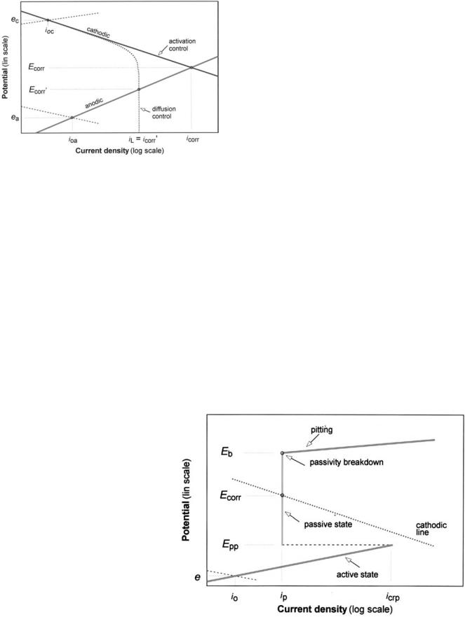
The vast majority of uses for metallic biomaterials in the human body are successful due to the phenomenon of passivity. In a passive state, these metals become covered with thin, protective films of stable, poorly soluble oxides or hydroxides when exposed to an aqueous electrolyte. Once this passivating film forms, the current density drops to a very low value and becomes much less dependent on the potential. The variation of the reaction current density with the potential can be illustrated in a polarization diagram. A schematic diagram in Fig. 1 shows some of the main reactions in corrosion and relevant parameters. A straight-line relationship in a semilogarithmic (E vs. log I) diagram indicates that an activation energy-controlled reaction is occurring. This electrochemical activity is known as Tafel behavior, and the slopes of the lines ( 50–150 mV per 10-fold change in the current or current density) are equal to the values of the Tafel constants. When the oxidation reaction of the metal shows this relationship at the corrosion potential, it indicates that the metal is actively corroding. If a metal forms a passivating film when the potential exceeds a critical value in the active corrosion region, then the current density drops from a value called the critical current density for passivation
(icrp) at a primary passivation potential (Epp) to a low current density in the passive state (ip). This behavior is
illustrated schematically in Fig. 2. For an electrode to maintain a stable passive state, the intersection of the oxidation (anodic) and reduction (cathodic) lines must occur in the region of passivity.
The polarization characteristics of a biomaterial can be experimentally determined using a device called a potentiostat, which maintains the sample potential at a set value
310 BIOMATERIALS CORROSION AND WEAR OF
Figure 1. Schematic polarization diagram showing oxidation (anodic) and reduction (cathodic) reactions of a corrosion process, for reactions controlled by activation energy and by mass transport (diffusion). In this figure, ea and ec refer to equilibrium potentials of the anodic and cathodic process, respectively, ioa and ioc refer to exchange current densities, Ecorr refers to mixed corrosion potential, and iL refers to limiting current density.
versus a reference electrode by passing current between the sample and an auxiliary counterelectrode. A scan generator can be used to vary the controlled potential over a range of interest, and the E–i relationship can be determined. The relationship of main interest is usually the oxidation rate as a function of the potential, which can be depicted in an anodic polarization diagram. Since only a net current (difference between the absolute values of the oxidation and reduction currents) can be measured, the experimental polarization curve shows a value approaching zero at the intersection of the anodic and cathodic polarization curves.
Experimental anodic polarization curves for passivating metals and alloys often do not exhibit the passivation peak
shown in Fig. 2, either because the metal forms an oxide in the electrolyte without undergoing active dissolution or because an oxide film already has formed as a result of exposure to air. More importantly for human body fluids and other chloride-containing electrolytes, the region of passivity is often limited by a localized passivation breakdown above a critical breakdown potential (Eb). When a breakdown occurs, intensive oxidation takes place within localized regions on the biomaterial surface, resulting in sometimes significant pit formation. In an experimental anodic polarization diagram, breakdown appears as a sharp increase in the measured current above the critical breakdown potential. Because of the destructive nature of surface pitting, the determination of critical breakdown potential is one of the most important ways of assessing the suitability of novel metallic biomaterials for use in medical devices.
The high current density in active pits is due to the absence of a passivating film, which results from local chemical and electrochemical reactions that change the electrolyte to become highly acidic and depleted in dissolved oxygen. A similar corrosion mechanism may occur in interstices known as crevices, where the transport of species to and from the localized corrosion cell is difficult. This process, known as crevice corrosion, does not require a potential exceeding the critical breakdown potential for the initiation of corrosion. Both pit and crevice corrosion cells may repassivate if the potential is lowered below a value needed for maintenance of a high oxidation rate on the bare (nonpassivated) metal surface. The potential below which active pits repassivate is called the repassivation or protection potential (Ep). The concept of a protection potential also applies to crevice corrosion. Experimentally, repassivation can be studied by reversing the anodic polarization scan and recording the potential at which the current returns to a passive state value (Fig. 3). Repassivation can also be examined by initiating pitting or crevice corrosion and lowering the potential in steps until the current
Figure 2. Schematic polarization diagram, showing a transition from active to passive state and a breakdown of passivity. In this figure, icrp refers to critical current density for passivation, ip refers to current density in the passive state, Epp refers to primary passivation potential, and Eb refers to breakdown potential.

Figure 3. Schematic experimental cyclic polarization diagram for a passivating electrode, showing passivity breakdown and repassivation after potential scan reversal. In this figure, Eprot refers to protection (repassivation) potential.
shows low values that decrease with time (standard test methods F2129 and F746, respectively, ASTM 2005) (4). The difficulty in finding a reliable protection potential value is due to the fact that the ease of repassivation depends on the extent of pitting or crevice corrosion damage that has occurred before the potential drop.
For some polyvalent metals (e.g., chromium), soluble species (e.g., CrO42 ) become thermodynamically stable as the valence changes (e.g., from 3 to 6 in chromium) at potentials above those for a stable oxide. This process may result in another region of active dissolution at high potentials in a phenomenon known as transpassivity.
When corrosion is relatively uniform throughout the biomaterial, the most important corrosion parameter is the average corrosion rate. For biomaterials with very low corrosion rates, the average corrosion rate is mostly determined by sensitive electrochemical techniques. The average corrosion current density (icorr ) is usually determined either from the results of the polarization scan by extrapolating the anodic and cathodic lines to the corrosion potential or from calculating the value of the polarization resistance. The polarization resistance (Rp) is defined as the slope of the polarization curve at zero current density ½Rp ¼ ðdE=dinÞin¼0&, in which in is the net (measured) current density.
When two or more dissimilar metals are placed in contact within an electrolyte, their interaction may cause galvanic corrosion. The oxidation degradation is enhanced for the metal with the lower individual corrosion potential, which becomes the anode of the cell. It is polarized towards a higher potential at the other electrode, which becomes the cathode. Since the oxidation current increase on the anode must be balanced by an identical reduction current increase on the cathode, a combination of a small anode with a large cathode is more detrimental than the reverse situation, since a larger increase in the oxidation current density is produced. In practical situations, resistance in the current path between the electrodes often reduces the galvanic effect. Differences in the concentrations of reaction species at different regions on the metal surface may
BIOMATERIALS CORROSION AND WEAR OF |
311 |
result in a potential difference, which leads to additional polarization. An increase in the oxidation current density, a difference in the equilibrium potentials, and a flow of current may result. Differences in concentrations of hydrogen ions, dissolved metal ions, or dissolved oxygen may result in concentration cell corrosion.
Metal parts subjected to mechanical loading in a corrosive environment may fail by environment-induced cracking (EIC). Stress corrosion cracking (SCC) may occur in some biomaterials when they are subjected to static loading under certain environmental conditions. Corrosion fatigue (CF) may result from variable loading in reactive environments. When the failure can be attributed to the entry of hydrogen atoms into the metal, the phenomenon is referred to as hydrogen induced cracking (HIC). Environment-induced cracking may be caused by complex combinations of mechanical, chemical, and electrochemical forces; however, the exact mechanisms of this behavior are subject to significant controversy. In these cases, mechanical factors may play important roles in crack propogation.
Intergranular corrosion occurs when dissolution is confined to a narrow region along the grain boundaries. This process is either due to precipitation of corrosion susceptible phases or due to depletion in elements that provide corrosion protection along the boundaries, which is caused by precipitation of phases rich in those elements. Some stainless steel and nickel-chromium alloys may be sensitized due to precipitation of chromium-rich carbides along grain boundaries when heated to a specific temperature range. Sensitization is normally prevented from occurring in stainless steels currently used in medical devices, which contain very low amounts of carbon.
Passivating films may also be mechanically destroyed in wear- , abrasion- , erosion- , and fretting-corrosion processes. Wear-corrosion involves materials in a friction contact that exhibit substantial relative movement. Fretting occurs in situations in which there are only small relative movements between materials that are essentially fixed with respect to one another. The resulting wear debris may cause abrasion–corrosion behavior. Wear-cor- rosion may occur in artificial joints, including the metal ball of a hip joint in contact with the polyethylene cup. Fretting may take place between the ball and the stem of multicomponent hip implants. In both forms of corrosion, the narrow gap between contacting surfaces creates crevice conditions. In addition, the destructive effect of friction and abrasion on the protective surface film is superimposed on the corrosion mechanism in the crevice cell. Erosion corrosion may occur on devices exposed to rapidly flowing fluids, including the surfaces of artificial heart valves.
A wide variety of metals and alloys have been used in medical devices. The three most commonly used alloys are stainless steel, cobalt alloys, and titanium alloys (5). Type 316 LVM (low carbon, vacuum-melted) stainless steel is less corrosion resistant than cobalt or titanium alloys, and it is most often used for temporary implants (5–8). This material is referred to as an austenitic steel, because it contains an iron carbide phase called austenite (g-iron). Implant-grade steel has a nominal composition of 18% chromium, 14% nickel, and 2.5% molybdenum; the
312 BIOMATERIALS CORROSION AND WEAR OF
compositional limits and properties are specified by ASTM standards F 138 and F 139 for wrought steel and F 745 for cast steel (ASTM, 2005) (4). Chromium serves to improve corrosion resistance through the formation of a highly protective surface film rich in chromium oxide. Implantgrade steel has a low carbon content in order to prevent sensitization and intergranular corrosion. Alloying with - molybdenum further improves the resistance, especially to crevice corrosion and pitting. Nickel serves to stabilize the face-centered cubic (fcc) structure. On the other hand, manganese sulfide inclusions, which contribute to initiation of pitting, are minimized.
The corrosion resistance of stainless steel greatly depends on the surface conditions, and stainless steel implants are almost always electropolished and prepassivated by exposure to nitric acid (standard practice F86, ASTM 2005) (4). The breakdown potential is usually around 0.4 V (SCE), with a large hysteresis loop and a low protection potential (9). Considering that the potential in the human body is not likely to exceed about 0.5 V (SCE) (see Eq. 3 and its equilibrium potential), a well polished and passivated 361 LVM stainless steel is not very susceptible to pitting in the human body, especially for unshielded and undisturbed implant surfaces. Once localized attack is initiated, however, repassivation is difficult. As a result, stainless steel implants are very susceptible to crevice corrosion, especially when the crevice situation is combined with destruction of the surface film (e.g., fretting of bone plates under the screw heads). Small single component stainless steel implants, such as balloonexpandable vascular stents, that are made of high purity precursor materials and are subjected to a high quality surface treatment and inspection can achieve a breakdown potential in excess of 0.8 V (SCE); these materials are considered very resistant to localized corrosion (10). Stainless steel bars [containing 22% chromium 12.5% nickel , 5% manganese, and 2.5% molybdenum (ASTM F 1586)] and wires [containing 22% chromium, 12.5% nickel, 5% manganese, and 2.5% molybdenum (ASTM F 1314)] strengthened with nitrogen have shown a higher breakdown potential than ASTM F 138 steel (4).
Vitallium and other cobalt–chromium alloys were developed as a corrosion resistant, high strength alternative to stainless steel alloys. These materials were first used in dentistry, and were later introduced to orthopedics and other surgical specialties. The cast cobalt–chromium alloy most commonly used in medical devices (ASTM F 75) contains 28% chromium and 6% molybdenum (4). This alloy was found to be suitable for investment casting into intricate shapes. In addition, it exhibited very good corrosion and excellent wear resistance; however, it possessed low ductility. Alloys with slightly modified compositions were later developed for forgings (ASTM F 799) and wrought bars, rods, and wire (ASTM F 1537) (4). Alloy F75 has shown corrosion resistance superior to stainless steel in the human body. Laboratory studies reported a breakdown potential of 0.5 V (SCE) and protection potential of 0.4 V (SCE) (6,7,9,11). These properties have made it possible to use cobalt–chromium alloys for permanent implants. Cobalt–chromium alloys with porous surfaces have been used for bone ingrowth, although they have
been superseded by even more crevice corrosion resistant and biocompatible titanium alloys. The excellent corrosion resistance of cobalt–chromium alloys can be attributed to a high chromium content and a protective surface film of chromium oxide. Concerns have been raised, however, regarding the release of biologically active hexavalent chromium ions (12). Other cobalt-based wrought surgical alloys include F90 (Co-Cr-W-Ni), F563 (Co-Ni-Cr-Mo-W- Fe), F563 (Co-Ni-Cr-Mo-W-Fe), F1058 (Co-Cr-Ni-Mo), and F688 (Co-Ni-Cr-Mo) (4). These alloys provide good to excellent corrosion behavior and a variety of mechanical properties, which depend on thermomechanical treatment. However, there is some concern regarding metal ion release in these alloys, which contain high nickel concentrations.
Titanium and titanium alloys have been used in orthopedic implants and other medical devices since the 1960s. Their popularity has rapidly increased because they possess high corrosion resistance, adequate mechanical properties, and relatively benign degradation products. Although titanium is thermodynamically one of the least stable structural metals in air and water, it acquires high resistance to corrosion due to a very protective titanium oxide film. Unalloyed titanium (ASTM F67 and F1341) and titanium-6 % aluminum, 4 % vanadium alloy (ASTM F136 and F1472 for wrought alloy and F1108 for castings) are commonly used in orthopedic prostheses (4). These materials exhibit a breakdown potential in body fluid substitutes well above the physiological range of potentials (several volts vs. SHE). In addition, they readily repassivate in biological fluids, which makes them highly resistant to pitting and crevice corrosion. The high crevice corrosion resistance and biocompatibility of titanium alloys have made it possible to create porous titanium surfaces that allow for bone ingrowth and cementless fixation of implants.
One shortcoming of titanium and titanium alloys is their relatively poor wear resistance (5). Since resistance to corrosion depends on the integrity of the protective oxide film, wear-corrosion remains a problem for titanium alloy prostheses. Surface treatments (including nitrogen diffusion hardening, nitrogen ion implantation, and thin-film deposition) may be used to provide more wear-resistant articulating surfaces. Another solution to titanium wear involves the use of multicomponent implants (e.g., implants that contain smooth surfaces made of cobalt– chromium alloy for articulating components and porous surfaces made out of titanium alloy for bone ingrowth and biological fixation). However, fretting corrosion may occur as a result of micromovement at the taper joints between the components, which may destroy the surface passivating films and increase overall corrosion rates (13–15). In spite of the very successful use of the Ti-6Al-4V alloy orthopedic implants, some concern remains regarding the possible toxicity of the aluminum and vanadium components within this alloy. A variety of vanadium-free or aluminum- , and vanadium-free alloys have been developed, including Ti-15Sn-4Nb-2Ta-0.2Pd, Ti-12Mo-6Zr-2Fe (TMZF), Ti-15Mo, and Ti-13Nb-13Zr (5). Ti-12Mo-6Zr-2Fe (TMZF) and Ti-13Nb-13Zr alloys exhibit lower elastic moduli and higher tensile properties. The alloying
elements also form highly protective oxides, which contribute to the excellent corrosion resistance of these materials (16).
An equiatomic nickel–titanium alloy (Nitinol) has received considerable interest as an implant material because of its shape memory and pseudoelasticity properties, the latter resulting in a very low apparent elastic modulus. This superelastic behavior has allowed the development of self-expandable vascular stents, bendable eyeglass frames, orthodontic dental archwire, and intracranial aneurysm clips. Several studies have shown good biocompatibility of Nitinol; however, clinical failures have also been reported (17–19). Laboratory studies have shown a wide variety of performance, with resistance to the breakdown of passivity ranging from poor to excellent (20–22). Resistance to the initiation of pitting critically depends on the surface conditions. A surface film that consists mostly of titanium oxide results in a high resistance to pitting; however, the presence of elemental nickel or nickel oxide reduces the breakdown potential. In addition, recent studies have shown that strained nickel–titanium alloy exhibits significant improved corrosion resistance over as-prepared materials. Other conditions that may affect corrosion resistance include surface roughness, the presence of inclusions, and the concentration of intermetallic species (23).
Another group of biomaterials is used in restorative dentistry and orthodontics. Materials for restorative dentistry must not only meet corrosion, wear, and compatibility considerations described earlier, but also satisfy aesthetic requirements and must have the capacity to be either precisely cast into intricate shapes or used to directly fill a prepared cavity in a tooth. Dental cast alloys can be roughly divided into three major groups of high noble alloys, seminoble alloys, and base alloys. The high noble alloys include those with a high percentage of gold or other noble metals (e.g., platinum), and derive their corrosion resistance mainly from a low thermodynamic tendency to react with the environment. Seminoble alloys often have complex compositions, and either possess a relatively low noble metal content or contain a significant concentration of silver. These materials possess a higher thermodynamic tendency to react than high noble alloys; however, their kinetics of aqueous corrosion in saliva is sufficiently slow, and allows these materials to provide adequate corrosion resistance under biological conditions. The main corrosion concern for seminoble alloys is their tendency to react with sulfur in food and drinks and form dark metallic sulfide film, resulting in the loss of aesthetic quality. Base dental cast alloys include cast titanium, titanium alloys, and nickel–chromium alloys. These materials lack the aesthetic qualities of noble alloys; however, they are resistant to sulfide tarnishing. Nickel–chromium alloys exhibit passivation behavior and some susceptibility to pitting and crevice corrosion. Cast titanium and titanium alloys exhibit highly protective passive films and high resistance to chloride corrosion; however, they demonstrate some susceptibility to fluoride attack, which is of some concern due to the prophylactic use of fluoride rinses and gels. Directfilling metallic materials include unalloyed gold and dental amalgams, which are alloys of mercury, silver, tin, copper,
BIOMATERIALS CORROSION AND WEAR OF |
313 |
and some other minor elements. Dental amalgams have a higher thermodynamic tendency for reaction with the oral environment than noble and seminoble cast dental alloys. In addition, these materials receive weaker protection by passivating surface films than implant alloys. However, these materials have shown adequate long-term clinical corrosion resistance. This property has been greatly improved by the transition from low copper amalgams, which contain a corrosion susceptible Sn–Hg structural phase, to high copper amalgams, which contain a more corrosion resistant Sn–Cu phase. Low copper amalgams exhibit breakdown of passivity and suffer from selective corrosion of the tin–mercury phase, which penetrates and weakens the structure. On the other hand, high copper amalgams do not show breakdown in laboratory testing and have demonstrated better clinical performance. The use of dental amalgam in dentistry has been on the decline as a result of concerns regarding the release of small amounts of toxic mercury and due to improvements in the performance of nonmetallic dental composites. Recent reviews on dental alloys and their corrosion behavior can be found in Refs. (24) and (25). Materials for orthodontic applications include cobalt–chromium alloys, titanium alloys, nickel–titanium alloys, which exhibit similar corrosion behavior in dental applications and medical applications.
Ceramic materials were first used in medical devices in the early 1970s. These materials are either crystalline or amorphous, and contain atoms linked by highly directional ionic bonds. Alumina (Al2O3) and zirconia (ZrO2) exhibit high passivation tendencies and resistance to breakdown properties. These materials exhibit better corrosion resistance, hardness, stiffness, wear resistance, and biocompatibility properties than metal alloys. Zirconia and alumina used in medical devices exhibit full-densities and uniformly controlled small grain sizes (<5 mm) (26). Full-den- sity ceramics are used in medical devices, because voids may increase stresses and degrade mechanical properties. Ceramics containing uniform small grains are used in order to minimize internal stresses from thermal contraction. In addition, ceramics with small grain sizes exhibit enhanced wear, hardness, and strength properties (27–31). Typical material combinations for ceramic hip prostheses include ceramic-on-ceramic; ceramic-on-metal; and ceramic-on-polymer wear couples.
A ceramic coating material that may provide corrosion resistance to an orthopedic prosthesis is diamond-like carbon (DLC). Diamond-like carbon refers to amorphous carbon materials that contain some component of sp3- hybridized atoms. Nanoor microcrystalline graphite regions may also be present within the amorphous matrix. Hydrogen-free diamond-like carbon exhibits atomic number densities >3.19 g atom cm 3. Hydrogenated diamondlike carbon (HDLC) contains up to 30 atomic percent hydrogen and up to 10 atomic percent oxygen within CH3 and OCH3 inclusions, which are surrounded by an amorphous carbon matrix. The density of hydrogenated coatings rarely exceeds 2.2 g cm 3. Hydogenated or hydrogen-free diamond-like carbon coatings may provide a medical device with an atomically smooth, low friction, wear resistant, corrosion resistant hermetic seal
314 BIOMATERIALS CORROSION AND WEAR OF
between the bulk biomaterial, and the surrounding tissues. Tiainen demonstrated extremely low corrosion rates for diamondlike carbon-coated metals (32). The hydrogen-free diamond-like carbon coated-cobalt–chromium–molybde- num alloy and cobalt–chromium–molybdenum alloy were placed in saline solution equivalent to placement in body fluid for 2 years at a temperature of 37 8C. The DLC-coated cobalt–chromium–molybdenum alloy exhibited 105 lower corrosion rate than cobalt–chromium–molybdenum alloy. Similarly, the corrosion rate of DLC-coated titanium– aluminum-vanadium alloy in saline solution has been shown to be extremely low.
Bioactive ceramic materials, which develop a highly adherent interface with bony tissue, have been developed for several medical and dental applications, including coatings for promoting bone ingrowth, grouting agents for hip arthroplasty, and replacements for autologous bone grafts. The most commonly used bioactive ceramics include hydroxyapatite, Ca10(PO4)6(OH)2, tricalcium phosphate, Ca3(PO4)2, and Na2OCaOP2O5SiO2 glasses (e.g., Bioglass). These materials undergo chemical–biochemical processes, which are dependent on several material properties. For example, 45S5 Bioglass, which contains 45 wt% SiO2 and 5:1 CaO:P2O5 ratio, forms SiOH bonds, hydrated silica gel, hydroxyl carbonate apatite layer, matrix, and bone at the material/tissue interface. Materials with high (>60 mol%) SiO2, low CaO/P2O5 ratios, and additions of Al2O3, ZrO2, or TiO2 are not highly reactive in aqueous media, and do not demonstrate bonding to bone. For example, Bioglass degradation is highly dependent on composition. The dissolution behavior of calcium phosphate ceramics depends on their composition, crystallinity, and processing parameters. For example, materials with larger surface areas (e.g., powders) and smaller grain sizes resorb more rapidly due to preferential degradation at grain boundaries. Phase is another important factor, with alpha-tricalcium phosphate and beta-tricalcium phosphate degrading more slowly than hydroxyapatite. Hydrated forms of calcium phosphate are more soluble than nonhydrated forms. In addition, ionic substitutions affect resorption rate; CO32 , Mg2þ, and Sr2þ increase and F decreases biodegradation. Finally, low pH conditions seen in infection and inflammation can result in locally active dissolution processes.
Polymers used in medicine include polyethylene, poly- (methyl methacrylate), poly(dimethylsiloxane), poly(tetrafluoroethylene), and poly(ethyleneterephylate). These structures contain primarily covalent atomic bonds, and many undergo several in vivo degradation processes. Water, oxygen, and lipids may be absorbed by the polymer, which may result in local swelling. Polyamides avidly absorb lipids and undergo a stress-cracking process known as crazing; these materials may swell up to five volume percent, and can serve as locking inserts for screws. Desorption (leaching) of low molecular weight species can occur due to release of species remaining from fabrication or from chain scission processes, including free radical depolymerization and hydrolysis. Hydrolyticand enzymatic-based degradation processes are also possible. Wettability also has a prominent effect on the degradation rate of polymers. Degradation of hydrophilic polymers occurs by surface recession, and may resemble uniform corrosion of metals. Hydrophobic poly-
mers may absorb water and other polar species. As a result, the amorphous regions may dissolve preferentially to crystalline ones, increasing the surface area and the effective dissolution rate. A process similar to inter-granular corrosion may result, with abrupt loss of integrity and small particle release.
WEAR
Wear is the loss of material as debris when two materials slide against one another, which may result in abrasion, burnishing, delamination, pitting, scratching, or embedding of debris. The study of wear, friction, and lubrication was integrated in a 1966 British Department of Education and Science report into a new branch of science known as tribology. The term biotribology was coined in 1973 by Dowson to describe wear, friction, and lubrication in biological systems (33). Over the past 30 years, biotribologists have considered the wear properties of orthopedic, dental, cardiovascular, ophthalmic, and urologic devices, including artificial joints, dental restorations, artificial vessels, prosthetic heart valves, and urinary catheters.
Much of biotribology research has focused on orthopedic prostheses, including devices that replace the function of the hip, knee, shoulder, and finger joints. Hip prostheses have provided control of pain and restoration of function for patients with hip disease or trauma, including osteoarthritis, rheumatoid arthritis, osteonecrosis, posttraumatic arthritis, ankylosing spondylitis, bone tumors, and hip fractures. Polymers, metals, ceramics, and composites have been used on the bearing surfaces of orthopedic prostheses. At present, there are three material combinations used in hip prostheses: a metallic head articulating with a polymeric acetabular ceramic cup; a metallic head articulating with a metallic acetabular metallic cup; a ceramic head articulating with a ceramic acetabular polymeric cup.
Osteolysis and aseptic loosening (loosening in the absence of infection) are the major causes of hip prosthesis failure. In 1994, the National Institutes of Health concluded that the major issues limiting hip prosthesis lifetime include the long-term fixation of the acetabular component, biological response due to wear debris, and problems related to revision surgery (34). Although problems with acetabular fixation have been significantly reduced in the intervening years, wear and the biological response to wear debris remain major problems that reduce the longevity of hip prostheses.
Wear may affect the longevity and the function of hip and other orthopedic prostheses. Clinical practices, patient-specific factors, design considerations, materials parameters, and tissue-biomaterial interaction all play significant roles in determining implant wear rates (35). The complex interaction between these parameters makes it difficult to determine a relationship between the in vitro properties of biomaterials and the in vivo wear performance for joint prostheses. For example, particles produced by wear may excite both local and systemic inflammatory responses. In addition, the function of prostheses may be affected by the shape changes that are
caused by uneven wear of surfaces. More effective collaboration among clinicians, material scientists, and biologists is necessary to understand the underlying biological, chemical, mechanical, and patient related parameters associated with wear of prostheses.
Wear may occur via adhesive, abrasive, fatigue, or corrosive mechanisms (30–33). The wear process for a given medical device is usually a combination of these mechanisms; however, one mechanism often plays a dominant role. The most important wear mechanism in orthopedic prostheses is adhesive wear. Adhesive wear is caused by adhesive forces that occur at the junction between rough surfaces. Adhesive wear may occur at asperities, or regions of unevenness, on opposing surfaces. Extremely large local stresses and cold welding processes may occur at the junctions between materials. Material may be transferred from one surface to the other as a result of relative motion at the junction. The transferred fragments may be either temporarily or permanently attached to the counterface surface. During this process, the volume of wear material produced is proportional to both the sliding distance acting on the device and the load. The volume of wear materials produced is also inversely proportional to the hardness of the material. For acetabular hip and tibial knee prostheses, adhesive wear is dependent on the large-strain deformation of polyethylene. For acetabular components under multiaxial loading conditions, plastic strain is locally accumulated until a critical strain is reached. Adhesive wear and submicron wear particle release occurs if this critical value is exceeded (30). Although adhesive wear is the most commonly occurring wear mechanism, it is also the most difficult one to prevent.
Abrasive wear takes place when a harder material ploughs into the surface of a softer material, resulting in the removal of material and the formation of depressions on the surface of the softer material. In general, materials that possess higher hardness values exhibit greater resistance to abrasive wear; however, the relationship between resistance to abrasive wear and hardness is not directly proportional. Abrasive wear is called two-body wear when asperities on one surface plough into and cause abrasion on the counterface surface (36). For example, hip prosthesis simulator testing has shown a positive correlation between the surface roughness of the metallic femoral head and the amount of wear damage to the polyethylene acetabular cup. Isolated scratches on a metallic counterface may also participate in abrasive wear. Three-body wear can also occur if hard, loose particles grind between two opposing surfaces that possess similar hardness values. These loose particles may arise from the material surfaces or from the immediate environment, and may become either trapped between the sliding surfaces or embedded within one of the surfaces. For example, metal, polymer, or tissue (e.g., bone) particles embedded in a polyethylene-bearing surface may act to produce third-body wear in orthopedic prostheses. The overall rate of abrasive wear in polyethylene, metal, and ceramic orthopedic prosthesis components depends both on the surface roughness of the materials and the presence of hard third-body particles.
Fatigue wear is caused by the fracture of materials that results from cyclical loading (fatigue) processes.
BIOMATERIALS CORROSION AND WEAR OF |
315 |
Surface cracks created by fatigue may lead to the generation of wear particles. Cracks deeper within the biomaterial may generate larger particles, in a process known as microcracking. This process typically occurs in metal components; however, has been observed in other materials (e.g., polyethylene) as well. Corrosive wear results from chemical or electrochemical reactions at a wear surface. For example, metals may react with oxygen at a wear surface (oxidation). The resulting oxide may have a lower shear strength than the underlying metal, and may exhibit a more rapid wear rate than the surrounding material. The rate of corrosive wear is governed by the reactivity of the biomaterial, the chemical properties of the implant site, and the mechanical activity of the medical device.
A film or layer of lubricant between the two bearing surfaces can serve to reduce frictional forces and wear. Lubrication can be divided into three regimes: full film (hydrodynamic) lubrication, boundary lubrication, and mixed lubrication. In full film lubrication, the sliding surfaces are entirely separated by a lubricant film that is greater in thickness than the roughness of the surfaces. In boundary lubrication, the surfaces are separated by an incomplete lubricant film, which does not prevent contact by asperities on the surfaces. A mixed lubrication is the one that encompasses aspects of full film and boundary lubrication, in which a region of the two surfaces exhibits boundary lubrication, and the remainder exhibits full film lubrication. The healthy synovial joint provides a low wear and low friction environment, which may exhibit combination of these lubrication modes. Under normal conditions, the hip, knee, and shoulder joints exhibit full film lubrication, in which the two opposing surfaces are entirely separated by a lubricant film of synovial fluid, which carries the load of the joint.
Wear testing is an important consideration during the development of novel biomaterials and medical devices. Any changes in biomaterial or implant design parameters, including composition, processing, and finishing, should be accompanied by studies that confirm that these changes provide either equivalent or improved wear performance to the implant under clinical conditions. As mentioned earlier, asperities on the contact surfaces generally have a significant effect on overall wear performance. In addition, wear has been described as an accumulative process, because overall wear behavior is highly dependent on the material and testing history. An isolated event during a wear test (e.g., the presence of a third-body wear particle) may have a significant impact on the behavior that is observed.
Wear can be assessed in several ways, including which involve changes in shape (dimensions), size, weight of the implant, weight of the debris, or location of radioactive tracers (37). A standard parameter, known as a wear factor, can be used to estimate the wear effects obtained from different wear tests. The wear factor (K) is defined as
K ¼ V=LX |
ð4Þ |
in which V is the volume of wear (mm3), L is the applied load (N), and X is the sliding distance (m). Many parameters can influence the results of wear testing,
316 BIOMATERIALS CORROSION AND WEAR OF
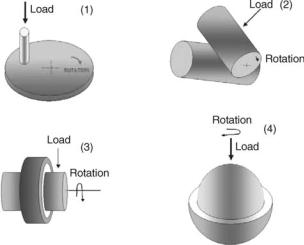
Figure 4. Schematic illustration of geometries in which wear phenomena are likely to occur: (1) pin-on-disk; (2) crossed cylinder;
(3) journal bearing; and (4) ball-and-socket bearing (After Ref. 31.)
including test lubricants, test duration, sliding velocity, contact area, alignment, and vibration.
Wear studies fall under three broad categories: (a) screening studies that involve testing of materials with simple geometries under well-controlled conditions; (b) simulator studies that involve testing of partial or complete prostheses; and (c) in vivo and retrieval studies of complete implanted devices. Screening studies may provide a basis for comparing novel materials against established materials; however, they can only provide estimations for wear of medical device components. Screening studies involve four general types of geometries: (1) pin-on-disk; (2) crossed cylinder; (3) journal bearing; and (4) spherical or ball and socket bearing. Geometries (3) and (4) are most similar to those encountered in orthopedic protheses (Fig. 4) (35). Simulator studies can be used to assess biomaterials and compare the wear characteristics of materials within a medical device. Various design and material combinations can be examined prior to animal studies and the clinical trials. Clinical assessments and implant retrieval studies also provide useful information for improving biomaterials, medical device design, and manufacturing protocols.
Much of current biotribology research focuses on the relationship between wear performance and biomaterial properties, including composition, processing, and finishing. However, other parameters have significant impact on the wear performance of the prostheses, including surgical and patient factors. The wear performance of polymer, metal, and ceramic biomaterials is discussed below.
Ultrahigh molecular weight polyethylene is commonly used in load-bearing components of total joint prostheses (38–45). The use of a polyethylene/cobalt–chromium wear couple in orthopedic prosthesis was first advocated by Charnley. In many contemporary total hip prosthesis designs, an ultrahigh molecular weight polyethylene acetabular cup slides against a cobalt–chromium alloy femoral ball. Significant numbers of submicron-sized ultrahigh
molecular weight polyethylene wear particles are commonly released from these prostheses with each movement of the joint. These particles may remain in the synovial fluid that serves to lubricate the joint (and contribute to third-body wear), embed in prosthesis surfaces, or enter lymphatic circulation. Immune cells (e.g., macrophages) may identify these particles as foreign materials and initiate an inflammatory response, which can lead to rapid bone loss (osteolysis), prosthesis loosening, or bone fracture (39). The volume and size of wear particles are critical factors that affect macrophage activation (40). These biological and physical effects of ultra-high molecular weight polyethylene wear particles are presently the leading cause of long term failure for metal-on-polyethylene hip prostheses (41,42).
Several mechanisms have been proposed to describe wear of ultrahigh molecular weight polyethylene prosthesis components. The wear mechanism of ultrahigh molecular weight polyethylene in hip prostheses has been described by Jasty et al. (43). They found that ultrahigh molecular weight polyethylene surfaces of retrieved implants contained numerous elongated fibrils, which were indicative of large strain deformation. This plastic deformation resulted from strain hardening of the material in the sliding direction and weakening of the material in the transverse direction. Once strain deformation of the surface has occurred, the surface will fragment during the relative motion, and micronand submicron-sized wear particles will be released. Subsurface cracking, pitting, and delamination caused by oxidative embrittlement and subsurface stresses are responsible for wear of ultra-high molecular weight polyethylene tibial knee inserts.
The wear resistance of ultrahigh molecular weight polyethylene can be improved by reducing the plastic-strain deformation and increasing the oxidization stability (30). The large-strain plastic deformation of ultrahigh molecular weight polyethylene can be diminished by increasing the number of covalent bonds between the long molecular chains of the polymer, which reduces the mobility of the polymer chain and minimizes the creep of the polymer. This process can be achieved by chemical methods (e.g., silane reactions) or, more commonly, by exposing polyethylene to ionizing radiation (46–50). Gamma-ray, e-beam, or X-ray radiation is used to cleave C¼C and C¼H bonds in polyethylene, which leads to the formation of species with unpaired electrons (free radicals). If the carboncarbon bond is cleaved (chain scission), the polymer molecular weight is reduced. Cross-linking can occur if free radicals from separate chains react with one another, and form an inter-chain covalent bond. If cross-linking occurs as a result of recombination by two radicals cleaved from C¼H bonds, it is referred to as an H-type cross-link. If one of the free radicals comes from the cleavage of the C¼C bond, it is referred to as a Y-type cross-link. The Y-type cross-linking process can increase the extent of polymer side chain branching (51). The yield of cross-linking processes has been estimated to be three times greater than the yield of chain scission processes for radiation/ultrahigh molecular weight polyethylene interaction. Cross-linking is most significant in amorphous regions of ultrahigh molecular weight polyethylene. An 83% reduction in wear rate has
been reported for ultrahigh molecular weight polyethylene surfaces treated with 5 Mrad radiation (38).
However, not all of the free radicals recombine with other free radicals. In crystalline regions, where the spatial separation between free radicals is large, the residual free radicals become trapped. These species are often confined to the crystalline-amorphous interfaces (52,53). Residual free radicals can cause long-term embrittlement through a series of complex cascade reactions. The residual free radicals first react with oxygen, leading to the formation of oxygen-centered radicals. The oxygen-centered radicals can take a hydrogen atom from a nearby chain to form a hydroperoxide species and another free radical on a chain. This additional free radical can repeat the process by generating another hydroperoxide and forming another free radical on a chain. These unstable species may decay into carbonyl species after exposure to high temperatures or after long periods of time, resulting in lower molecular weights and recrystallization. These processes result in increased stiffness, which is highly undesirable for biotribological applications.
Significant research has been done on reducing the concentration of residual free radicals and limiting the brittleness of irradiated ultrahigh molecular weight polyethylene. One cross-linking postprocessing treatment involved annealing the polymer above its melting transition, which allowed the residual free radicals to be removed through recombination reactions. The polymer recrystallized on cooling; however, the covalent bonds obtained during cross-linking were maintained. Unfortunately, the ultrahigh molecular weight polyethylene exhibited slightly lower crystallinity after this treatment. Another treatment involved annealing the cross-linked polymer at a temperature below the peak melting transition. One advantage of this technique is that a greater degree of crystallinity is retained; however, only a partial reduction in the number of residual free radicals is achieved. Other treatments for residual free radicals include irradiation at room temperature followed by annealing at temperatures below the melting transition; irradiation at room temperature with gamma or electron beams followed by melting; or irradiation at high temperatures followed by melting (34).
The physical properties of the ultrahigh molecular weight polyethylene can be significantly altered by crosslinking and annealing treatments. The effect of these treatments is dependent on the cross-linking parameters (e.g., technique, radiation source, dose, temperature during irradiation) and the annealing parameters (e.g., annealing temperature, annealing time). For example, the ultimate elongation (<45%) and the work to failure for ultrahigh molecular weight polyethylene are reduced as the radiation dose level is increased. Large radiation doses also reduce the yield strength (<30%) and the modulus (<27%) of ultrahigh molecular weight polyethylene (34). In addition, toughness decreases as the radiation dose level is increased, since the energy absorption before failure decreases as the chain mobility is reduced (54).
One alternative to the use of ultrahigh molecular weight polyethylene involves the use of so-called metal-on-metal prostheses, which contain two metallic load-bearing components. The primary motivation for use of these implants
BIOMATERIALS CORROSION AND WEAR OF |
317 |
is friction; metal-on-metal bearings generate less frictional torque during simulated gait than metal-on-polyethylene bearings (55–59). A stainless steel metal-on-metal hip prosthesis design was attempted by Wiles in 1938. Cobalt–chromium alloy/cobalt–chromium alloy prostheses designs were later developed by McKee and Watson-Ferrar (55,56). Although many of these early cobalt–chromium alloy metal-on-metal hip prostheses failed relatively soon after implantation, others have remained in place for >20 years (35). These first-generation metal-on-metal prostheses were displaced by ultrahigh molecular weight poly- ethylene/cobalt-chromium alloy prostheses in the 1970s for several reasons, including seizure of the cast metal surfaces (56). In the 1980s, second generation cobalt– chromium alloy/cobalt–chromium alloy prostheses were developed, which have again attracted interest from biomaterials researchers and prosthesis manufacturers (58). Earlier problems with seizing have been minimized through the use of wrought alloys, which are prepared using a thermal-mechanical forming process. Scholes et al. recently demonstrated using a hip simulator system that the mode of lubrication in metal-on-metal hip prostheses is strongly influenced by the diameter of the femoral head and diameter clearance (42). In small diameter joints, the wear rate increased as the diameter of the femoral head was increased. These results were attributed to the development of mixed lubrication in this system.
Alumina and zirconia have also been considered for use in orthopedic prostheses. Alumina exhibits very high hardness and elastic modulus values of 1900 kgf mm 2 (Vickers hardness) and 380 GPa, respectively (60). This material is polished to provide an extremely smooth finish; surface roughness values <0.005 mm are routinely obtained. In addition, alumina surfaces are hydrophilic and may provide prostheses with full film lubrication (35). Fracture toughness and wear resistance can be improved by lowering grain size, increasing grain uniformity, increasing purity, and lowering porosity.
Alumina prostheses have demonstrated wear rates <1 mm million 1 cycles during simulator testing (35). In addition, the in vivo wear rate for early alumina-on- alumina hip prostheses was shown to be as low as 1 mm year (61). However, retrieval studies involving early alumina-on-alumina hip prostheses found high rates of wear on some prostheses. Microseparation of the head and cup was shown to be responsible for this in vivo wear behavior (62). Insley et al. examined alumina-on-alumina prostheses with a laboratory simulator, and found that many very small ( 40 nm) and some large (100–3000 nm) particles were generated under microseparation conditions (35). Zirconia is harder than alumina, and is used to fabricate smaller components that can withstand higher stresses. Deformation-induced phase transformation has a significant effect on the mechanical properties of zirconia (63). The crystalline phase of a pure zirconium changes from monoclinic to tetragonal during deformation, which is accompanied by volume expansion of 3–4%. The addition of either yttrium oxide (Y2O3) or magnesium oxide (MgO) stabilizes the tetragonal phase at room temperature. However, aging can cause zirconia to return the more stable monoclinic phase, and can limit the lifespan of zirconia
318 BIOMATERIALS CORROSION AND WEAR OF
prostheses (64,65). In addition, Sato et al. showed that the tetragonal-monoclinic transformation on the surface of zirconia prostheses can be promoted by the presence of water molecules in the environment (66). The resulting volume change can lead to the generation of surface microcracks and an increase in surface roughness. The failure of some zirconia prostheses has been attributed to this microcracking process. Finally, in vivo fracture of some zirconia prostheses has been attributed to variations in sintering.
Thermal oxidation of zirconium alloys has also been used to create biocompatible, corrosionand wear-resistant surfaces for orthopedic prostheses (67). Wrought zirconium- 2.65 weight % niobium alloy contains a two-phase microstructure, which consists of elongated hexagonal alphazirconium grains that are bordered by cubic beta-zirconium grains. This material is oxidized for up to 8 h in air at temperatures near 620 8C (the eutectoid temperature). The resulting 5 mm thick monoclinic ZrO2 surface contains 40 nm wide 200 nm long grains that are arranged in a brickwork pattern, which is resilient to grain pull-out and lateral fracture. At the interface between the alloy and the surface oxide, regions of unoxidized niobium in beta-zirco- nium second-phase grains continue from the alloy into the oxide and serve to anchor the oxide to the alloy. The outermost portion of the oxide surface is burnished to create a smooth bearing surface. The oxide surface provides excellent wear behavior against polyethylene components, with reduced wear particle generation and inflammation.
Diamond-like carbon coatings on orthopedic prostheses can exhibit a wide range of elastic modulus and hardness values, which can be correlated with the fraction of sp3- hybridized atoms within the coating (68–71). Collins et al. developed a relation between sp3 fraction and Vickers hardness values for hydrogen-free diamond-like carbon coatings. They found that an sp3 fraction of 10% corresponded to a hardness value of 2000–3000 Hv, an sp3 fraction of 50% corresponded to a hardness value of 7000-8000 Hv, and an sp3 fraction of 100% corresponded to a hardness value of 10,000 Hv (72). Schneider et al. found that hydrogen-free and diamond-like carbon films with sp3 fractions between 0 and 90% provided elastic modulus values between 300 and 800 GPa. In contrast, typical hardness and elastic modulus values for hydrogenated diamond-like carbon films are <17 and <200 GPa, respectively (73).
The coefficients of friction values for diamond-like carbon coatings depend on ambient humidity, topology, and sliding partner (74). The most important parameter determining the coefficient of friction for hydrogenated dia- mond-like carbon thin films is relative humidity. Friction values for hydrogenated diamond-like carbon films can be as low as 0.01–0.3 in vacuum conditions, but greatly increase under humid conditions due to incomplete formation of the graphitic transfer surface. This variation in coefficient of friction values can be correlated with hydrogen/carbon ratio in the precursor material. As the hydrogen content in the precursor material increases, the friction coefficient demonstrates a greater positive correlation with ambient humidity. For example, hydrogenated diamond-like carbon films produced from hydrogen-diluted methane demonstrate lower friction coef-
ficients under high humidity conditions than other hydrogenated diamond-like carbon films. On the other hand, hydrogen-free diamond-like carbon thin films maintain low friction coefficients (<0.1) under low and high humidity conditions (75,76).
The combination of high hardness and low coefficient of friction values allows diamond-like carbon coatings to provide significant wear protection to a bulk implant material (71). Hirvonen et al. found that the wear resistance of diamond-like carbon coatings is superior to that of silicon carbide, tungsten carbide–cobalt, silicon nitride, or alumina by factors of 40, 60, 230, and 290, respectively (77). Hydrogen-free diamond-like carbon thin films exhibit wear rates of 10 9 mm3 N 1 m 1, these values are 100 times lower than those for hydrogenated diamond-like carbon thin films (10 7 mm3 N 1 m 1) under similar testing conditions (78). Many diamond-like carbon substrate materials are significantly softer than the diamond-like carbon coatings; high contact pressures can initiate substrate deformation and coating failure. Nitriding processes can harden the substrate surface, reduce subsurface deformation, and extend diamond-like carbon coating lifetimes (79).
The friction and wear properties of diamond-like carbon-coated metal hip prostheses against diamond-like carbon-coated polyethylene cups have been determined using both screening and simulator techniques (80). For example, Tiainen et al. demonstrated extremely low coefficients of friction for prostheses coated with hydrogen-free diamond-like carbon using a pulsed arc discharge method. They demonstrated coefficients of friction for diamond-like carbon/diamond-like carbon and metal–metal pairs of 0.05 and 0.14, respectively. In addition, they found that wear rate in the diamond-like carbon-coated metal/diamond-like carbon-coated metal wear couple was 105–106 times lower than that observed in conventional metal–polyethylene and metal–metal wear couples. They also observed that wear of polyethylene in the diamond-like carbon-coated metal/ultrahigh molecular weight polyethylene wear couple was 10–600 times lower than that observed in conventional metal/ultrahigh molecular weight polyethylene wear couples. On the other hand, other investigators have found little difference in wear rates between diamond-like carbon-coated prosthesis materials and conventional prosthesis materials. For example, Sheeja et al. found little difference in wear rates between cobalt-chromium–molyb- denum alloy/ultrahigh molecular weight polyethylene and multilayered diamond-like carbon-coated cobalt– chromium–molybdenum alloy/ultrahigh molecular weight polyethylene wear couples (81,82). The seemingly contradictory results suggest other factors, such as use of lubricant, may play a significant role in determining wear rates (83–85). For example, physiologic lubricants may not allow graphitic layers to form on the surfaces of the test materials. In addition, diamond-like carbon coatings that contain particulates and pits may increase adhesive wear (86).
Adhesion of diamond-like carbon coatings is dependent on several factors, including film stress, film–substrate chemical bonding, and substrate topology (87,88). Large internal compressive stresses (as high as 10 GPa) are typically observed in
diamond-like carbon coatings. These stresses limit maximum diamond-like carbon coating thickness to 0.1–0.2 mm and prevent widespread medical use. Lifshitz et al. attributed these stresses to subplantation (low energy subsurface implantation) of carbon ions during coating formation (89). They suggested that carbon ions with energies between 10 and 1000 eV undergo shallow implantation to depths of 1–10 nm during growth of diamond-like carbon coatings. Carbon species are trapped in subsurface sites due to restricted mobility, leading to the development of very large internal compressive stresses.
BIOMATERIALS CORROSION AND WEAR OF |
319 |
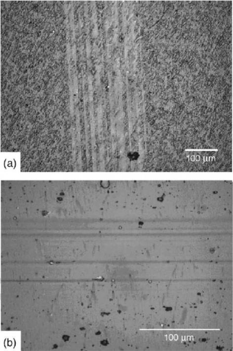
Diamond-like carbon can be alloyed with metals in order to reduce internal compressive stresses and promote specific biological responses (90,91). Diamond-like carbon– metal composite coatings retain hardness and wear properties similar to those of unalloyed diamond-like carbon films, and exhibit excellent adhesion to metal alloy substrates (Fig. 5). The metal component can provide additional biological functionality to the implant surface; for example, silver has been shown to possess a wide antimicrobial spectrum against a broad range of Gramnegative bacteria (including Pseudomonas aeruginosa),
Figure 5. Wear testing of diamondlike carbon–metal composite coatings using a linear tribometer (screening wear test). (a) Wear track of functionally gradient diamondlike carbon–silver composite film after 10,000 cycles, (b) Wear track of functionally gradient diamondlike carbon–titanium composite film after 10,000 cycles.
320 BIOMATERIALS CORROSION AND WEAR OF
Gram-positive bacteria (including methicillin-resistant Staphylococcus aureus), fungi, viruses, and yeasts. Films containing both silver and platinum may demonstrate enhanced antimicrobial activity due to formation of a galvanic couple that accelerates silver ion release. Narayan et al. showed that diamond-like carbon–silver–platinum nanocomposite films reduce bacterial colonization rates by 90% compared to uncoated silicon substrates (92).
Corrosion and wear are critical parameters that affect overall success of a biomaterial or a medical device design. The complicated interaction between material-, device-, surgical-, patient-specific parameters has made it difficult to predict clinical behavior of implanted medical devices. In addition, the large variation in measurement techniques for corrosion and wear has led problems in interpreting and comparing the work performed in the biomaterials community. If these issues can be successfully resolved, significant advances in these areas may be achieved in the coming years.
BIBLIOGRAPHY
Cited References
1.Burke DR. The Composition and Function of Body Fluids, 3rd ed. St. Louis: C. V. Mosby; 1980.
2.Lentner C, editor. Geigy Scientific Tables, Vol. 1: Units of Measurement, Body Fluids, Composition of the Body, Nutrition. Basel: CIBA-Geigy; 1981.
3.Lentner C, editor. Geigy Scientific Tables, Vol. 3: Physical Chemistry, Composition of Blood, Hematology, Somatometric Data. Basel: CIBA-Geigy; 1984.
4.ASTM, Annual Book of ASTM Standards. West Conshohocken (PA): ASTM. International; 1977.
5.Pilliar RM. Metals and irthopaedic implants — past successes, present limitations, future challenges. Shrivastava S, editor. Proceedings of the Materials & Processes for Medical Devices Conference. Materials Park (OH): ASM International; 2004.
6.Hoar TP, Mears DC. Corrosion-resistant alloys in chloride solutions: materials for surgical implants. Proc R Soc London 1966;294:486–510.
7.Sury P, Semlitsch M. Corrosion behavior of cast and forged cobalt-based alloys for double-alloy joint endoprostheses. J Biomed Mater Res 1978;12:723–741.
8.Steinemann S. Corrosion of surgical implants – in vivo and in vitro tests. In: Winder GD, editor. Evaluation of Biomaterials. Chichester: John Wiley & Sons Inc.; 1980.
9.Cahoon JR, Bandyopadhya R, Tennese L. The concept of protection potential applied to the corrosion of metallic orthopedic implants. J Biomed Mater Res 1975;9:259–264.
10.Bandy CR. Effects of composition on the electrochemical behavior of austenitic stainless steel in Ringer’s solution. Corrosion (Houston) 1977;33:204–208.
11.Scales JT, Winter GD, Shirley HT. Corrosion of orthopaedic implants. J Bone Joint Surg 1959;41B:810–820.
12.Merritt K, Brown SA. Release of hexavalent chromium from corrosion of stainless steel and cobalt-chromium alloys. J Biomed Mater Res 1995;29:627–633.
13.McKellop HA, Sarmiento A, Brien W, Park SH. Interface corrosion of a modular head total hip prosthesis. J Arthroplasty 1992;7:291–294.
14.Brown SA, et al. Fretting corrosion accelerates crevice corrosion of modular hip tapers. J Appl Biomater 1995;6:19–26.
15.Kawale JS, Brown SA, Payer JH, Merritt K. Mixed-metal fretting corrosion of Ti6Al4V and wrought cobalt alloy. J Biomed Mater Res 1995;29:867–873.
16.Metikos-Hukovic M, Kwokal A, Piljac J. The influence of niobium and vanadium on passivity of titanium-based implants in physiological solution. Biomaterials 2003;24: 3765–3775.
17.Cutright DE, et al. Tissue reaction to nitinol wire alloy. Oral Surg Oral Med Oral Pathol 1973;35:578–584.
18.Kapanen A, Ryha¨nen J, Danilov A, Tuukkanen J. Effect of nickel-titanium shape memory metal alloy on bone formation. Biomaterials 2001;22:2475–2480.
19.Ryha¨nen J, et al. Bone healing and mineralization, implant corrosion, and trace metals after nickel-titanium shape memory metal intramedullary fixation. J Biomed Mater Res 1999;47:472–480.
20.Villermaux F, et al. Corrosion resistance improvement of NiTi osteosynthesis staples by plasma polymerized tetrafluorethylene coating. Biomed Mater Eng 1996;6:241–254.
21.Rondelli G, Vincentini B. Localized corrosion behavior in human body fluids of commercial NiTi orthodontic wires. Biomaterials 1999;20:785–792.
22.Carroll W, Kelly M, Brien B. Corrosion behavior of Nitinol wires in body fluid environment. Int Conf Shape Memory Superelastic Technol 1999; 240–249.
23.Montero-Ocampo C, Lopez H, Salinas RA. Effect of compressive straining on corrosion resistance of a shape memory NiTi alloy in Ringer’s solution. J Biomed Mater Res 1996;32: 583–591.
24.Shabalovskaya SA. Surface, corrosion and biocompatibility aspects of Nitinol as an implant material. Bio-Med Mater Eng 2002;12:69–109.
25.Niinomi M. Recent research and development in titanium alloys for biomedical applications and healthcare goods. Sci Technol Adv Mat 2003;4:445–454.
26.Senda T, Yasuda E, Kaji M, Bradt RC. Effect of Grain Size on the Sliding Wear and Friction of Alumina at Elevated Temperatures. J Am Ceram Soc 1999;82:1505–1511.
27.Dogan CP, Hawk JA. Role of composition and microstructure in the abrasive wear of high-alumina ceramics. Wear 1999; 225:1050–1058.
28.Rodr´ıguez J, et al. Sliding wear of alumina/silicon carbide nanocomposites. J Am Ceram Soc 1999;55:2252–2306.
29.Webster TJ, Siegel RW, Bizios R. Osteoblast adhesion on nanophase ceramics. Biomaterials 1999;20:1221–1227.
30.Morsi K, Keshavan H, Bal S. Hot pressing of graded ultrafinegrained alumina bioceramics. Mater Sci Eng A 2004;386:384– 389.
31.Kingery WD. Introduction to Ceramics. New York: John Wiley & Sons Inc.; 1976.
32.Tiainen VM. Amorphous Carbon as a Bio-mechanical coat- ing-mechanical properties and biological applications. Diamond Related Mater 2001;10:153–160.
33.Hutchings IM. Biotribology-A Personal View. In: Hutchings IM, editor. Friction, Lubrication and Wear of Artificial Joints. Bury St. Edmunds (UK): Professional Engineering Publishing Ltd.; 2003.
34.Wright TM, Goodman SB, editors. Implant Wear in Total Joint Replacement: Clinical and Biologic Issues, Material and Design Considerations. Rosemont (IL): American Academy of Orthopaedic Surgeons; 2001.
35.Hutchings IM, editor. Friction, Lubrication and Wear of Artificial Joints. Bury St. Edmunds (UK): Professional Engineering Publishing Ltd; 2003.
36.Schmalzreid TP, Callaghn JJ. Current concepts review: Wear in total hip and knee replacement. J Bone Joint Surg Am 1999;81:115–136.
37.Clarke IC, McKellop HA. Wear Testing. In: von Recum AF, editor. Handbook of biomaterials evaluation: scientific, technical, and clinical testing of implant materials. New York: Macmillan; 1986.
38.McKellop H, Shen FW, DiMaio W, Lancaster JG. Wear of gamma-crosslinked polyethylene acetabular cups against roughened femoral balls. Clin Orthop 1999;369:73–82.
39.Unsworth A, Dowson D, Wright V. Some new evidence on human joint lubrication. Ann Rheum Dis 1975;34:277–285.
40.Green TR, et al. Effect of size and dose on bone resorption activity of macrophages by in vitro clinically relevant ultra high molecular weight polyethylene particles. J Biomed Mater Res 2000;B53:490–497.
41.Ingham E, Fisher J. Biological reactions to wear debris in total joint replacement. Proc Inst Mech Eng H 2000;214:21–37.
42.Scholes C, Unsworth A. Comparison of friction and lubrication of different hip prostheses. Proc Inst Mech Eng H 2000;214:49–57.
43.Jasty MJ, et al. Wear of polyethyene acetabular components in total hip arthroplasty: an analysis of 128 components retrieved at autopsy or revision operation. J Bone Joint Surg Am 1977;79:349–358.
44.Charnley JC. Arthroplasty of the hip: a new operation. Lancet 1961;280:1129–1132.
45.Charnley JC. Tissue reaction to polytetrafluorethylene. Lancet 1963;285:1379.
46.Collier JP, et al. Overview of polyethylene as a bearing material: comparison of sterilization methods. Clin Orthop Rel Res 1996;333:76–86.
47.Wang A, Sun DC, Stark C, Dumbleton JH. Wear Mechanisms of UHMWPE in total joint replacements. Wear 1998;181– 183:241–249.
48.Collier JP, et al. Results of implant retrieval from postmortem specimens in patients with well-function, long term total hip replacement. Clin Orthop 1992;274:97–112.
49.Cameron HU. Tibial component wear in total knee replacement. Clin Orthop 1994;309:29–32.
50.Atkinson JR, Cicek RZ. Silane crosslinked polyethylene for prosthetic applications: Part II. Creep and wear behavior and a preliminary molding test. Biomaterials 1984; 5:326–335.
51.Muratoglu OK, et al. Larger diameter femoral heads used in conjunciton with a highly cross-linked ultra high molecular weight polyethylene: A New Concept. J Arthroplasty 2001;16: 24–30.
52.Pearson RW. Mechanism of the radiation crosslinking of polyethylene. J Polym Sci 1957;25:189–200.
53.Zhu QR, Horii F, Kitamaru R, Yamaoka H. C-13-NMR study of cross-linking and long-chain branching in linear polyethylene induced by Co-60 gamma-ray irradiation at different temperatures. J Polym Sci, Part A: Polym Chem 1990;28: 2741–2751.
54.Premnath V, Harris WH, Jasty M, Merrill EW. Gamma sterilization of UHMWPE articular implants: an analysis of the oxidation problem. Biomaterials 1996;17:1741–1753.
55.Steinberg DR, Steinberg ME. The early history of arthroplasty in the United States. Clin Orthop Relat Res 2000;374:55–89.
56.McKee GK, Watson-Farrar, J. Replacement of arthritic hip by the McKee-Farrar prostheses. J Bone Joint Surg 1966;48B: 245–259.
57.Wimmer MA, et al. The acting wear mechanisms on metal- on-metal hip joint bearings: in vitro results. Wear 2001; 250:129–139.
58.Dorr LD, et al. Total hip arthroplasty with use of the metasul metal-on-metal articulation: 4–7-year results. J Bone Joint Surg Am 2000;82:789–798.
BIOMATERIALS CORROSION AND WEAR OF |
321 |
59.Poggie RA, Afflitto R, St John K. The wear performance of precision Co–Cr–Mo alloy metal-on-metal hip bearings. Proc Conf Trans 12th Ann Int Symp Technol Arthroplasty 1999;11: 1–2.
60.Boutin P, et al. The use of dense aluminaalumina ceramic combination in total hip replacement. M J Biomed Mater Res 1988;22:1203–1232.
61.Willmann G. Development in medical-grade alumina during the past two decades. J Mater Process Technol 1996;56:168–176.
62.Nevelos J, et al. Microseparation of the centers of aluminaalumna artificial hip joints during simulator testing produces clinically relevant wear and patterns. J Arthroplasty 2000;15: 793–795.
63.Christel P, et al. Mechanical properties and short-term In vivo evaluation of yttrium-oxide-partially-stabilized zirconia. J Biomed Mater Res 1989;23:45–61.
64.Chevalier J, et al. Critical effect of cubic phase on aging in 3 mol% yttria-stabilized zirconia ceramics for hip replacement prosthesis. Biomaterials 2004;25:5539–5545.
65.Gremillard L, et al. Modeling the aging kinetics of zirconia ceramics. J Eur Ceram Soc 2004;24:3483–3489.
66.Sato T, Ohtaki S, Endo T, Shimada M. Science and technology of zirconia. Advances in Ceramics. Westerville (OH): American Ceramic Society; 1988.
67.Hobbs LW, et al. Oxidation microstructures and interfaces in the oxidized zirconium knee. Int J Appl Ceram Technol 2005;2:221–246.
68.Enke K, Dimigen H, Hubsch H. Frictional properties of diamondlike carbon layers. Appl Phys Lett 1980;36:291–292.
69.Pharr GM, et al. Hardness, elastic modulus, and structure of very hard carbon films produced by cathodic-arc deposition with substrate pulse-biasing. Appl Phys Lett 1996;68:779– 781.
70.Ronkainen H, Varjus S, Koskinen J, Holmberg K. Differentiating the tribological performance of hydrogenated and hydrogen-free DLC coatings. Wear 2001;249:260–266.
71.Holmberg K, Mathews A. Coatings tribology: a concept, critical aspects, and future directions. Thin Solid Films 1994;253:173–178.
72.Collins CB, et al. Noncrystalline films with the chemistry, bonding, and properties of diamond. J Vac Sci Technol B 1993;11:1936–1941.
73.Schneider D, Schwarz T, Scheibe HJ, Panzner M. Nondestructive evaluation of diamond and diamond-like carbon films by laser induced surface acoustic waves. Thin Solid Films 1997;295:107–116.
74.Erdemir A, Bindal C, Pagan J, Wilbur P. Characterization of Transfer layers on Surfaces Sliding Against Diamond-like Hydrocarbon Films in Dry Nitrogen. Surf Coatings Technol 1995;76–77:559–563.
75.Liu Y, Erdemir A, Meletis EI. An investigation of the relationship between graphitization and frictional behavior of DLC coatings. Surface Coatings Technol 1996;86:564–568.
76.Oguri K, Arai T. Friction mechanisms of Diamond-like carbon with silicon coatings formed by plasma-assisted chemical vapor-deposition. J Mater Res 1992;7:1313–1316.
77.Hirvonen JP, Koskinen J, Lappalainen R, Anttila A. Preparation and properties of high density hydrogen free hard carbon films with direct ion beam or arc discharge deposition. Mater Sci Forum 1990;52:197.
78.Voevodin AA, Donley MS, Zabinski JS, Bultman JE. Mechanical and tribological properties of diamond-like carbon coatings prepared by pulsed laser deposition. Surface Coatings Technol 1995;77:534–539.
79.Voevodin AA, Phelps AW, Zabinski JS, Donley MS. Friction induced phase transformation of pulsed laser deposited diamondlike carbon. Diamond Related Mater 1996;5:1264–1269.