
- •VOLUME 1
- •CONTRIBUTOR LIST
- •PREFACE
- •LIST OF ARTICLES
- •ABBREVIATIONS AND ACRONYMS
- •CONVERSION FACTORS AND UNIT SYMBOLS
- •ABLATION.
- •ABSORBABLE BIOMATERIALS.
- •ACRYLIC BONE CEMENT.
- •ACTINOTHERAPY.
- •ADOPTIVE IMMUNOTHERAPY.
- •AFFINITY CHROMATOGRAPHY.
- •ALLOYS, SHAPE MEMORY
- •AMBULATORY MONITORING
- •ANALYTICAL METHODS, AUTOMATED
- •ANALYZER, OXYGEN.
- •ANESTHESIA MACHINES
- •ANESTHESIA MONITORING.
- •ANESTHESIA, COMPUTERS IN
- •ANGER CAMERA
- •ANGIOPLASTY.
- •ANORECTAL MANOMETRY
- •ANTIBODIES, MONOCLONAL.
- •APNEA DETECTION.
- •ARRHYTHMIA, TREATMENT.
- •ARRHYTHMIA ANALYSIS, AUTOMATED
- •ARTERIAL TONOMETRY.
- •ARTIFICIAL BLOOD.
- •ARTIFICIAL HEART.
- •ARTIFICIAL HEART VALVE.
- •ARTIFICIAL HIP JOINTS.
- •ARTIFICIAL LARYNX.
- •ARTIFICIAL PANCREAS.
- •ARTERIES, ELASTIC PROPERTIES OF
- •ASSISTIVE DEVICES FOR THE DISABLED.
- •ATOMIC ABSORPTION SPECTROMETRY.
- •AUDIOMETRY
- •BACTERIAL DETECTION SYSTEMS.
- •BALLOON PUMP.
- •BANKED BLOOD.
- •BAROTRAUMA.
- •BARRIER CONTRACEPTIVE DEVICES.
- •BIOCERAMICS.
- •BIOCOMPATIBILITY OF MATERIALS
- •BIOELECTRODES
- •BIOFEEDBACK
- •BIOHEAT TRANSFER
- •BIOIMPEDANCE IN CARDIOVASCULAR MEDICINE
- •BIOINFORMATICS
- •BIOLOGIC THERAPY.
- •BIOMAGNETISM
- •BIOMATERIALS, ABSORBABLE
- •BIOMATERIALS: AN OVERVIEW
- •BIOMATERIALS: BIOCERAMICS
- •BIOMATERIALS: CARBON
- •BIOMATERIALS CORROSION AND WEAR OF
- •BIOMATERIALS FOR DENTISTRY
- •BIOMATERIALS, POLYMERS
- •BIOMATERIALS, SURFACE PROPERTIES OF
- •BIOMATERIALS, TESTING AND STRUCTURAL PROPERTIES OF
- •BIOMATERIALS: TISSUE-ENGINEERING AND SCAFFOLDS
- •BIOMECHANICS OF EXERCISE FITNESS
- •BIOMECHANICS OF JOINTS.
- •BIOMECHANICS OF SCOLIOSIS.
- •BIOMECHANICS OF SKIN.
- •BIOMECHANICS OF THE HUMAN SPINE.
- •BIOMECHANICS OF TOOTH AND JAW.
- •BIOMEDICAL ENGINEERING EDUCATION
- •BIOSURFACE ENGINEERING
- •BIOSENSORS.
- •BIOTELEMETRY
- •BIRTH CONTROL.
- •BLEEDING, GASTROINTESTINAL.
- •BLADDER DYSFUNCTION, NEUROSTIMULATION OF
- •BLIND AND VISUALLY IMPAIRED, ASSISTIVE TECHNOLOGY FOR
- •BLOOD BANKING.
- •BLOOD CELL COUNTERS.
- •BLOOD COLLECTION AND PROCESSING
- •BLOOD FLOW.
- •BLOOD GAS MEASUREMENTS
- •BLOOD PRESSURE MEASUREMENT
- •BLOOD PRESSURE, AUTOMATIC CONTROL OF
- •BLOOD RHEOLOGY
- •BLOOD, ARTIFICIAL
- •BONDING, ENAMEL.
- •BONE AND TEETH, PROPERTIES OF
- •BONE CEMENT, ACRYLIC
- •BONE DENSITY MEASUREMENT
- •BORON NEUTRON CAPTURE THERAPY
- •BRACHYTHERAPY, HIGH DOSAGE RATE
- •BRACHYTHERAPY, INTRAVASCULAR
- •BRAIN ELECTRICAL ACTIVITY.
- •BURN WOUND COVERINGS.
- •BYPASS, CORONARY.
- •BYPASS, CARDIOPULMONARY.
216BIOINFORMATICS
109.Gersing E. Impedance spectroscopy on living tissue for determination of the state of organs. Bioelectrochem Bioenerget 1998;45:145–149.
110.Owens L, et al. Correlation of ischemia-induced extracellular and intracellular ion changes to cell-to-cell electrical uncoupling in isolated blood-perfused rabbit hearts. Circulation 1996;94:10–13.
111.Schafer M, Gebhard Gersing E. Characterization of organ tissue during the transition between life and death: Cardiac and skeletal muscle. Med Biol Eng Comput 1999;37:100– 101.
112.Dzwonczyk R, et al. Myocardial electrical impedance responds to ischemia and reperfusion in humans. IEEE Trans Biomed Eng 2004;51(12):2206–2209.
113.Howie M, Dzwonczyk R, McSweeney T. An evaluation of a new two-electrode myocardial electrical impedance monitor for detecting myocardial ischemia. Anesthesia Analgesia 2001;92:12–18.
114.Sezer M, et al. New support for clarifying the relation between ST segment resolution and microvascular function: Degree of ST segment resolution correlates with the pressure derived collateral flow index. Heart 2004;90: 146–150.
115.Leung J, et al. Automated electrocardiograph ST segment trending monitors: Accuracy in detecting myocardial ischemia. Anesthesia Analgesia 1998;87:4–10.
116.Leung J, et al. Electrocardiographic ST-segment changes during acute, severe isovolemic hemodilution in humans. Anesthesiology 2000;93:1004–1010.
117.Katz A. Physiology of the Heart. 2nd ed. New York: Raven Press; 1992. p 609–637.
118.Garrido H, et al. Bioelectrical tissue resistance during various methods of myocardial preservation. Ann Thorac Surg 1983;36:143–151.
119.Gebhard M, et al. Impedance spectroscopy: A method for surveillance of ischemia tolerance of the heart. Thorac Cardiovasc Surg 1987;35:26–32.
120.Mueller J, et al. Electric impedance recording: A noninvasive method of rejection diagnosis. J Extra Corpor Technol 1992;23:49–55.
121.Matthie J, et al. Development of commercial complex bioimpedance spectroscopic system for determining intracellular and extracellular water volumes. Proceedings of the 8th International Conference on Electrical Bioimpedance, Kupio, Finland, 1992. p 203–205.
122.Van Loan M, et al. Use of bioimpednace spectroscopy to determine extracellular fluid, intracellular fluid, total body water, and fat-free mass. In: Human body composition: In vivo methods, models and assessment. New York: Plenum; 1993. p 67–70.
123.Van Marken Lichtenbelt W, et al. Validation of bioelectric impedance measurements as a method to estimate body water compartments. Am J Clin Nutr 1994;60: 159–166.
124.Van Loan M, et al. Fluid changes during pregnancy: Use of bioimpedance spectroscopy. J Appl Physiol 1995;27:152– 158.
125.Jaffrin M, et al. Continuous monitoring of plasma, interstitial, and intracellular fluid volumes in dialyzed patients by bioimpedance and hematocrit measurements. ASAIO J 2002;48:326–333.
126.Cox-Reijven P, et al. Role of bioimpedance spectroscopy in assessment of body water compartments in hemodialysis patients. Am J Kidney Dis 2001; 38(4):832–838.
127.Mancini A, et al. Nutritional status in hemodialysis patients and bioimpedance vector analysis. J Renal Nutrition 2003;13(3):199–204.
128.DeVries P, et al. Measurement of transcellular fluid shifts during hemodialysis. Med Biol Eng Comput 1989;27:152– 158.
129.Sinning W, et al. Monitoring hemodialysis with bioimpedance: What do we really measure? ASAIO J 1993;39:M584– M589.
130.Scanferla F, et al. On-line bioelectric impedance during hemodialysis: Monitoring of body fluids and cell membrane status. Nephrol Dial Transplant 1990; 5(Suppl 1):167– 170.
131.Jaffrin M, et al. Extracellular and intracellular fluid volume during dialysis by multifrequency impedancemetry. ASAIO J 1996;42:M533–M537.
132.Hanai T. Electrical properties of emulsions. In: Emulsions Science. London: Academic Press; 1968. p 354–477.
133.Foley K, et al. Use of single-frequency bioimpedance at 50 kHz to estimate total body water in patients with multiple organ failure and fluid overload. Crit Care Med 1999;27(8):1472–1477.
134.Ward L, Elia M, Cornish B. Potential errors in the application of mixture theory to multifrequency bioelectrical impedance analysis. Physiol Meas 1998;19:53–60.
135.Zhu F, et al. Estimation of body fluid changes during peritoneal dialysis by segmental bioimpedance analysis. Kidney Int 2000;57:299–306.
136.Patterson R, et al. Measurement of body fluid volume change using multisite impedance measurements. Med Biol Eng Comput 1988;26:33–37.
137.Raaijmakers E, et al. Estimation of non-cardiogenic pulmonary edema using dual-frequency electrical impedance. Med Biol Eng Comput 1998;36:461–466.
Further Reading
Cole KS, Cole RH. Dispersion and absorption in dielectrics. J Chem Phys 1941;9:341–351.
See also ELECTROCARDIOGRAPHY, COMPUTERS IN; EXERCISE STRESS TESTING; FLOWMETERS, ELECTROMAGNETIC; IMPEDANCE PLETHYSMOGRAPHY;
NEONATAL MONITORING; PHONOCARDIOGRAPHY.
BIOINFORMATICS
ALI ABBAS
LEI LIU
University of Illinois
Urbana, Illinois
INTRODUCTION
The past two decades have witnessed revolutionary changes in biomedical research and biotechnology and an explosive growth of biomedical data. High throughput technologies developed in automated DNA sequencing, functional genomics, proteomics, and metabolomics enable production of such high volume and complex data that the data analysis becomes a big challenge. Consequently, a promising new field, bioinformatics has emerged and is growing rapidly. Combining biological studies with computer science, mathematics, and statistics, bioinformatics develops methods, solutions, and software to discover patterns, generate models, and gain insight knowledge of complex biological systems.
BIOINFORMATICS 217
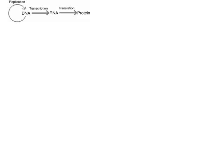
Figure 1. Central dogma of molecular biology.
Before bioinformatics is discussed further, a brief review of the basic concepts in molecular biology, which are the foundations for bioinformatics studies, is provided. The genetic information is coded in DNA sequences. The physical form of a gene is a fragment of DNA. A genome is the complete set of DNA sequences that encode all the genetic information for an organism, which is often organized into one or more chromosomes. The genetic information is decoded through complex molecular machinery inside a cell composed of two major parts, transcription and translation, to produce functional protein and RNA products. These molecular genetic processes can be summarized precisely by the central dogma shown in Fig. 1. The proteins and active RNA molecules combined with other large and small biochemical molecules, organic compounds, and inorganic compounds form the complex dynamic network systems that maintain the living status of a cell. Proteins form complex 3D structures that carry out functions. The 3D structure of a protein is determined by the primary protein sequence and the local environment. The protein sequence is decoded from the DNA sequence of a gene through the genetic codes as shown in Table 1. These codes have been shown to be universal among all living forms on earth.
The high throughput data can be generated at many different levels in the biological system. The genomics data are generated from the genome sequencing that deciphers the complete DNA sequences of all the genetic information in an organism. We can measure the mRNA levels using microarray technology to monitor the gene expression of all the genes in a genome known as transcriptome. Proteome is the complete set of proteins in a cell at a certain stage, which can be measured by high throughput 2D gel electrophoresis and mass spectrometry. We also can monitor all the metabolic compounds in a cell known as metabolome in a high throughput fashion. Many new terms ending with ‘‘ome’’ can be viewed as the complete set of entities in a cell. For example, the ‘‘interactome’’ refers to the complete set of protein-protein interactions in a cell.
Bioinformatics is needed at all levels of high throughput systematic studies to facilitate the data analysis, mining, management, and visualization. But more importantly, the major task is to integrate data from different levels and prior biological knowledge to achieve system-level understanding of biological phenomena. As bioinformatics touches on many areas of biological studies, it is impossible to cover every aspect in a short chapter. In this chapter, the authors will provide a general overview of the field and focus on several key areas, including sequence analysis, phylogenetic analysis, protein structure, genome analysis, microarray analysis, and network analysis.
Sequence analysis often refers to sequence alignment and pattern searching in DNA and protein sequences. This area can be considered classic bioinformatics, which can be dated back to 1960s, long before the word bioinformatics appeared. It deals with the problems such as how to make an optimal alignment between two sequences and how to
Table 1. The Genetic Code
|
|
Second Position |
|
|
|
|
|
|
|
|
|
|
|
|
|
|
|
|
|
|
|
|
|
First Position |
T |
C |
A |
G |
Third Position |
||
|
|
|
|
|
|
|
|
T |
TTT Phe [F] |
TCT Ser [S] |
TAT Tyr [Y] |
TGT Cys [C] |
|
T |
|
|
TTC Phe [F] |
TCC Ser [S] |
TAC Tyr [Y] |
TGC Cys [C] |
|
C |
|
|
TTA Leu [L] |
TCA Ser [S] |
TAA Stop[end] |
TGA Stop[end] |
|
A |
|
|
TTG Leu [L] |
TCG Ser [S] |
TAG Stop[end] |
TGG Trp [W] |
|
G |
|
C |
CTT Leu [L] |
CCT Pro [P] |
CAT His [H] |
CGT Arg [R] |
|
T |
|
|
CTC Leu |
CCC Pro [P] |
CAC His [H] |
CGC Arg [R] |
|
C |
|
|
CTA Leu [L] |
CCA Pro [P] |
CAA Gln [Q] |
CGA Arg [R] |
|
A |
|
|
CTG Leu [L] |
CCG Pro [P] |
CAG Gln [Q] |
CGG Arg [R] |
|
G |
|
A |
ATT Ile [I] |
ACT Thr [T] |
AAT Asn [N] |
AGT Ser [S] |
|
T |
|
|
ATC Ile [I] |
ACC Thr [T] |
AAC Asn [N] |
AGC Ser [S] |
|
C |
|
|
ATA Ile [I] |
ACA Thr [T] |
AAA Lys [K] |
AGA Arg [R] |
|
A |
|
|
ATG Met [M] |
ACG Thr [T] |
AAG Lys [K] |
AGG Arg [R] |
|
G |
|
G |
GTT Val [V] |
GCT Ala [A] |
GAT Asp [D] |
GGT Gly [G] |
|
T |
|
|
GTC Val [V] |
GCC Ala [A] |
GAC Asp [D] |
GGC Gly [G] |
|
C |
|
|
GTA Val [V] |
GCA Ala [A] |
GAA Glu [E] |
GGA Gly [G] |
|
A |
|
|
GTG Val [V] |
GCG Ala [A] |
GAG Glu [E] |
GGG Gly [G] |
|
G |
|
|
|
|
|
|
|
|
|
218 BIOINFORMATICS
(a) |
Sequence 1 |
C |
O |
U |
N |
T |
I |
N |
G |
|
|
Sequence 2 |
N |
T |
I |
G |
|
|
|
|
|
(b) |
Sequence 1 |
C |
O |
U |
N |
T |
I |
N |
G |
Possible Alignment |
|
Sequence 2 |
- |
- |
- |
N |
T |
I |
G |
- |
(Shifting Sequence 2) |
(c) |
Sequence 1 |
C |
O |
U |
N |
T |
I |
N |
G |
Possible Alignment |
|
Sequence 2 |
- |
- |
- |
N |
T |
- |
I |
G |
(Shifting Sequence 2 and inserting a gap) |
(d) |
Sequence 1 |
C |
O |
U |
N |
T |
I |
N |
G |
Possible Alignment |
|
Sequence 2 |
- |
- |
- |
N |
T |
I |
- |
G |
(Shifting Sequence 2 and inserting a gap) |
Figure 2. Possible alignments of two sequences.
search sequence databases quickly with an unknown sequence. Phylogenetic analysis is closely related to sequence alignment. The idea is to use DNA or protein sequence comparison to infer evolution history. The first step in this analysis is to perform multiple sequence alignment. Then, a phylogenetic tree is built based on the multiple alignments. The protein structure analysis involves the prediction of protein secondary and tertiary structures from the primary sequences. So far, the analyses focus on individual sequences or a handful of sequences. The next three areas are involved in systemwide analysis. Genome analysis mainly deals with the sequencing of a complete or partial genome. The problems include genome assembly, gene structure prediction, gene function annotation, and so on. Many techniques of sequence analysis are used in genome analysis, but many new methods were developed for the unique problems. Microarray technologies provide an opportunity for biologists to study the gene expression at a system level. The problems faced in the analysis are completely different from sequence analysis. Many statistical and data mining techniques are applied in the field. Network analysis is another system level study of the biological system. Biological networks can be divided into three categories: metabolic network, protein-protein interaction network, and genetic network. The questions in this area include network modeling, network inference from high throughput data, such as microarray, and network properties study. In the following several sections, the authors will provide a more in-depth discussion of each area.
SEQUENCE ALIGNMENT
Pair-Wise Sequence Alignment
Sequence alignment can be described by the following problem. Given two strings of text, X and Y (which may be DNA or amino acid sequences), find the optimal way of
inserting dashes into the two sequences so as to maximize a given scoring function between them. The scoring function depends on both the length of the regions of consecutive dashes and the pairs of characters that are in the same position when gaps have been inserted. The following example from Abbas and Holmes (1) illustrates the idea of sequence alignment for two strings of text. Consider the two sequences, COUNTING and NTIG, shown in Fig. 2a. Figures 2b, 2c, and 2d show possible alignments obtained by inserting gaps (dashes) at different positions in one of the sequences. Figure 2d shows the alignment with the highest number of matching elements. The optimal alignment between two sequences depends on the scoring function that is used. As shall be shown, an optimal sequence alignment for a given scoring function may not be.
Now that what is meant by an optimal sequence alignment has been discussed, the motivation for doing so must be explained. Sequence alignment algorithms can detect mutations in the genome that lead to genetic disease and also provide a similarity score, which can be used to determine the probability that the sequences are evolutionarily related. Knowledge of evolutionary relation between a newly identified protein sequence and a family of protein sequences in a database may provide the first clues about its 3D structure and chemical function. Furthermore, by aligning families of proteins that have the same function (and may have very different sequences), a common subsequence of amino acids can be observed that is key to its particular function. These subsequences are termed protein motifs. Sequence alignment is also a first step in constructing phylogenetic trees that relate biological families of species.
A dynamic programming approach to sequence alignment was proposed by Needleman and Wunsch (2). The idea behind the dynamic programming approach can be explained using the two sequences, CCGAT and CA-AT, of Fig. 3a. If this alignment is broken into two parts (Fig. 3b),
C C G A T |
C C G A |
T |
C C G A T |
|||
| |
| | |
| |
| + |
| |
| |
| | |
C A _ A T |
C A _ A |
T |
C _ A A T |
|||
Figure 3. Overview of the dynamic program- |
(a) |
|
(b) |
|
|
(c) |
|
|
|
|
|
|
|
ming approach.
two alignments exist: the left is the alignment of the two sequences CCGA and CA-A, and the right is the alignment of the last elements T-T. If the scoring system is additive, then the score of the alignment of Fig. 3b is the sum of the scores of the four base-alignment on the left plus the score of the alignment of the pair T-T on the right. If the alignment in Fig. 3a is optimal, then the four-base alignment in the left-hand side of Fig. 3b must also be optimal. If this were not the case (e.g., if a better alignment would be obtained by aligning A with G), then the optimal alignment of Fig. 3c would lead to a higher score than the alignment shown in Fig. 3a. The optimal alignment ending at any stage is therefore equal to the total (cumulative) score of the optimal alignment at the previous stage plus the score assigned to the aligned elements at that current stage.
The optimal alignment of two sequences ends with either the last two symbols aligned, the last symbol of one sequence aligned to a gap, or the last symbol of the other sequence aligned to a gap. In the author’s analysis, xi refers to the ith symbol in sequence 1 and yi refers to the jth symbol in sequence 2 before any alignment has been made. The authors will use the symbol S(i,j) to refer to the cumulative score of the alignment up until symbols xi and yj, and the symbol s(xi,yj) to refer to the score assigned to matching elements xi and yj. The authors will use d to refer to the cost associated with introducing a gap.
1. If the current stage of the alignment matches two symbols, xi and yj, then the score, S(i,j), is equal to the previous score, S(i–1,j–1), plus the score assigned to aligning the two symbols, s(xi,yj).
2. If the current match is between symbol xi in sequence 1 and a gap in sequence 2, then the new score is equal to the score up until symbol xi–1 and the same symbol yj, S(i–1, j), plus the penalty associated with introducing a gap, –d
3. If the current match is between symbol yj in sequence 2 and a gap in sequence 1, then the new score is equal to the previous score up until symbol yj–1 and the same symbol xi, S(i,j–1), plus the gap penalty –d
The optimal cumulative score at symbols xi and yj is:
8
><Sði 1; j 1Þ þ sðxi; y jÞ
Sði; jÞ ¼ max>Sði 1; jÞ d :Sði; j 1Þ d
The previous equation determines the new elements at each stage in the alignment by successive iterations from the previous stages. The maximum at any stage may not be unique. The optimal sequence alignment (s) is the one that provides the highest score, which is usually performed using a matrix representation, where the cells in the matrix are assigned an optimal score, and the optimal alignment is determined by a process called trace back (3,4).
BIOINFORMATICS 219
The optimal alignment between two sequences depends on the scoring function that is used, which brings the need for a score that is biologically significant and relevant to the phenomenon being analyzed. Substitution matrices present one method of achieving this alignment using a ‘‘logodds’’ scoring system. One of the first substitution matrices used to score amino acid sequences was developed by Dayhoff et al. (5). Other matrices such as the BLOSUM50 matrix (6) were also developed and use databases of more distantly related proteins.
The Needleman–Wunsch (N–W) algorithm and its variation (3) provide the best global alignment for two given sequences. Smith and Waterman (7) presented another dynamic programming algorithm that deals with finding the best local alignment for smaller subsequences of two given sequences rather than the best global alignment of the two sequences. The local alignment algorithm identifies a pair of subsegments, one from each of the given sequences, such that no other pair of subsegments exist with greater similarity.
Heuristic Alignment Methods
Heuristic search methods for sequence alignment have gained popularity and extensive use in practice because of the complexity and large number of calculations in the dynamic programming approach. Heuristic approaches search for local alignments of subsegments and use these alignments as ‘‘seeds’’ in which to extend out to longer sequences. The most widely used heuristic search method available today is BLAST (Basic Local Alignment Search Tool) by Altschul et al. (8). BLAST alignments define a measure of similarity called MSP (Maximal Segment Pair) as the highest scoring pair of identical length subsegments from two sequences. The lengths of the subsegments are chosen to maximize the MSP score.
Multiple Sequence Alignments
Multiple sequence alignments are alignments of more than two sequences. The inclusion of additional sequences can improve the accuracy of the alignment, find protein motifs, identify related protein sequences in a database, and predict protein secondary structure. Multiple sequence alignments are also the first step in constructing phylogenetic trees.
The most common approach for multiple alignments is progressive alignment, which involves choosing two sequences and performing a pairwise alignment of the first to the second. The third sequence is then aligned to the first and the process is repeated until all the sequences are aligned. The score of the multiple alignment is the sum of scores of the pairwise alignments. Pairwise dynamic programming can be generalized to perform multiple alignments using the progressive alignment approach; however, it is computationally impractical even when only a few sequences are involved (9). The sensitivity of progressive alignment was improved for divergent protein sequences using CLUSTAL-W (10) (available at http://clustalw.genome.ad.jp/).
Many other approaches to sequence alignment have been proposed in the literature. For example, a Bayesian

220 BIOINFORMATICS
approach was suggested for adaptive sequence alignments (11,12). The data that is now available from the human genome project has suggested the need for aligning whole genome sequences where large-scale changes can be studied as opposed to single-gene insertions, deletions, and nucleotide substitutions. MuMMer (12) follows this direction and performs alignments and comparisons of very large sequences.
PHYLOGENETIC TREES
Biologists have long built trees to classify species based on morphological data. The main objectives of phylogenetic tree studies are (1) to reconstruct the genealogical ties between organisms and (2) to estimate the time of divergence between organisms since they last shared a common ancestor. With the explosion of genetic data in the last few years, tree building has become more popular, where molecular-based phylogenetic studies have been used in many applications, such as the study of gene evolution, population subdivisions, analysis of mating systems, paternity testing, environmental surveillance, and the origins of diseases that have transferred species.
From a mathematical point of view, a phylogenetic tree is a rooted binary tree with labeled leaves. A tree is binary if each vertex has either one or three neighbors. A tree is rooted if a node, R, has been selected and termed the root. A root represents an ancestral sequence from which all other nodes descend. Two important aspects of a phylogenetic tree are its topology and branch length. The topology refers to the branching pattern of the tree, and the branch length is used to represent the time between the splitting events (mutations). Figure 4a shows a rooted binary tree with six leaves. Figure 4b shows all possible distinct rooted topologies for a tree with three leaves.
The data that is used to construct trees is usually in the form of contemporary sequences and is located at the leaves. For this reason, trees are represented with all their leaves ‘‘on the ground level’’ rather than at different levels.
The tree-building analysis consists of two main steps. The first step, estimation, uses the data matrix to produce a
tree, ~, that estimates the unknown tree, . The second
T T
step provides a confidence statement about the estimator
~, which is often performed by bootstrapping methods.
T
Tree-building techniques can generally be classified into one of four types: distance-based methods, parsimony methods, maximum likelihood methods, and Bayesian methods. For a detailed discussion of each of these methods, see Li (13).
Figure 4. (a) Rooted tree with six leaves. (b) All possible topologies for three leaves.
Tree-building methods can be compared using several criteria such as accuracy (which method gives the true tree, T, when we know the answer?), consistency (when the number of characters increases to infinity, do the trees provided by the estimator converge to the true tree?), efficiency (how quickly does a method converge to the correct solution as the data size increases?), and robustness (is the method stable when the data does not fulfill the necessary assumptions?). To clarify some of these issues, read Holmes (14), where a geometric analysis of the problem is provided and these issues are further discussed.
The second part of the tree-building analysis is concerned with how close we believe the estimated tree is to the true tree. This analysis builds on a probability distribution on the space of all trees. The difficult part of this problem is that, exponentially, many possible trees exist. A nonparametric approach using a multinomial probability model on the whole set of trees would not be feasible as the number of trees is (2N-3)!!. The Bayesian approach defines parametric priors on the space of trees, and then computes the posterior distribution on the same subset of the set of all trees. This analysis enables confidence statements in a Bayesian sense (15).
PROTEIN FOLDING, SIMULATION, AND STRUCTURE PREDICTION
The main motivation for this study is that the structure of a protein greatly influences its function. Knowledge of protein structure and function can help determine the chemical structure of drugs needed to reverse the symptoms that develop due to its malfunction.
The structure of a molecule consists of atoms connected together by bonds. The bonds in a molecular structure contribute to its overall potential energy. The authors shall neglect all quantum mechanical effects in the following discussion and consider only the elements that contribute largely to the potential energy of a structure [as suggested by Levitt and Lifson (16)].
1.Pair Bonds: A bond that exists between atoms physically connected by a bond and separated by a distance b. It is like a spring action where energy is
stored above and below an equilibrium distance, b0.
The energy associated with this bond is UðbÞ ¼ 12 Kb ðb b0Þ2, where b0 can be determined from X rays and Kb can be determined from spectroscopy.
2.Bond Angles: This bond exists when an angular
deviation from an equilibrium angle, u0, occurs between three atoms. The bond angle energy associated with the triplet is UðuÞ ¼ 12 Kuðu u0Þ2.
3.Torsion Angles: This bond exists when a torsion angle, f, exists between the first and fourth atoms on the axis of the second and third atoms. The energy associated with this bond is UðfÞ ¼ Kfð1 cosðnfþ dÞÞ, where u is an initial torsion angle.
4.Nonbonded pairs: Bonds also exist between atoms that are not physically connected in the structure. These bonds include:

a. Van der Waal forces, which exist between
nonbonded pairs and contribute to energy, UðrÞ ¼ e½ðrr0Þ12 2ðrr0Þ6&, r0 is an equilibrium dis-
tance and e a constant.
b.Electrostatic interactions, which contribute to an energy of UðrÞ ¼ a qirq j; and
c.Hydrogen bonds, which result from van Der Waals forces and the geometry of the system, and contribute to the potential energy of the structure.
The total potential energy function of a given structure can thus be determined by the knowledge of the precise position of each atom. The three main techniques that are used for protein structure prediction are homology (comparative modeling), fold recognition and threading, and ab initio folding.
Homology or Comparative Modeling. Comparative modeling techniques predict the structure of a given protein sequence based on its alignment to one or more protein sequences of known structure in a protein database. The approach uses sequence alignment techniques to establish a correspondence between the known structure ‘‘template’’ and the unknown structure. Protein structures are archived for public use in an Internet-accessible database known as the Protein Data Bank (http://www.rcsb.org/pdb/) (17).
Fold Recognition and Threading. When the two sequences exhibit less similarity, the process of recognizing which folding template to use is more difficult. The first step, in this case, is to choose a structure from a library of templates in the protein databank, called fold recognition. The second step ‘‘threads’’ the given protein sequence into the chosen template. Several computer software programs are available for protein structure prediction using the fold recognition and threading technique such as PROSPECT (18).
Ab Initio (New Fold) Prediction. If no similarities exist with any of the sequences in the database, the ab initio prediction method is used. This method is one of the earliest structure prediction methods, and uses energy interaction principles to predict the protein structure (16,19,20). Some of these methods include optimization where the objective is to find a minimum energy structure (a local minimum in the energy landscape has zero forces acting on the atoms and is therefore an equilibrium state).
Monte Carlo sampling is one of the most common techniques for simulating molecular motion. The algorithm starts by choosing an initial structure, A, with potential energy, U(A). A new structure, B, is then randomly generated. If the energy of the new structure is less than that of the old structure, the new structure is accepted. If the energy of the new structure is higher than the old structure, then we generate a random number, RAND, from a uniform distribution U(0,1). The new structure is
accepted if e DKTE > RAND, where DE ¼ EB EA is the difference in energy levels, K is Boltzman’s constant, and T is
the temperature in kelvins. Otherwise, the new structure
BIOINFORMATICS 221
is rejected. Another random structure is then generated (either from the new accepted structure or from the old structure if the first one was rejected) and the process is repeated until some termination condition is satisfied (e.g., the maximal number of steps has been achieved).
Another type of analysis uses molecular dynamics uses equations of motion to trace the position of each atom during folding of the protein (21). A single structure is used as a starting point for these calculations. The force acting on each atom is the negative of the gradient of the potential energy at that position. Accelerations, ai, are related through masses, mi, to forces, Fi, via Netwon’s second law (Fi ¼ miai). At each time step, new positions and velocities of each of the atoms are determined by solving equations of motion using the old positions, old velocities, and old accelerations. Beeman (22) showed that new atomic positions and velocities could be determined by the following equations of motion
xðt þ DtÞ ¼ xðtÞ þ vðtÞDt þ ½4aðtÞ aðt þ DtÞ&
ðDtÞ2
6
vðt þ DtÞ ¼ vðtÞ þ ½2aðt þ DtÞ þ 5aðtÞ aðt DtÞ& D6t
where x(t) ¼ position of the atom at time t, v(t) ¼ velocity of
the atom at time t, a(t) ¼ |
acceleration at time t, and |
||
|
15 |
s for the simulation |
|
Dt ¼ time step in the order of 10 |
|
||
to be accurate.
In 1994, the first large-scale experiment to assess protein structure prediction methods was conducted. This experiment is known as CASP (Critical Assessment of techniques for protein Structure Prediction). The results of this experiment were published in a special issue of Proteins in 1995. Further experiments were developed to evaluate the fully automatic web servers for fold recognition. These experiments are known as CAFASP (Critical Assessment of Fully Automated Structure Prediction). For a discussion on the limitations, challenges, and likely future developments on the evaluation of the field of protein folding and structure prediction, the reader is referred to Bourne (23).
GENOME ANALYSIS
Analysis of completely sequenced genomes has been one of the major driving forces for the development of the bioinformatics field. The major challenges in this area include genome assembly, gene prediction, function annotation, promoter region prediction, identification of single nucleotide polymorphism (SNP), and comparative genomics of conserved regions. For a genome project, one must ask several fundamental questions: How can we put the whole genome together from many small pieces of sequences? where are the genes located on a chromosome? and what are other features we can extract from the completed genomes?
Genome Assembly
The first problem is pertaining to the genome mapping and sequence assembly. During the sequencing process, large DNA molecules with millions of base pairs, such as a
222 BIOINFORMATICS
human chromosome, are broken into smaller fragments ( 100 kb) and cloned into vector such as bacterial artificial chromosome (BAC). These BAC clones can be tiled together by physical mapping techniques. Individual BACs can be further broken down into smaller random fragments of 1– 2 kb. These fragments are sequenced and assembled based on overlapping fragments. With more fragments sequenced, enough overlaps will exist to cover most of the sequence. This method is often referred as ‘‘shotgun sequencing’’. Computer tools were developed to assemble the small random fragments into large contigs based on the overlapping ends among the fragments using similar algorithms as the ones used in the basic sequence alignment. The widely used ones include PHRAP/Consed (24,25) and CAP3 (26). Most of the prokaryotic genomes can be sequenced directly by the shotgun sequencing strategy with special techniques for gap closure. For large genomes, such as the human genome, two strategies exist. One is to assemble large contigs first and then tile together the contigs based on the physical map to form the complete chromosome (27). Another strategy is called Whole Genome Shotgun Sequencing (WGS) strategy, which assemble the genome directly from the shotgun sequencing data in combination with mapping information (28). WGS is a faster strategy to finish a large genome, but the challenge of WGS is how to deal with the large number of repetitive sequences in a genome. Nevertheless, WGS has been successfully used in completing the Drosophila and human genomes (29,30).
Genome Annotation
The second problem is related to deciphering the information coded in a genome, which is often called genome annotation. The process includes the prediction of gene structures and other features on a chromosome and the function annotation of the genes. Two basic types of genes exist in a genome: RNA genes and protein encoding genes. RNA genes produce active RNA molecules such as ribosomal RNA, tRNA, and small RNA. The majority of genes in a genome are protein encoding genes. Therefore, the big challenge is how to find the protein encoding region in a genome. The simplest way to search for a protein encoding region is to search for open reading frames (ORF), which is a contiguous set of codons between two stop codons. Six possible reading frames for a given DNA sequence exist, three of which start at the first, second, and third base. The other three reading frames are at the complementary strand. The longest ORFs between the start codon and the stop codon in the same reading frame provide good, but not sufficient, evidence of a protein encoding region. Gene prediction is generally easier and more accurate in prokaryotic than eukaryotic organisms due to the intron/exon structure in eukaryote genes. Computational methods of gene prediction based on the Hidden Markov Model (HMM) have been quite successful, especially in prokaryote genome. These methods involve training a gene model to recognize genes in a particular organism. As a result of the variations in codon usage, a model must be trained for each new genome. In a prokaryote genome, genes are packed densely with relatively short intergenic sequences.
The model reads through a sequence with unknown gene composition and find the regions flanked by start and stop codons. The codon composition of a gene is different from that of an intergenic region and can be used as a discriminator for gene prediction. Several software tools, such as GeneMark (31) and Glimmer (32) are widely used HMM methods in prokaryotic genome annotation. Similar ideas are also applied to eucaryote gene prediction. As a result of the intron/exon structure, the model is much more complex with more attention on the boundary of intron and exon. Programs such as GeneScan (33) and GenomeScan (34) are HMM methods for eukaryote gene prediction. Neural net- work-based methods have also been applied in eukaryote gene prediction, such as Grial (35). Additional information for gene prediction can be found using expressed sequence tags (ESTs), which are the sequences from cDNA libraries. As cDNA is derived from mRNA, a match to an EST is a good indication that the genomic region encodes a gene. Functional annotation of the predicted genes is another major task in genome annotation. This process can be also viewed as gene classification with different functional classification systems such as protein families, metabolic pathways, and gene ontology. The simplest way is to infer annotation from the sequence similarity to a known gene (e.g., BLAST search against a well-annotated protein database such as SWISS-PROT). A better way can be a search against protein family databases [e.g., Pfam (36)], which are built based on profile HMMs. The widely used HMM alignment tools include HMMER (37) and SAM (38). All automated annotation methods can produce mistakes. More accurate and precise annotation requires manual checking and a combination of information from different sources.
Besides the gene structures, other features such as promoters can be better analyzed with a finished genome. In prokaryotic organisms, genes involved in the same pathway are often organized in an operon structure. Finding operons in a finished genome provides information on the gene regulation. For eukaryotic organisms, the completed genomes provide upstream sequences for promoter search and prediction. Promoter prediction and detection has been a very challenging bioinformatics problem. The promoter regions are the binding sites for transcription factors (TF). Promoter prediction is to discover the sequence patterns that are specific for TF binding. Different motif finding algorithms have been applied including scoring matrix method (39), Gibbs sampling (40), and Multiple EM for Motif Elicitation (MEME) (41). The results are not quite satisfactory. Recent studies using comparative genomics methods on the problem have produced some promising results and demonstrated that the promoters are conserved among closely related species (42). In addition, microarray studies can provide additional information for promoter discoveries (see the section on microarray analysis).
Comparative Genomics
With more and more genomes being completely sequenced, comparative analysis becomes increasingly valuable and provides more insights of genome organization and
evolution. One comparative analysis is based on the orthologous genes, called clusters of orthologous groups (COG) (43). Two genes from two different organisms are considered orthologous genes if they are believed to come from a common ancestor gene. Another term, paralogous genes, refers to genes in one organism and are related to each other by gene duplication events. In COG, proteins from all completed genomes are compared. All matching proteins in all the organisms are identified and grouped into orthologous groups by speciation and gene duplication events. Related orthologous groups are then clustered to form a COG that includes both orthologs and paralogs. These clusters correspond to classes of functions. Another type of comparative analysis is based on the alignment of the genomes and studies the gene orders and chromosomal rearrangements. A set of orthologous genes that show the same gene order along the chromosomes in two closely related species is called a synteny group. The corresponding region of the chromosomes is called synteny blocks (44). In closely related species, such as mammalian species, the gene orders are highly conserved. The gene orders are changed by chromosomal rearrangements during evolution including the inversion, translocation, fusion, and fission. By comparing completely sequenced genomes, for example, human and mouse genomes, we can reveal the rearrangement events. One challenging problem is to reconstruct the ancestral genome from the multiple genome comparisons and estimate the number and types of the rearrangements (45).
MICROARRAY ANALYSIS
Microarray technologies allow biologists to monitor genome-wide patterns of gene expression in a high throughput fashion. Gene expression refers to the process of transcription. Gene expression for a particular gene can be measured as the fluctuation of the amount of messenger RNA produced from the transcription process of that gene in different conditions or samples.
DNA microarrays are typically composed of thousands of DNA sequences, called probes, fixed to a glass or silicon substrate. The DNA sequences can be long (500–1500 bp) cDNA sequences or shorter (25–70 mer) oligonucleotide sequences. The probes can be deposited with a pin or piezoelectric spray on a glass slide, known as spotted array technology. Oligonucleotide sequences can also be synthesized in situ on a silicon chip by photolithographic technology (i.e., Affymetrix GeneChip). Relative quantitative detection of gene expression can be carried out between two samples on one array (spotted array) or by single samples comparing multiple arrays (Affymetrix GeneChip). In spotted array experiments, samples from two sources are labeled with different fluorescent molecules (Cy3 and Cy5) and hybridized together on the same array. The relative fluorescence between each dye on each spot is then recorded and a composite image may be produced. The relative intensities of each channel represent the relative abundance of the RNA or DNA product in each of the two samples. In Affymetrix GeneChip experiments, each sample is labeled with the same dye and hybridized to different
BIOINFORMATICS 223
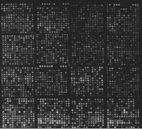
Figure 5. An image from a spotted array after laser scanning. Each spot on the image represents a gene and the intensity of a spot reflects the gene expression.
arrays. The absolute fluorescent values of each spot may then be scaled and compared with the same spot across arrays. Figure 5 gives an example of a composite image from one spotted array.
Microarray analyses usually include several steps including: image analysis and data extraction, data quantification and normalization, identification of differentially expressed genes, and knowledge discovery by data mining techniques such as clustering and classification. Image analysis and data extraction is fully automated and mainly carried out using a commercial software package or a freeware depending on the technology platforms. For example, Affymetrix developed a standard data processing procedure and software for its GeneChips (for detailed information, see http://www.affymetrix.com); GenePix is widely used image analysis software for spotted arrays. For the rest of the steps, the detailed procedures may vary depending on the experiment design and goals. We will discuss some of the procedures below.
Statistical Analysis
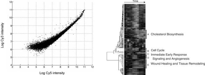
The purpose of normalization is to adjust for systematic variations, primarily for labeling and hybridization efficiency, so that the true biological variations can be discover as defined by the microarray experiment (46,47). For example, as shown in the self-hybridization scatter plot (Fig. 6) for a two-dye spotted array, variations (dye bias) between dyes is obvious and related to spot intensities. To correct the dye bias, one can apply the following model:
log2ðR=GÞ ! log2ðR=GÞ cðAÞ
where R and G are the intensities of the dyes; A is the signal strength (log2(R G)/2); M is the logarithm ratio (log2(R/G)); c(A) is the locally weighted polynomial regression (LOWESS) fit to the MA plot (48,49).
After correction of systematic variations, we want to determine which genes are significantly changed during the experiment and to assign appropriately adjusted p values to the genes. For each gene, we wish to test the null hypothesis that the gene is not differentially expressed. The P value is the probability of finding a result
224 BIOINFORMATICS
Figure 6. Self-hybridization scatter plot. The y axis is the intensity from one dye; the x axis is the intensity from the other dye. Each spot is a gene.
by chance. If P value is less than a cut-off (e.g., 0.05), one would reject the null hypothesis and state that the gene is differentially expressed (50). Analysis of variance (ANOVA) is usually used to model the factors for a particular experiment. For example,
logðmi jkÞ ¼ m þ Ai þ D j þ Vk þ ei jk
where mijk is the ratio of intensities from the two dyelabeled samples for a gene; m is the mean of ratios from all replicates; A is the effect of different arrays; D is the dye effects; and V is the treatment effects (51). Through F test, it will be determined if the gene exhibits differential expression between any Vk. For a typical microarray, thousands of genes exist. We need to perform thousands of tests in an experiment at the same time, which introduce the statistical problem of multiple testing and adjustment of p value. False discovery rate (FDR) (52) has been commonly adopted for this purpose.
For Affymetrix GeneChips analysis, even though the basic steps are the same as spotted microarrays, because of the difference in technology, different statistical methods were developed. Besides the statistical methods provided by Affymetrix, several popular methods are packaged into software such as dChip (53) and RMA (54) in Bioconductor (http://www.bioconductor.org). With rapid accumulation of microarray data, one challenging problem is how to compare microarray data across different technology platforms. Some recent studies on data agreements have provided some guidance (55–57).
Clustering and Classification
Once a list of significant genes is obtained from the statistical test, different data mining techniques would be applied to find interesting patterns. At this step, the microarray dataset is organized as a matrix. Each column represents a condition; each row represents a gene. An entry is the expression level of the gene under the corresponding condition. If a set of genes exhibit the similar fluctuation
Figure 7. Hierarchical clustering of microarry data. Rows are genes. Columns are RNA samples at different time points. Values are the signals (expression levels) that are represented by the color spectrum. Green represents down-regulation whereas red represents up-regulation. The color bars beside the dendrogram show the clusters of genes that exhibit similar expression profiles (patterns). The bars are labeled with letters and description of possible biological processes involving the genes in the clusters. [Reprinted from Eisen et al. (58).]
under all of the conditions, it may indicate that these genes are co-regulated. One way to discover the co-regulated genes is to cluster genes with similar fluctuation patterns using various clustering algorithm. Hierarchical clustering was the first clustering method applied to the problem (58). The result of hierarchical clustering forms a 2D dendrogram as shown in Fig. 7. The measurement used in the clustering process can be either a similarity, such as Pearson’s correlation coefficient, or a distance, such as Euclidian distance.
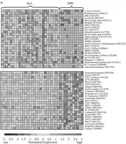
Many different clustering methods have been applied later on, such as k means (59), self-organizing map (60), and support vector machine (61). Another type of microarray study involves classification techniques. For example, we can use the gene expression profile to classify cancer types. Golub et al. (62) first reported using classification techniques to classify two different types of leukemia as shown in Fig. 8. Many commercial software packages (e.g., GeneSpring and Spotfire) offer the use of these algorithms for microarray analyses.
COMPUTATIONAL MODELING AND ANALYSIS OF BIOLOGICAL NETWORKS
The biological system is a complex system involving hundreds of thousands of elements. The interaction among the elements forms an extremely complex network. With the development of high throughput technologies in functional genomics, proteomics, and metabolomics, one can start looking into the system-level mechanisms governing the interactions and properties of biological networks. Network modeling has been used extensively
BIOINFORMATICS 225
in social and economical fields for many years (63). Many methods can be applied to biological network studies.
The cellular system involves complex interactions between proteins, DNA, RNA, and smaller molecules and can be categorized in three broad subsystem, metabolic network or pathway, protein network, and genetic or gene regulatory network. Metabolic network represents the enzymatic processes within the cell, which provide energy and building blocks for cells. It is formed by the combination of a substrate with an enzyme in a biosynthesis or degradation reaction. Considerable information about metabolic reactions has been accumulated through many years and organized into large databases, such as KEGG (64), EcoCyc (65), and WIT (66). Protein network refers to the signaling networks where the basic reaction is between two proteins. Protein-protein interactions can be determined systematically using techniques such as yeast two-hybrid system (67) or derived from the text mining of literatures (68). Genetic network or regulatory network refers to the functional inference of direct causal gene interactions (69). One can conceptualize gene expression as a genetic feedback network. The network can be inferred from the gene expression data generated from microarray
Figure 8. An example of microarray classification. Genes distinguishing acute myeloid leukemia (AML) and acute lymphoblastic leukemia (ALL). The 50 genes most highly correlated with the ALL-AML class distinction are shown. Each row corresponds to a gene, with the columns corresponding to expression levels in different samples. Expression levels for each gene are normalized across the samples such that the mean is 0 and the SD is 1. The scale indicates SDs above or below the mean. The top panel shows genes highly expressed in ALL, the bottom panel shows genes more highly expressed in AML. [Reprinted from Golub et al. (62).]
or proteomics studies in combination with computation modeling.
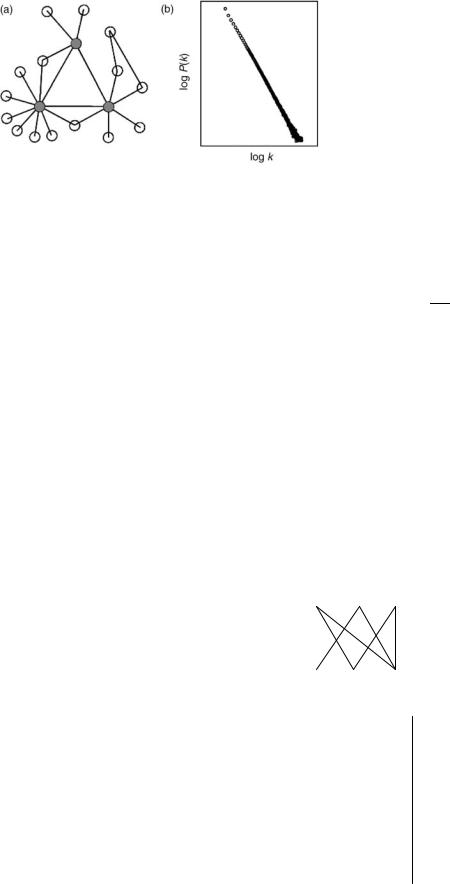
Metabolic network is typically represented as a graph with the vertex being all the compounds (substrates) and the edges being reactions linking the substrates. With such representation, one can study the general properties of the metabolic network. It has been shown that metabolic network exhibits typical property of small world or scale-free network (70,71). The distribution of compound connectivity follows a power law as shown in Fig. 9. Nodes serving as hubs exist in the network. Such property makes the network quite robust to random deletion of nodes, but vulnerable to selected deletion of nodes. For example, deletion of hub nodes will cause the network collapse very quickly. A recent study also shows that the metabolic network can be organized in modules based on the connectivity. The connectivity is high within modules, but low between modules (72).
Flux analysis is another important aspect in metabolic network study. Building on the stoichiometric network analysis, which only uses the well-characterized network topology, the concept of elementary flux modes was introduced (73,74). An elementary mode is a minimal set of enzymes that could operate at steady state, with the
226 BIOINFORMATICS
Figure 9. a. In the scale-free network, most nodes have only a few links, but a few nodes, called hubs (filled circle), have a very large number of links. b. The network connectivity can be characterized by the probability, P(k), that a node has k links. P(k) for a scale-free network has no well-defined peak, and for large k, it decays as a power-law, P(k) k-g, appearing as a straight line with slope -g on a log–log plot. [Reprinted from Jeong et al. (70).]
enzymes weighted by the relative flux they need to carry out the mode to function. The total number of elementary modes for given conditions has been used as a quantitative measure of network flexibility and as an estimate of faulttolerance (75,76).
A system approach to model regulatory networks is essential to understand their dynamics. Recently, several high-level models have been proposed for the regulatory network including Boolean models, continuous systems of coupled differential equations, and probabilistic models. Boolean networks assume that a protein or a gene can be in one of two states, active or inactive, represented by 1 or 0. This binary state varies in time and depends on the state of the other genes and proteins in the network through a discrete equation:
Xiðt þ 1Þ ¼ Fi½X1ðtÞ; . . . ; XNðtÞ& |
ð4Þ |
Thus, the function Fi is a Boolean function for the update of the ith element as a function of the state of the network at time t (69). Figure 10 gives a simple example.
Gene expression patterns contain much of the state information of the genetic network and can be measured experimentally. We are facing the challenge of inferring or reverse engineering the internal structure of this genetic network from measurements of its output. Genes with similar temporal expression patterns may share common genetic control processes and may, therefore, be related functionally. Clustering gene expression patterns according to a similarity or distance measure is the first step toward constructing a wiring diagram for a genetic network (78).
Differential equations can be an alternative model to the Boolean network and applied when the state variables X are continuous and satisfy a system of differential equations of the form
dXdti ¼ Fi½X1ðtÞ; . . . ; XNðtÞ; IðtÞ&
where the vector I(t) represents some external input into the system. The variable Xi can be interpreted as representing concentrations of proteins or mRNAs. Such a model has been used to model biochemical reactions in the metabolic pathways and gene regulation (69).
Bayesian networks are provided by the theory of graphical models in statistics. The basic idea is to approximate a complex multidimensional probability distribution using a product of simpler local probability distributions. Generally, a Bayesian network model is based on a directed acyclic graph (DAG) with N nodes. In genetic network, the nodes may represent genes or proteins and the random variables Xi levels of activity. The parameters of the model are the local conditional distributions of each random variable given the random variables associated with the
(a) |
|
|
(b) |
A |
B |
C |
A’= B |
|
|
|
|
|
|
|
B’= A OR C |
|
|
|
C’= (A AND B) OR (B AND C) OR (A AND C) |
A’ |
B’ |
C’ |
|
Figure 10. Target Boolean network for reverse engineering. (a) The network wiring and (b) logical rules determine (c) the dynamic output. The challenge lies in inferring (a) and (b) from (c). [Reprinted from Liang et al. (77).]
(c)
|
Input |
|
|
Output |
|
|
|
|
|
|
|
A |
B |
C |
A’ |
B’ |
C’ |
|
|
|
|
|
|
0 |
0 |
0 |
0 |
0 |
0 |
0 |
0 |
1 |
0 |
1 |
0 |
0 |
1 |
0 |
1 |
0 |
0 |
0 |
1 |
1 |
1 |
1 |
1 |
1 |
0 |
0 |
0 |
1 |
0 |
1 |
0 |
1 |
0 |
1 |
1 |
parent nodes |
|
PðX1; . . . ; XNÞ ¼ YPðXijX j : j 2 N ðiÞÞ |
ð4Þ |
i |
|
where N (i) denotes all the parents of vertex i. Given a dataset D representing expression levels derived using DNA microarray experiments; it is possible to use learning techniques with heuristic approximation methods to infer the network architecture and parameters. As data from microarray experiments are still limited and insufficient to completely determine a single model, people have developed heuristics for learning classes of models rather than single models, for instance, for a set of co-regulated genes (69). Bayesian networks have recently been shown to combine heterogeneous datasets, for instance, microarray data with functional annotation and mutation data to produce an expert system (79).
In this chapter, some major development in the field of bioinformatics were reviewed and some basic concepts in the field were introduced covering six areas: sequence analysis, phylogenetic analysis, protein structure analysis, genome analysis, microarray analysis, and network analysis. Due to the limited space, some topics have been left out. One such topics is text mining, which uses Natural Language Processing (NLP) techniques to extract information from the vast amount of literature in biological research. Text mining has become an integral part in bioinformatics. With the continuing development and maturing of new technologies in many system-level studies, the way that biological research is conducted is undergoing revolutionary change. Systems biology is becoming a major theme and driving force. The challenges for bioinformatics in the post-genomics era lie on the integration of data and knowledge from heterogeneous sources and system-level modeling and simulation providing molecular mechanism for physiological phenomena.
BIBLIOGRAPHY
Cited References
1.Abbas A, Holmes S. Bioinformatics and management science. Some common tools and techniques. Operations Res 2004; 52(2):165–190.
2.Needleman SB, Wunsch CD. A general method applicable to the search for similarities in amino acid sequence of two proteins. J Mol Biol 1970;48:443–453.
3.Gotoh O. An improved algorithm for matching biological sequences. J Mol Biol 1982;162:705–708.
4.Durbin S, Eddy S, Krogh A, Mitchison G. Biological Sequence Analysis: Probabilistic Models of Proteins and Nucleic Acids. Cambridge, (UK): Cambridge University Press; 1998.
5.Dayhoff MO, Schwartz RM, Orcutt BC. A model of evolutionary change in proteins. Atlas of Protein Sequence and Structure. vol. 5, supplement 3. National Biomedical Research Foundation. Washington, (DC): 1978. p 345–352.
6.Henikoff S, Henikoff JG. Amino acid substitution matrices from protein blocks. Proc Natl Acad Sci USA 1992;89:10915– 10919.
7.Smith TF, Waterman MS. Identification of common molecular subsequences. J Mol Biol 1981;147:195–197.
8.Altschul SF, Gish W, Miller W, Myers E, Lipman J. Basic local alignment search tool. J Molec Biol 1990;215:403– 410.
BIOINFORMATICS 227
9.Lipman JD, Altschul SF, Kececioglu JD. A tool for multiple sequence alignment. Proc Natl Acad Sci 1989;86:4412–4415.
10.Thompson JD, Higgins DG, Gibson TJ. CLUSTAL W: Improving the sensitivity of progressive multiple sequence alignment through sequence weighting, position specific gap penalties and weight matrix choice. Nucleic Acids Res 1994;22: 4673–4680.
11.Lawrence CE, Altschul SF, Boguski MS, Liu JS, Neuwald AN, Wootton J. Detecting subtle sequence signals: A Gibbs sampling strategy for multiple alignment. Science 1993;262:208–214.
12.(a) Delcher et al. 2002. (b) Zhu J, Liu JS, Lawrence CE. Bayesian adaptive sequence alignment algorithms. Bioinformatics 1998; 14:25–39.
13.Li WH. Molecular Evolution. Boston, MA: Sinauer Associates; 1997.
14.Holmes S. Bootstrapping phylogenetic trees. To appear in Statistical Science. Submitted in (2002).
15.Li S, Pearl DK, Doss H. Phylogenetic tree construction using MCMC. J Am Statist Assoc 2000;95:493–503.
16.Levitt M, Lifson S. Refinement of protein confirmations using a macromolecular energy minimization procedure. J Mol Biol 1969;46:269–279.
17.Berman HM, Westbrook J, Feng Z, Gilliland G, Bhat TN, Weissig H, Shindyalov IN, Bourne PE. The protein data bank. Nucleic Acids Res 2000;28:235–242.
18.Xu Y, Xu D. Protein threading using PROSPECT: Design and evaluation. Proteins Structure, Function, and Genetics 2000;40:343–354.
19.Levitt M, Warshel A. Computer simulation of protein folding. Nature 1975;253:694–698.
20.Nemethy G, Scheraga HA. Theoretical determination of sterically allowed conformations of a polypeptide chain by a computer method. Biopolymers 1965;3:155–184.
21.Levitt M. Molecular dynamics of native protein: Computer simulation of the trajectories. J Mol Biol 1983;168:595– 620.
22.Beeman D. Some multi-step methods for use in molecular dynamics calculations. J Comput Phys 1976;20:130–139.
23.Bourne PE. CASP and CAFASP experiments and their findings. Methods Biochem Anal 2003;44:501–507.
24.Gordon D, Abajian C, Green P. Consed: A graphical tool for sequence finishing. Genome Res 1998;8(3):195–202.
25.Gordon D, Desmarais C, Green P. Automated finishing with autofinish. Genome Res 2001;11(4):614–625.
26.Huang X, Madan A. CAP3: A DNA sequence assembly program. Genome Res 1999;9(9):868–877.
27.Waterston RH, Lander ES, Sulston JE. On the sequencing of the human genome. Proc Natl Acad Sci USA 2002; 99(6):3712–3716.
28.Myers EW, et al. A whole-genome assembly of Drosophila 2000;287(5461):2196–2204.
29.Adams MD, et al. The genome sequence of Drosophila melanogaster Science 2000;287(5461):2185–2195.
30.Venter JC, et al. The sequence of the human genome. Science 2001;29:1304–1351.
31.Lukashin AV, Borodovsky M. GeneMark.hmm: New solutions for gene finding. Nucleic Acids Res 1998;26(4):1107– 1115.
32.Delcher AL, Harmon D, Kasif S, White O, Salzberg SL. Improved microbial gene identification with GLIMMER. Nucleic Acids Res 1999;27(23):4636–4641.
33.Burge C, Karlin S. Prediction of complete gene structures in human genomic DNA. J Mol Biol 1997;268:78–94.
34.Yeh R-F, Lim LP, Burge CB. Computational inference of homologous gene structures in the human genome. Genome Res 2001;11:803–816.
228BIOINFORMATICS
35.Xu Y, Uberbacher CE. Automated gene identification in largescale genomic sequences. J Comp Biol 1997;4:325–338.
36.Bateman A, Coin L, Durbin R, Finn RD, Hollich V, GriffithsJones S, Khanna A, Marshall M, Moxon S, Sonnhammer ELL, Studholme DJ, Yeats C, Eddy SR. The Pfam Protein Families Database. Nucleic Acids Res 2004;32:D138–D141.
37.Eddy S. Profile hidden Markov models. Bioinformatics 1998;14:755–763.
38.Krogh A, Brown M, Mian IS, Juolander K, Haussler D. Hidden Markov models in computational biology applications to protein modeling. J Mol Biol 1994;235: 1501–1531.
39.Stomo GD, Hartzell GW. Identifying protein-binding sites from unaligned DNA fragments. Proc Natl Acad Sci 1989;86:1183–1187.
40.Lawrence CE, Reilly AA. An expectation maximization (EM) algorithm for the identification and characterization of common sites in unaligned biopolymer sequences. Proteins Struct Funct Genet 1990;7:41–51.
41.Bailey LT, Elkan C. Fitting a mixture model by expectation maximization to discover motifs in biopolymers. Proceedings of the Second International Conference on Intelligent Systems for Molecular Biology 1994; 28–36.
42.Kellis M, Birren BW, Lander ES. Proof and evolutionary analysis of ancient genome duplication in the yeast Saccharomyces cerevisiae. Nature 2004;428:617–624.
43.Tatusov RL, Koonin EV, Lipman DJ. A genomic perspective on protein families. Science 1997; 631–637.
44.O’Brien SJ, Menotti-Raymond M, Murphy WJ, Nash WG, Wienberg J, Stanyon R, Copeland NG, Jenkins NA, Womack JE, Graves JAM. The promise of comparative genomics in mammals. Science 1999;286:458–481.
45.Bourque G, Pevzner AP. Genome-scale evolution: Reconstructing gene orders in the ancestral species. Genome Res 2002;12:26–36.
46.Bolstad BM, Irizarry RA, Astrand M, Speed TP. A comparison of normalization methods for high density oligonucleotide array data based on bias and variance. Bioinformatics 2003;19(2):185–193.
47.Bajesy et al. 2005.
48.Yang YH, Dudoit S, Luu P, Lin DM, Peng V, Ngai J, Speed TP. Normalization for cDNA microarray data: A robust composite method addressing single and multiple slide systematic variation. Nucleic Acids Res 2002;30(4):e15.
49.Yang YH, Thorne N. Normalization for two-color cDNA microarray data. Science and statistics: A festschrift for terry speed. In: Goldstein D, ed. IMS Lecture Notes, Monograph Series. Vol. 40; 2003. p 403–418.
50.Smyth GK, Yang YH, Speed TP. Statistical issues in microarray data analysis. In: Brownstein MJ, Khodursky AB, eds. Functional Genomics: Methods and Protocols. Methods in Molecular Biology. vol. 224. Totowa, (NJ): Humana Press; 2003. p 111–136.
51.Kerr M, Churchill G. Analysis of variance for gene expression microarray data. J Comp Biol 2000;7:819–837.
52.Benjamini Y, Hochberg Y. Controlling the false discovery rate: A practical and powerful approach to multiple testing. J Royal Statist Soc B 1995;57(1):289–300.
53.Li C, Wong WH. Model-based analysis of oligonucleotide arrays: Expression index computation and outlier detection. Proc Natl Acad Sci 2001;98:31–36.
54.Bolstad BM, Irizarry RA, Astrand M, Speed TP. A comparison of normalization methods for high density oligonucleotide array data based on bias and variance. Bioinformatics 2003;19(2):185–193.
55.Wang H, He X, Band M, Wilson C, Liu L. A study of inter-lab and inter-platform agreement of DNA microarray data. BMC Genomics 2005;6(1):71.
56.Jarvinen A, Hautaniemi S, Edgren H, Auvinen P, Saarela J, Kallioniemi O, Monni O. Are data from different gene expression microarray platforms comparable? Genomics 2004;83: 1164–1168.
57.Culhane AC, Perriere G, Higgins DG. Cross-platform comparison and visualisation of gene expression data using coinertia analysis. BMC Bioinformatics 2003;4:59.
58.Eisen MB, Spellman PT, Brown PO, Botstein D. Cluster analysis and display of genome-wide expression patterns. Proc Natl Acad Sci USA 1998;95(25):14863–14868.
59.Ben-Dor A, Shamir R, Yakhini Z. Clustering gene expression patterns. J Comp Biol 1999;6(3/4):281–297.
60.Tamayo P, Solni D, Mesirov J, Zhu Q, Kitareewan S, Dmitrovsky E, Lander ES, Golub TR. Interpreting patterns of gene expression with self-organizing maps: Methods and application to hematopoietic differentiation. Proc Natl Acad Sci USA 1999;96(6):2907–2912.
61.Alter O, Brown PO, Bostein D. Singular value decomposition for genome-wide expression data processing and modeling. Proc Natl Acad Sci USA 2000;97(18):10101– 10106.
62.Golub TR, Slonim DK, Tamayo P, Huard C, Gaasenbeek M, Mesirov JP, Coller H, Loh ML, Downing JR, Caligiuri MA, Bloomfield CD, Lander ES. Molecular classification of cancer: Class discovery and cass prediction by gene expression monitoring. Science 1999;286:531–537.
63.Sole RV, Ferrer-Cancho R, Montoya JM, Valverde S. Selection, tinkering, and emergence in complex networks. Complexity 2003;8:20–33.
64.Kanehisa M. A database for post-genome analysis. Trends Genet 1997;13:375–376.
65.Keseler IM, Collado-Vides J, Gama-Castro S, Ingraham J, Paley S, Paulsen IT, Peralta-Gil M, Karp PD. EcoCyc: A comprehensive database resource for Escherichia coli. Nucleic Acids Res 2005;33:D334–D337.
66.Overbeek R, Larsen N, Pusch GD, D’Souza M, Selkov E, Kyrpides N, Fonstein M, Maltsev N, Selkov E. WIT: Integrated system for high-throughput genome sequence analysis and metabolic reconstruction. Nucleic Acids Res 2000;28(1): 123–125.
67.Fields S, Song OK. A novel genetic system to detect proteinprotein interactions. Nature 1989;340:245–246.
68.Daraselia N, Yuryev A, Egorov S, Novichkova S, Nikitin A, Mazo I. Extraction of human protein interactions from MEDLINE using full-sentence parser. Bioinformatics 2003;19:1– 8.
69.Baldi P, Hatfield GW. Microarrays and Gene Expression. Cambridge, (UK): Cambridge University Press; 2001.
70.Jeong H, Tombor B, Albert1 R, Oltvai ZN, Baraba´si AL. The large-scale organization of metabolic networks. Nature 2000; 407:651–654.
71.Wagner A, Fell DA. The small world inside large metabolic networks. Proc Royal Soc Lond B 2001;268:1803–1810.
72.Guimera R, Nunes Ameral AL. Functional cartography of complex metabolic networks. Nature 2005;433:895–900.
73.Schuster S, Hilgetag C, Woods JH, Fell DA. Reaction routes in biochemical reaction systems: algebraic properties, validated calculation procedure and example from nucleotide metabolism. J Math Biol 2002;45(2):153–181.
74.Schuster S, Fell DA, Dandekar T. A general definition of metabolic pathways useful for systematic organization and analysis of complex metabolic networks. Nature 2000;18: 326–332.
75.Stelling J, Klamt S, Bettenbrock K, Schuster S, Gilles ED. Metabolic network structure determines key aspects of functionality and regulation. Nature 2002;420:190–193.
76.Cakir T, Kirdar B, Ulgen KO. Metabolic pathway analysis of yeast strengthens the bridge between transcriptomics and metabolic networks. Biotechnol Bioeng 2004;86:251–260.
77.Liang S, Fuhrman S, Somogyi R. REVEAL, a general reverse engineering algorithm for inference of genetic network architectures. Pacific Symp Biocomput 1998;3:18–29.
78.Somogyi R, Fuhrman S, Wen X. Genetic network inference in computational models and applications to large-scale gene expression data. Cambridge, (MA): MIT Press; 2001.
79.Troyanskaya OG, Dolinski K, Owen AB, Altman RB, Botstein D. A Bayesian framework for combining heterogeneous data sources for gene function prediction (in Saccharomyces cerevisiae). Proc Natl Acad Sci USA 2003;100: 8348–8353.
Further Reading
Altschul SF, Madden TL, Schaffer AA, Zhang J, Zhang Z, Miller W, Lipman DJ. Gapped BLAST and PSI-BLAST: A new generation of protein database search programs. Nucleic Acids Res 1997;25:3389–3402.
Baldi P, Chauvin Y, Hunkapillar T, McClure M. Hidden Markov models of biological primary sequence information. Proc Natl Acad Sci USA 1994;91:1059–1063.
Baldi P, Brunak S. Bioinformatics: The Machine Learning Approach. 2nd ed. Cambridge, (MA): MIT Press; 2001.
Bork P, Dandekar T, Diaz-Lazcoz Y, Eisenhaber F, Huynen M, Yuan Y. Predicting function: From genes to genomes and back. J Mol Biol 1998;283:707–725.
Bower J, Bolouri H. Computational Modeling of Genetic and Biochemical Networks. Cambridge, (MA): MIT Press; 2001.
Bray N, Dubchak I, Pachter L. AVID: A global alignment program. Genome Res 2003;13(1):97–102.
Brown PO, Botstein D. Exploring the new world of the genome with DNA microarrays. Nature Genetics 1999;21:33–37.
Brudno M, CB Do, Cooper GM, Kim MF, Davydov E, Green ED, Sidow A, Batzoglou A. LAGAN and Multi-LAGAN: efficient tools for large-scale multiple alignment of genomic DNA. Genome Res 2003;13:(4):721–731.
Brudno M, Malde S, Poiakov A, Do C, Couronne O, Dubchak I, Batzoglou A. Glocal alignment: Finding rearrangements during alignment. Bioinformatics Special Issue on the Proceedings of the ISMB 2003;19:54i–62i.
Bryant SH, Altschul SF. Statistics of sequence-structure threading. Curr Opin Structur Biol 1995;5:236–244.
Cohen FE. Protein misfolding and prion diseases. J Mol Biol 1999;293:313–320.
Diaconis P, Holmes S. Random walks on trees and matchings. Electron J Probabil 2002;7:1–17.
Doyle JC. Robustness and dynamics in biological networks. In: The First International Conference on Systems Biology. New York: Japan Science and Technology Corporation, MIT Press; 2000.
Dudoit S, Fridlyand J, Speed TP. Comparison of discrimination methods for the classification of tumors using gene expression data. J Am Statistic Assoc 2002;97:77–87.
Eddy S, Mitchison G, Durbin R. Maximum discrimination hidden Markov models of sequence consensus. J Comput Biol 1995; 2:9–23.
Eddy SR. Non-coding RNA genes and the modern RNA world. Nature Rev Genet 2001;2:919–929.
Efron B, Halloran EE, Holmes S. Bootstrap confidence levels for phylogenetic trees. Proc Natl Acad Sci 1996;93:13429–13434.
Farris JS. The logical basis of phylogenetic analysis. In: Platnick N, Funk V, eds. Advances in Cladistics. vol. 2. 1983. p 7–36.
Fedorov AN, Baldwin TO. Contranslational protein folding. Biol Chem 1997;272(52):32715–32718.
BIOINFORMATICS 229
Felsenstein J. Evolutionary trees from DNA sequences: A maximum likelihood approach. J Mol Evol 1981;17(6):368– 376.
Felsenstein J. 1993. (Phylogeny Inference Package) version 3.5c. Department of Genetics, University of Washington, Seattle, WA. Available http://evolution.genetics.washington.edu/ phylip.html.
Fischer D, Barret C, Bryson K, Elofsson A, Godzik A, Jones D, Karplus KJ, Kelley LA, MacCallum RM, Pawowski K, Rost B, Rychlewski L, Sternberg M. CAFASP-1: Critical assessment of fully automated structure prediction methods. Proteins 1999;3:209–217.
Fitch WM, Margoliash E. Construction of phylogenetic trees. Science 1967;155:279–284.
Foulds LR, Graham RL. The Steiner problem in Phylogeny is NPcomplete. Adv Appl Math 1982;3:43–49.
Friedman N, Linial M, Nachman I, Peter D. Using Bayesian networks to analyze expression data. J Comp Bio 2000;7: 601–620.
Gardner M. The Last Recreations. New York: Copernicus-Springer Verlag; 1997.
Geman S, Geman D. Stochastic relaxation, Gibbs distribution and the Bayesian restoration of images. IEEE Trans Pattern Anal Machine Intell 1984;6:721–741.
Gibson KD, Scheraga HA. Revised algorithms for the build-up procedure for predicting protein conformations by energy minimization. J Comp Chem 1987;9:327–355.
Goloboff PA. SPA. 1995. (S)ankoff (P)arsimony (A)nalysis, version 1.1. Computer program distributed by J. M. Carpenter, Department of Entomology, American Museum of Natural History, New York.
Gribaldo S, Cammarano P. The root of the universal tree of life inferred from anciently duplicated genes encoding components of the protein-targeting machinery. J Mol Evol 1998;47(5):508– 516.
Haeckel E. Morphologie der Organismen: Allgemeine Grundzuge der Organischen FormenWissenschaft, Mechanisch Begrundet durch die von Charles Darwin Reformirte Descendenz-Theorie. Berlin: Georg Riemer; 1866.
Hannenhalli S, Pevzner PA. Transforming cabbage into turnip: Polynomial algorithm for sorting signed permutations by reversals. STOC 1995; 178–189.
Helden JV, Andre B, Collado-Vides J. Extracting regulatory sites from the upstream region of yeast genes by computational analysis of oligonucleotide frequencies. J Mol Bio 1998;281: 827–842.
Hooper E. The River. Boston, (MA): Little, Brown; 1999. Huelsenbeck J, Ronquist F. 2002. Mr. Bayes. Bayesian inference of
phylogeny. Available at http://morphbank.ebc.uu.se/mrbayes/ links.php.
Jukes T, Cantor C. Evolution of protein molecules. In: eds. Munro HN, Mammalian Protein Metabolism. New York: Academic Press; 1969. p 21–132.
Karlin S, Altschul SF. Methods for assessing the statistical significance of molecular sequences features by using general scoring schemes. Proc Natl Acad Sci USA 1990;87(6):2264– 2268.
Keith JM, Adams P, Bryant D, Kroese DP, Mitchelson KR, Cochran DAE, Lala GH. A simulated annealing algorithm for finding consensus sequences. Bioinformatics 2002;18: 1494–1499.
Kent WJ. BLAT–the BLAST-like alignment tool. Genome Res 2002;12(4):656–664.
Kirkpatrick S, Gelatt CD Jr, Vecchi MP. Optimization by simulated annealing. Science 1983;220:671–680.
Korf I, Flicek P, Duan D, Brent MR. Integrating genomic homology into gene structure prediction. Bioinformatics 2001;17:S140– S148.
