
- •VOLUME 1
- •CONTRIBUTOR LIST
- •PREFACE
- •LIST OF ARTICLES
- •ABBREVIATIONS AND ACRONYMS
- •CONVERSION FACTORS AND UNIT SYMBOLS
- •ABLATION.
- •ABSORBABLE BIOMATERIALS.
- •ACRYLIC BONE CEMENT.
- •ACTINOTHERAPY.
- •ADOPTIVE IMMUNOTHERAPY.
- •AFFINITY CHROMATOGRAPHY.
- •ALLOYS, SHAPE MEMORY
- •AMBULATORY MONITORING
- •ANALYTICAL METHODS, AUTOMATED
- •ANALYZER, OXYGEN.
- •ANESTHESIA MACHINES
- •ANESTHESIA MONITORING.
- •ANESTHESIA, COMPUTERS IN
- •ANGER CAMERA
- •ANGIOPLASTY.
- •ANORECTAL MANOMETRY
- •ANTIBODIES, MONOCLONAL.
- •APNEA DETECTION.
- •ARRHYTHMIA, TREATMENT.
- •ARRHYTHMIA ANALYSIS, AUTOMATED
- •ARTERIAL TONOMETRY.
- •ARTIFICIAL BLOOD.
- •ARTIFICIAL HEART.
- •ARTIFICIAL HEART VALVE.
- •ARTIFICIAL HIP JOINTS.
- •ARTIFICIAL LARYNX.
- •ARTIFICIAL PANCREAS.
- •ARTERIES, ELASTIC PROPERTIES OF
- •ASSISTIVE DEVICES FOR THE DISABLED.
- •ATOMIC ABSORPTION SPECTROMETRY.
- •AUDIOMETRY
- •BACTERIAL DETECTION SYSTEMS.
- •BALLOON PUMP.
- •BANKED BLOOD.
- •BAROTRAUMA.
- •BARRIER CONTRACEPTIVE DEVICES.
- •BIOCERAMICS.
- •BIOCOMPATIBILITY OF MATERIALS
- •BIOELECTRODES
- •BIOFEEDBACK
- •BIOHEAT TRANSFER
- •BIOIMPEDANCE IN CARDIOVASCULAR MEDICINE
- •BIOINFORMATICS
- •BIOLOGIC THERAPY.
- •BIOMAGNETISM
- •BIOMATERIALS, ABSORBABLE
- •BIOMATERIALS: AN OVERVIEW
- •BIOMATERIALS: BIOCERAMICS
- •BIOMATERIALS: CARBON
- •BIOMATERIALS CORROSION AND WEAR OF
- •BIOMATERIALS FOR DENTISTRY
- •BIOMATERIALS, POLYMERS
- •BIOMATERIALS, SURFACE PROPERTIES OF
- •BIOMATERIALS, TESTING AND STRUCTURAL PROPERTIES OF
- •BIOMATERIALS: TISSUE-ENGINEERING AND SCAFFOLDS
- •BIOMECHANICS OF EXERCISE FITNESS
- •BIOMECHANICS OF JOINTS.
- •BIOMECHANICS OF SCOLIOSIS.
- •BIOMECHANICS OF SKIN.
- •BIOMECHANICS OF THE HUMAN SPINE.
- •BIOMECHANICS OF TOOTH AND JAW.
- •BIOMEDICAL ENGINEERING EDUCATION
- •BIOSURFACE ENGINEERING
- •BIOSENSORS.
- •BIOTELEMETRY
- •BIRTH CONTROL.
- •BLEEDING, GASTROINTESTINAL.
- •BLADDER DYSFUNCTION, NEUROSTIMULATION OF
- •BLIND AND VISUALLY IMPAIRED, ASSISTIVE TECHNOLOGY FOR
- •BLOOD BANKING.
- •BLOOD CELL COUNTERS.
- •BLOOD COLLECTION AND PROCESSING
- •BLOOD FLOW.
- •BLOOD GAS MEASUREMENTS
- •BLOOD PRESSURE MEASUREMENT
- •BLOOD PRESSURE, AUTOMATIC CONTROL OF
- •BLOOD RHEOLOGY
- •BLOOD, ARTIFICIAL
- •BONDING, ENAMEL.
- •BONE AND TEETH, PROPERTIES OF
- •BONE CEMENT, ACRYLIC
- •BONE DENSITY MEASUREMENT
- •BORON NEUTRON CAPTURE THERAPY
- •BRACHYTHERAPY, HIGH DOSAGE RATE
- •BRACHYTHERAPY, INTRAVASCULAR
- •BRAIN ELECTRICAL ACTIVITY.
- •BURN WOUND COVERINGS.
- •BYPASS, CORONARY.
- •BYPASS, CARDIOPULMONARY.
hydroxyapatite) as part of a circumferential fusion. Spine 2002;27:E518–E525.
107.Delecrin J, Takahashi S, Gouin F, Passuti N. A synthetic porous ceramic as a bone graft substitute in the surgical management of scoliosis: A prospective, randomized study. Spine 2000;25:563–569.
108.McAndrew MP, Gorman PW, Lange TA. Tricalcium phosphate as a bone graft substitute in trauma: Preliminary report. J Orthop Trauma 1988;2:333–339.
109.Bucholz RW, Carlton A, Holmes RE. Hydroxyapatite and tricalcium phosphate bone graft substitutes. Orthop Clin North Am 1987;18:323–334.
110.Holmes R, Mooney V, Bucholz R, Tencer A. A coralline hydroxyapatite bone graft substitute. Preliminary report. Clin Orthop 1984; 252–262.
111.Finn RA, Bell WH, Brammer JA. Interpositional ‘‘grafting’’ with autogenous bone and coralline hydroxyapatite. J Maxillofac Surg 1980;8:217–227.
112.Holmes RE. Bone regeneration within a coralline hydroxya-
patite implant. Plast Reconstr Surg 1979;63:626– 633.
113.Holmes RE, Salyer KE. Bone regeneration in a coralline hydroxyapatite implant. Surg Forum 1978;29:611–612.
114.Jamali A, Hilpert A, Debes J, Afshar P, Rahban S, Holmes R. Hydroxyapatite/calcium carbonate (HA/CC) vs. plaster of Paris: A histomorphometric and radiographic study in a rabbit tibial defect model. Calcif Tissue Int 2002;71:172– 178.
115.Kenny SM, Buggy M. Bone cements and fillers: A review. J Mater Sci Mater Med 2003;14:923–938.
116.Horstmann WG, Verheyen CC, Leemans R. An injectable calcium phosphate cement as a bone-graft substitute in the treatment of displaced lateral tibial plateau fractures. Injury 2003;34:141–144.
117.Kamano M, Honda Y, Kazuki K, Yasudab M. Palmar plating with calcium phosphate bone cement for unstable Colles’ fractures. Clin Orthop 2003; 285–290.
118.Zimmermann R, Gabl M, Lutz M, Angermann P, Gschwentner M, Pechlaner S. Injectable calcium phosphate bone cement Norian SRS for the treatment of intra-articular compression fractures of the distal radius in osteoporotic women. Arch Orthop Trauma Surg 2003;123:22–27.
119.Schildhauer TA, Bauer TW, Josten C, Muhr G. Open reduction and augmentation of internal fixation with an injectable skeletal cement for the treatment of complex calcaneal fractures. J Orthop Trauma 2000;14:309–317.
See also DRUG DELIVERY SYSTEMS; MATERIALS AND DESIGN FOR ORTHOPEDIC DEVICES; POROUS MATERIALS FOR BIOLOGICAL APPLICATIONS.
BIOMATERIALS: AN OVERVIEW
BRANDON L. SEAL
ALYSSA PANITCH
Arizona State University
Tempe, Arizona
INTRODUCTION
Biomaterials are materials that are used or that have been designed for use in medical devices or in contact with the body. Traditionally, they consist of metallic, ceramic, or synthetic polymeric materials, but more recent develop-
BIOMATERIALS: AN OVERVIEW |
267 |
ments in biomaterials design have attempted to incorporate materials derived from or inspired by biological materials (e.g., silk and collagen). Often, the use of biomaterials focuses on the augmentation, replacement, or restoration of diseased or damaged tissues and organs. The prevalence of biomaterials within society is most evident within medical and dental offices, pharmacies, and hospitals. However, the influence of biomaterials has reached into many households with examples ranging from increasingly common news media coverage of medical breakthroughs to the availability of custom color noncorrective contact lenses.
The evolving character of the discipline of biomaterials is evidenced by how the term biomaterial has been defined. In 1974, the Clemson Advisory Board, in response to a request by the World Health Organization (WHO), stated that a biomaterial is a ‘‘systemically pharmacologically inert substance designed for implantation within or incorporation with living tissue’’ (1). Dr. Jonathan Black further modified this definition to state that a biomaterial is ‘‘any pharmacologically inert material, viable or nonviable, natural product or manmade, that is part of or is capable of interacting in a beneficial way with a living organism’’ (1). An National Institute of Health (NIH) consensus definition appeared in 1983 and defined biomaterials as ‘‘any substance (other than a drug) or combination of substances, synthetic or natural in origin, which can be used for any period of time, as a whole or as a part of a system that treats, augments, or replaces any tissue, organ, or function of the body’’ (2). Thus, relatively newer definitions of the term biomaterial recognize that more modern medical and diagnostic devices will rely increasingly upon direct biological interaction between biological molecules, cells, and tissues and the materials from which these devices are manufactured.
HISTORY OF BIOMATERIALS
Compared with the much larger field of materials science, the field of biomaterials is relatively new. Although there exist recorded cases of glass eyes and metallic or wooden dental implants (some of which can be dated back to ancient Egypt), the modern age of biomaterials could not have existed without the adoption of aseptic surgical techniques pioneered by Sir Joseph Lister in the midnineteenth century and indeed, did not fully emerge as an industry or discipline until after the development of synthetic polymers just prior to, during, and following World War II. Prior to World War II, implanted biomaterials consisted primarily of metals (e.g., steel, used in pins and plates for bone fixation, joint replacements, and the covering of bone defects). In the late 1940s, Harold Ridley observed that shards of poly(methyl methacrylate) (PMMA), from airplane cockpit windshields, embedded within the eyes of World War II aviators did not provoke much of an inflammatory response (3). This observation led not only to the development of PMMA intraocular lenses, but also to greater experimentation of available materials, especially polymers, as biomaterials that could be placed in direct contact with living tissue.
268 BIOMATERIALS: AN OVERVIEW
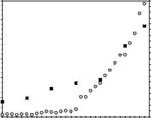
As the fields of cellular, molecular, and developmental biology began to grow during the 1970s and 1980s, new insights into the organization, function, and properties of biological systems, tissues, and interactions led to a greater understanding of how cells respond to their environment. This wealth of biological information allowed the field of biomaterials to undergo a paradigm shift. Instead of focusing primarily on replacing an organ or a tissue with a synthetic, usually nondegradable biomaterial, a new branch of biomaterials would attempt to combine biologically active molecules, therapeutics, and motifs into existing and novel biomaterial systems derived from both synthetic and natural sources (4–6). Although there exist many examples of successful, commercially available biomaterials consisting of metallic and ceramic bases, the focus of biomaterials research has shifted to the development of polymeric or composite materials with biologically sensitive or environmentally controlled properties. This change has resulted largely due to the reactivity and variety of chemical moieties that are found in or that can be engineered into natural and synthetic polymers. Indeed, by viewing biomaterials as materials designed to interact with biology rather than being inert substances, the field of biomaterials has exploded with innovative designs that promote cell attachment, encapsulation, proliferation, differentiation, migration, and apoptosis, and that allow the biomaterial to polymerize, swell, and degrade under a variety of environmental conditions and biological stimuli. Evidence of this polymer and composite revolution is the dramatic increase in the number of publications relating to biomaterials research. Figure 1 shows a plot of the number of journal articles with biomaterial or biomaterials in their title, abstract, or keyword as a function of publication year as searched in the Web of Science database. As seen in Fig. 1, publications matching the search criteria have increased exponentially starting around the early 1990s and continuing until the present. The number of scientific journals, shown in Table 1, related
PublicationsofNumber |
1200 |
|
|
|
|
|
100 |
DepartmentsBMEofNumber |
1000 |
|
|
|
|
|
80 |
||
|
|
|
|
|
|
|
||
|
|
|
|
|
|
|
|
|
|
800 |
|
|
|
|
|
60 |
|
|
|
|
|
|
|
|
|
|
|
600 |
|
|
|
|
|
|
|
|
400 |
|
|
|
|
|
40 |
|
|
|
|
|
|
|
|
|
|
|
200 |
|
|
|
|
|
20 |
|
|
|
|
|
|
|
|
|
|
|
0 |
|
|
|
|
|
0 |
|
|
1975 |
1980 |
1985 |
1990 |
1995 |
2000 |
2005 |
|
Year
Figure 1. A plot of the number of publications (*) containing the word biomaterial or biomaterials in the title, abstract, or keywords, as searched in the Web of Science database, as a function of the publication year as well as a plot of the number of bioengineering or biomedical engineering departments (BME departments) within the United States as a function of time (&).
to research in the field of biomaterials has also grown. Although the number of journal articles related to biomaterials research may have resulted primarily from the large increase in the number of biomaterials-related scientific journals, the exponential growth of biomaterials is evidenced further by the growth of the number of bioengineering or biomedical engineering departments (the department in which most biomaterials programs reside) established at universities throughout the United States (Fig. 1).
MARKET SIZE AND TYPES OF APPLICATIONS
The field of biomaterials, by nature, is interdisciplinary. Successful biomaterial designs have involved talents, knowledge, and expertise provided by physicians and clinicians, materials scientists, engineers, chemists, biologists, and physicists. As a result, it is not surprising that the biomaterials industry is both relatively young and very diversified. The diversity of this industry has resulted from the types of products created and marketed, the size and location of involved companies, and the types of regulatory policies imposed by government agencies and third party reimbursement organizations. Specifically, the biomaterials industry is part of the Medical Device and Diagnostic Industry, a multibillion dollar industry comprised of organizations that design, fabricate, and/or manufacture materials that are used in the health and life science fields. The end use applications are medical and dental devices, prostheses, personal hygiene products, diagnostic devices, drug delivery vehicles, and biotechnology systems. Some examples of these applications include full and hybrid artificial organs, biosensors, vascular grafts, pacemakers, catheters, insulin pumps, cochlear implants, contact lenses, intraocular lenses, artificial joints and bones, burn dressings, and sutures. Table 2 shows a list of some common medical devices that require various biomaterials, and Table 3 displays a list of the prevalence and market potential of a few of these applications (7).
GOVERNMENT REGULATION
Within the United States, in a research only environment, biomaterials by themselves do not necessarily require government regulation. However, if any biomaterial is used within a medical or diagnostic device designed and destined for commercialization, the biomaterials used within the medical device (as well as the device itself) are subject to the jurisdiction of the U.S. Food and Drug Administration (FDA) as set forth in the Federal Food, Drug, and Cosmetic Act of 1938, the Medical Device Amendments of 1976, and the Food and Drug Administration Modernization Act of 1997. These laws have empowered the FDA to regulate conditions involving premarket controls, postmarket reporting, production involving Good Manufacturing Practices, and the registration and listing of medical devices. Any biomaterial within a marketed medical device prior to the Medical Device Amendments of 1976 were grandfathered and are considered approved materials. Modifications to these materials or

|
|
BIOMATERIALS: AN OVERVIEW |
269 |
Table 1. A List of Journals with Publications Related to the Field of Biomaterialsa |
|
|
|
Name of Journal |
Name of Journal |
Name of Journal |
|
|
|
|
|
Advanced Drug Delivery |
Biosensors and Bioelectronics (1985) |
Journal of Biomaterials Science: Polymer |
|
Reviews (1987) |
|
Edition (1990) |
|
American Journal of Drug |
Cells and Materials (1991) |
Journal of Biomedical Materials |
|
Delivery (2003) |
|
Research (1967) |
|
American Society of Artificial |
Cell Transplantation (1992) |
Journal of Controlled Release (1984) |
|
Internal Organs Journal (1955) |
|
|
|
Annals of Biomedical |
Clinical Biomechanics (1986) |
Journal of Drug Targeting (1993) |
|
Engineering (1973) |
|
|
|
Annual Review of Biomedical |
Colloids and Surfaces |
Journal of Long Term Effects of |
|
Engineering (1999) |
B: Biointerfaces (1993) |
Medical Implants (1991) |
|
Artificial Organs (1977) |
Dental Materials (1985) |
Journal of Nanobiotechnology (2003) |
|
Artificial Organs Today (1991) |
Drug Delivery (1993) |
Macromolecules (1968) |
|
Biomacromolecules (2000) |
Drug Delivery Systems and Sciences (2001) |
Materials in Medicine (1990) |
|
Biofouling (1985) |
Drug Delivery Technology (2001) |
Medical Device and Diagnostics |
|
|
|
Industry (1996) |
|
Biomedical Engineering |
e-biomed: the Journal of |
Medical Device Research Report (1995) |
|
OnLine (2002) |
Regenerative Medicine (2000) |
|
|
Bio-medical Materials and |
European Cells and Materials (2001) |
Medical Device Technology (1990) |
|
Engineering (1991) |
|
|
|
Biomaterial-Living System |
Federation of American Societies |
Medical Plastics and Biomaterials (1994) |
|
Interactions (1993) |
for Experimental Biology |
|
|
|
Journal (1987) |
|
|
Biomaterials (1980) |
Frontiers of Medical and |
Nanobiology |
|
|
Biological Engineering (1991) |
|
|
Biomaterials, Artificial Cells |
IEEE Transactions on |
Nanotechnology (1990) |
|
and Artificial Organs (1973) |
Biomedical Engineering (1954) |
|
|
Artificial Cells, Blood Substitutes, |
International Journal of |
Nature: Materials (2002) |
|
and Immobilization |
Artificial Organs (1976) |
|
|
Biotechnology (1973) |
|
|
|
Biomaterials Forum (1979) |
Journal of Bioactive and Compatible |
Tissue Engineering (1995) |
|
Biomedical Microdevices (1998) |
Polymers (2002) Journal of |
Trends in Biomaterials and Artificial |
|
|
Biomaterials Applications (2001) |
Organs (1986) |
|
aThe date of first publication is listed in parentheses following the name of each journal.
new materials are subject to controls established by the FDA. These controls consist of obtaining an Investigational Device Exemption for the medical device, including the biomaterials used within the device, prior to conducting clinical trials.
In addition, biomaterials can be considered part of a Class I, II, or III device depending on FDA classifications and depending on whether or not the biomaterial is considered to be part of a biologic, drug, or medical device. Class I devices are generally considered those devices needing the least amount of regulatory control since they do not present a great risk for a patient. Examples include tongue depressors and surgical drills. Class II devices represent a moderate risk to patients and require additional regulation (e.g., mandatory performance standards, additional labeling requirements, and postmarket surveillance). Some examples include X-ray systems and cardiac mapping catheters. Class III devices (e.g., cardiovascular stents and heart valves), represent those devices with the highest risk to patients and require extensive regulatory control. Usually, for biomaterials in direct contact with tissue within the body, devices are considered Class III devices and are subject to a Premarket Approval process before they can be sold within the United States. In general, for most biomaterials, some of the tests the FDA reviews to evaluate biomaterial safety includes tests
Table 2. Some Common Uses for Biomaterials
Organ/Procedure |
Associated Medical Devices |
|
|
Bladder |
Catheters |
Bone |
Bone plates, joint replacements |
|
(metallic and ceramic) |
Brain |
Deep brain stimulator, |
|
hydrocephalus shunt, |
|
drug eluting polymers |
Cardiovascular |
Polymer grafts, metallic stents, |
|
drug eluting grafts |
Cosmetic enhancement |
Breast implants, injectable collagen |
Eye |
Intraocular lenses, contact lenses |
Ear |
Artificial cochlea, artificial stapes |
Heart |
Artificial heart, ventricular assist |
|
devices, heart valves, pacemakers |
Kidney |
Hemodialysis instrumentation |
Knee |
Metallic knee replacements |
Lung |
Blood oxygenator |
Reproductive system |
Hormone replacement patches, |
|
contraceptives |
Skin |
Artificial skin, living skin equivalents |
Surgical |
Scalpels, retractors, drills |
Tissue repair |
Sutures, bandages |
|
|
270 BIOMATERIALS: AN OVERVIEW
Table 3. A Summary of the Prevalence and Economic Cost of Some of the Healthcare Treatments Requiring Biomaterials for the Year 2000
|
Incident |
Prevalent |
Total Therapy |
Medical |
Patient |
Patient |
Cost (Billions |
Applicationa |
Populationa |
Populationa |
of US Dollars)a |
|
|
|
|
Dialysis |
188,000 |
1,030,000 |
$67 |
Cardiovascular |
|
|
|
Bypass grafts |
733,000 |
6,000,000 |
$65 |
Valves |
245,000 |
2,400,000 |
$27 |
Pacemakers |
670,000 |
5,500,000 |
$44 |
Stents |
1,750,000 |
2,500,000 |
$48 |
Joint replacement |
1,285,000 |
7,000,000 |
$41 |
Hips |
610,000 |
|
|
Knees |
675,000 |
|
|
aAll data taken from that reported by Lysaght and O’Loughlin (7).
involving cellular toxicity (both direct and indirect), acute and chronic inflammation, debris and degradation byproducts and associated clearance events, carcinogenicity, mutagenicity, fatigue, creep, tribology, and corrosion. Further information regarding FDA approval for medical devices can be found on the FDA webpage, www.fda.gov.
Many FDA approved biomaterials continue to be monitored for efficacy and safety in an effort not only to protect patients, but also to improve biocompatibility and reduce material failure. Perhaps the best known example of an FDA regulated biomaterial is silicone. Silicone had been used since the early 1960s in breast implants. As a result, silicone breast implants were grandfathered into the Medical Device Amendments of 1976. During the 1980s, some concerns regarding the safety of silicone breast implants arose and prompted the FDA to request, in 1990, additional safety data from manufacturers of breast implants. Due to fears of connective tissue disease, multiple sclerosis, and other ailments resulting from ruptured silicone implants, the FDA banned silicone breast implants in 1992. Recently, however, manufacturers (e.g., the Mentor Corporation) have applied for and received premarket approval for the sale of silicone breast implants contingent on the compliance of various conditions (8). Thus, silicone is a good example of the complexity surrounding the testing of both efficacy and safety for biomaterials.
TYPES OF BIOMATERIALS
Similar to the field of materials science, the field of biomaterials focuses on four major types of materials: metals, ceramics, polymers, and composites. Examples of a few selected medical devices made from these materials are shown in Fig. 2. The materials selected for any particular application depend on the properties desired for a particular function or set of functions. In all materials applications, the structure, properties, and processing of the selected material will affect performance. As a result, physicians, scientists and engineers who design biomaterials need to understand not only mechanical and physical properties of materials, but also biological properties of materials. Mechanical and physical properties include strength, fatigue, creep resistance, flexibility, permeability
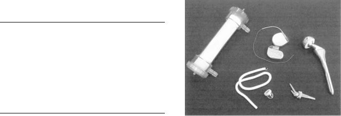
Figure 2. A representation of a few medical devices made from various biomaterials. From the upper left corner and moving clockwise, this picture shows a kidney hemodialyzer, two pacemakers, a hip replacement, an articulating wrist joint, a heart valve, and a vascular graft.
to gases and liquids, thermal and electrical properties, chemical reactivity, and degradation. Biological properties of materials largely focus on biocompatibility issues related to toxicity, immune system reactivity, thrombus formation, tribiology, inflammation, carcinogenic and teratogenic potential, integration with tissues and cells, and the ability to be sterilized. Regardless of the material, recent approaches to biomaterials research has focused on directing specific tissue interaction by using materials to introduce chemical bonds with the surrounding tissue, to act as scaffolds for tissue ingrowth, to introduce an inductive signal that will influence the behavior of surrounding cells or matrix, or to form new tissue when incubated or presented to transplanted cells.
Metals
Metals have been used as biomaterials for centuries. Although some fields (e.g., dentistry) continue to use amalgams, gold, and silver, most modern metallic biomaterials consist of iron, cobalt, titanium, or platinum bases. Since they are strong, metals are most often employed as biomaterials in orthopedic or fracture fixation medical devices; however, metals are also excellent conductors, and are therefore used for electrical stimulation of the heart, brain, nerves, muscle, and spinal cord. The most common alloys for orthopedic applications include stainless steel, cobalt, and titanium alloys. These alloys have enjoyed frequent use in medical procedures related to the function of joints and load-bearing. For example, metal alloys are commonly found in medical devices for knee replacement as well as in the femoral stem used in total hip replacements. Since all metals are subject to corrosion, especially in the salty, aqueous environment within the body, metals used as biomaterials often require an external oxide layer to protect against pitting and corrosion. These electrochemically inert oxide layers consist of Cr2O3 for stainless steel, Cr2O3 for cobalt alloys, and TiO2 for

BIOMATERIALS: AN OVERVIEW |
271 |
Figure 3. Photographs of stainless steel (a), cobalt–chromium (b), and titanium alloy (Ti6Al4V) (c) hip implants. (All three photographs are used with permission from the Department of Materials at Queen Mary University of London.)
titanium alloys. Figure 3 displays examples of three types of metallic hip replacements.
Stainless Steel Alloys. The stainless steel most commonly used as orthopedic biomaterials is classified 316L by the American Iron and Steel Institute. This particular austenitic alloy contains a very low carbon content (a maximum of 0.03%) and chromium content of 17–20%. The added chromium will react with oxygen to produce a corrosion-resistant chromium oxide layer. The 316L grade of stainless steel is a casting alloy, and its relatively high ductility makes this alloy amenable to extensive postcasting mechanical processing. Compared to cobalt and titanium alloys, stainless steel has a moderate yield and ultimate strength, but high ductility. Furthermore, it may be fabricated by virtually all machining and finishing processes and is generally the least expensive of the three major metallic alloys (4,5,9).
Cobalt Alloys. Cobalt alloys have been used since the early twentieth century as dental alloys and in heavily loaded joint applications. For use as a biomaterial, cobalt alloys are either cast (i.e., primarily formed within a mold) or wrought (i.e., worked into a final form from a large ingot). Two examples of cobalt alloys include Vitallium (designated F 75 by ASTM International), a cast alloy that consists of 27–30% chromium and >34% cobalt, and the wrought cobalt alloy MP35N (designated F 563 by ASTM International), which consists of 18–22% chromium, 15–25% nickel, and >34% cobalt. Compared to Vitallium, the MP35N alloy has demonstrated superior fatigue resistance, larger ultimate tensile strength, and a higher degree of corrosion resistance to chlorine. Consequently, this particular alloy is good for applications requiring long service life without fracture or stress fatigue. Compared to stainless steel alloys, cobalt-based alloys have slightly higher tensile moduli, but lower ductility. In addition, they are more expensive to manufacture and more difficult to machine. However, relative to stainless steel and titanium, cobalt-based alloys can offer the most useful balance of corrosion resistance, fatigue resistance, and strength (4,5,9).
Titanium Alloys. The most recent of the major orthopedic metallic alloys to be employed as biomaterials are titanium alloys. Although pure titanium is relatively weak and ductile, titanium can be stabilized by adding elements (e.g., aluminum and vanadium) to the alloy. Often, pure titanium (designated F 67 by ASTM International) is pri-
marily used as a surface coating for orthopedic medical devices. For load-bearing applications, the alloy Ti6Al4V (designated F 136 by ASTM International) is much more widely used in implant manufacturing. As in the case of stainless steel and cobalt alloys, titanium contains an outer oxide layer, composed of TiO2, that protects the implant from corrosion. In fact, of the three major orthopedic alloys, titanium shows the lowest rate of corrosion. Moreover, the density of titanium is almost half that of stainless steel and cobalt alloys. As a result, implants made from titanium are lighter and reduce patient awareness of the implant; however, titanium alloys are among the most expensive metallic biomaterials. Relative to stainless steel and cobalt alloys, titanium has a lower Young’s modulus, which can aid in reducing the stresses around the implant by flexing with the bone. Titanium has a lower ductility than the other alloys, but does demonstrate high strength. These properties allow titanium alloys to play a diverse role as a biomaterial. Titanium alloys are used in parts for total joint replacements, screws, nails, pacemaker cases, and leads for implantable electrical stimulators (4,5,9).
Despite the reduced weight and improved mechanical match of titanium alloy implants to bone relative to stainless steel and chromium alloy implants, titanium alloy implants still exhibit issues with regard to mechanical mismatch. This problem stems from the large differences in properties (e.g., elastic moduli) between bone, metals, and polymers used as acetabular cups. For example, metals have elastic moduli ranging from 100 to 200 GPa, ultrahigh molecular weight polyethylene has an elastic modulus of 1–2 GPa, and the elastic modulus of cortical bone is 12 GPa (10). In addition, it is difficult to produce a titanium implant surface that is conducive to bone ingrowth or attachment. Novel titanium foams have been investigated as a method for reducing implant weight, better matching tissue mechanics, and improving bone ingrowth. The process involves mixing titanium powder with ammonium hydrogen carbonate powder and compressing and heating the mixture to form foams with densities varying from 0.2 to 0.65 times the density of solid titanium. These densities are close to those of cancellous bone (0.2–0.3 times the density of solid titanium) and cortical bone (0.5–0.65 times the density of solid titanium) (11). While they are preliminary, studies with novel materials such as these titanium foams illustrate a trend toward the development of materials that better mimic the properties of the native tissue they are designed to replace.
272 BIOMATERIALS: AN OVERVIEW
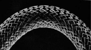
Figure 4. A photograph of the SMARTeR nitinol stent developed by the Cordis Corporation. (Reprinted from Ref. 12, with permission from Royal College of Radiologists.)
Other Metals. Besides stainless steel, cobalt alloys, and titanium alloys, there exist other examples of metals used as biomaterials. Some examples include nitinol, a singlephase nickel/titanium shape memory alloy, tantalum, a very dense, chemically inert, weak, but fatigue-resistant metal, and platinum, a very expensive metal used by itself or with iridum as a corrosion-resistant electrical conductor for electrode applications. Nitinol stents (e.g., that seen in Fig. 4) (12) and drug-eluting nitinol stents used for cardiovascular applications recently have seen enormous medical and commercial success. Indeed, metallic stents have significantly changed the way coronary blockages are treated (4,5,9).
Ceramics
Of the major types of materials used as biomaterials, ceramics have not been used as frequently as metals, polymers, or composites. However, ceramics continue to enjoy widespread use in certain bone-related applications (e.g., dentistry and joint replacement surgeries), due to their high compressive strength, high degree of hardness, excellent biocompatibility, superior tribological properties, and chemical inertness. Although they are very strong in compression, ceramics are susceptible to mechanical and thermal loading, have lower tensile strengths relative to other materials, and are very brittle in tension; this brittleness limits potential biomaterials applications.
Ceramics consist of a network of metal and nonmetal ions, with the general structure XmYn, arranged in a repeating structure. This structure depends on the relative size of the ions as well as the number of counterions needed to balance total charge. For example, if m ¼ n ¼ 1, and both ions are approximately the same size, then the structure would be of a simple cubic nature (e.g., CsCl or CsI); if the anion is much larger than the cation, then typically, a face centered cubic (fcc) structure would emerge (e.g., ZnS or CdS). If m ¼ 2 and n ¼ 3, as is the case with oxide ceramics (e.g., Al2O3), then a hexagonal closed pack structure would often result (13).
Ceramics used as biomaterials can be classified by processing–manufacturing methods, by chemical reactivity, or by ionic composition. Regarding chemical reactivity, ceramics can be bioinert, bioactive, or bioresorbable. Bio-
inert or nonresorbable ceramics are either porous or nonporous and are essentially not affected by the environment at the implant site. Bioactive or reactive ceramics are designed with specific surface properties that are intended to react with the local host environment and to elicit a desired tissue response. Bioresorbable ceramics dissolve over some prescribed period of time in vivo mediated by physiochemical processes. If one considers the application of bone replacement, then there would be about four ways for ceramics to interact with and attach to bone. First, a nonporous, inert ceramic material could be attached via glues, surface irregularities, or press-filling methods. Second, a porous, inert ceramic could be designed to have an optimal pore size, which promotes direct mechanical attachment of bone through bone ingrowth. Third, a nonporous, inert ceramic with a reactive surface could direct bone attachment via chemical bonding. Fourth, a nonporous or porous, resorbable ceramic could eventually be replaced by bone. When describing real examples of ceramics used as biomaterials, it is more useful to classify the ceramics based on ionic composition. This type of classification reveals a few major bioceramic groups: oxide ceramics, multiple oxides of calcium and phosphorus, glasses and glass ceramics, and carbon.
Oxide Ceramics. As their name implies, oxide ceramics consist of oxygen bound to a metallic species. Oxide ceramics are chemically inert, but can be nonporous or porous. One example of a nonporous oxide ceramic used as a biomaterial is aluminum oxide, Al2O3. Highly pure aluminum oxide (F 603 as designated by ASTM International), or alumina, has high corrosion resistance, good biocompatibility, high wear resistance, and good mechanical properties due to high density and small grain size. Aluminum oxide has been manufactured as an acetabular cup for total hip replacement. In comparison with metal or ultrahigh molecular weight polyethylene, Al2O3 provides better tribological properties by greatly decreasing friction within the joint and substantially increasing wear resistance. Recently, the FDA approved ceramic on ceramic hip replacements made from alumina and marketed by companies such as Wright Medical Technology and Stryker Osteonics. This ceramic on ceramic design is very resistant to wear and results in a much smaller amount of wear debris than traditional metal–polymer joints. With better wear properties and longer useful lifespan, ceramic on ceramic hip replacements likely will provide an attractive alternative to other biomaterial options, especially for younger patients that need better long-term solutions for joint replacements (4,5,9).
Ceramic oxides can also be porous. In bone formation, these pores are useful for allowing bone ingrowth, which will stabilize the mechanical properties of the implant without sacrificing the chemical inertness of the ceramic material. In general, there are three ways to make a porous ceramic oxide. First, a soluble metal or salt can be mixed with the ceramic and etched away. Second, a foaming agent that evolves gases during heating (e.g., calcium carbonate) can be mixed with the ceramic powder prior to firing. Third, the microstructure of corals can be used as a template to create a ceramic with a high degree
BIOMATERIALS: AN OVERVIEW |
273 |
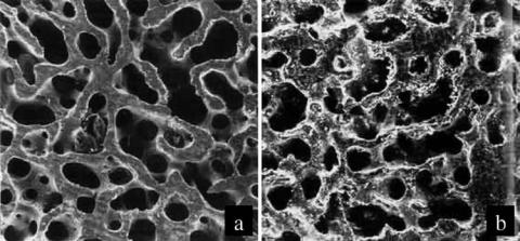
of interconnectivity and uniform pore size. In this third approach, coral is machined into the desired shape. Then, the coral is heated up to drive off carbon dioxide. The remaining calcium oxide provides a scaffold around which the ceramic material is deposited. After firing, the calcium oxide can be dissolved using hydrochloric acid. This dissolved calcium oxide will leave behind a very uniform and highly interconnected porous structure. Interestingly, the type of coral used will affect the pore size of the resulting ceramic. For example, if the genus Porites is used, then the pore size will range from 140 to 160 mm; the genus Goniopora will result in a pore size of 200–1000 mm (5). Porous ceramics do have many advantages for bone ingrowth, especially since the porous structure more closely mimics that of cancellous bone (see Fig. 5). However, the porous structure does result in a loss of strength and a tremendous increase in surface area that interacts with an in vivo saline environment.
Multiple Oxides of Calcium and Phosphorus. Aside from many types of proteins, the extracellular environment of bone contains a large concentration of organic mineral deposits known as hydroxyapatite. Chemically, hydroxyapatite generally has the following composition: Ca10(PO4)6(OH)2. Since hydroxyapatite is a naturally occurring ceramic produced by osteoblasts, it seemed reasonable to apply hydroxyapatite as filler or as a coating to allow better integration with existing bone. Coatings of hydroxyapatite have been applied (usually by plasma spraying) to metallic implants used in applications requiring bone ingrowth to provide a tight fit between bone and the implanted device, to minimize loosening over time, and to provide some measure of isolation from the foreign body response. Although hydroxyapatite is the most commonly used bioceramic containing calcium and phosphorus, there do exist other forms of calcium and phosphorus oxides including tricalcium phosphate, Ca3(PO4)2, and octacalcium phosphate, Ca8H2PO4 5H2O (4,5,9,14).
Glasses and Glass Ceramics. Just as in the case of traditional glass, glass ceramics used as biomaterials contain large amounts of silica, SiO2. Glass ceramics are formed using controlled crystallization techniques during
Figure 5. Photographs of (a) a cross-section of human cancellous bone and (b) coral of the genus Porites. These images illustrate how biologically derived materials (e.g., coral) can be used as scaffolds to create ceramic biomaterials that mimic the structure and porosity of natural bone. (Both photographs are used with permission from Biocoral, Inc.)
which silica is cooled down at rate slow enough to allow the formation of a hexagonal crystal structure with small, crystalline grains ( 1 mm) surrounded by an amorphous phase. Bioactive glass ceramics have been studied as biomaterials because they can attach directly to tissue via chemical bonds, they have a low thermal coefficient of expansion, they have good compressive mechanical strength, the mechanical strength of the glass–tissue interface is close to that of tissue, and they resist scratching and abrasion. Unfortunately, as with all ceramics, bioactive glasses are very brittle. Two well-known examples of commercially available glass ceramics include Bioglass, which consists of SiO2, Na2O, CaO, and P2O5, and Ceravital, which contains SiO2, Na2O, CaO, P2O5, K2O, and MgO. Relative to traditional soda lime glass, bioactive glass ceramics contain lower amounts of SiO2 and higher amounts of Na2O and CaO. The high ratio of CaO to P2O5 in bioactive ceramics allows the rapid formation of a hydroxycarbonate apatite (HCA) layer at alkaline pH. For example, a 50 nm layer of HCA can form from Bioglass 45S5 after 1 h. The release of calcium, phosphorus, and sodium ions from bioactive ceramics also allows the formation of a water-rich gel near the ceramic surface. This cationic-rich environment creates a locally alkaline pH that helps to form HCA layers and provide areas of adhesion for biological molecules and cells (4,5,9).
Carbon. Processed carbon has been used in biomaterials applications as a bioceramic coating. Although carbon can exist in several forms (e.g., graphite, diamond), bioceramic carbons consist primarily of low temperature isotropic (LTI) and ultralow temperature isotropic (ULTI) carbon. This form of carbon is synthesized through the pyrolysis of hydrocarbon gases resulting in the deposition of isotropic carbon in a layer 4 mm thick. Advantages to LTI and ULTI carbon include high strength, an elastic modulus close to that of bone, resistance to fatigue compared with other materials, excellent resistance to thrombosis, superior tribological properties, and excellent bond strength with metallic substances. The LTI carbon has been used as a coating for heart valves; however, applications remain limited primarily to coatings due to processing methods (4,5,9).
274 BIOMATERIALS: AN OVERVIEW
Polymers
Since the early to mid-twentieth century, the discovery of organic polymerization schemes and the advent of new polymeric species have fueled an incredible interest in the research of biomaterials. The popularity of polymers as potential biomaterials likely stems from the fact that polymers exist in a seemingly endless variety, can be easily fabricated into many forms, can be chemically modified or synthesized with chemically reactive moieties that interact with biological molecules or living tissues and cells, and can have physical properties that resemble that of natural tissues. Some disadvantages to polymeric biomaterials include relatively low moduli, instability following certain forms of sterilization, lot-to-lot variability, a lack of well-defined standards related to manufacturing, processing, and evaluating, and, for some polymers, hydrolytic instability, the need to add potentially toxic polymerization catalysts, and tissue biocompatibility of both the polymer and potential degradation byproducts. There also exist some characteristics of polymers that can be advantageous or disadvantageous depending on the application and type of polymer. Some of these characteristics include polymer degradation, chemical reactivity, polymer crystallinity, and viscoelastic behavior. Early examples of polymeric biomaterials included nylon for sutures and cellulose for kidney dialysis membranes, but more recent developments in the design of polymeric biomaterials are leading the field of biomaterials to embrace cellular and tissue interactions in order to directly induce tissue repair or regeneration.
Polymers consist of an organic backbone from which other pendant molecules extend. As their name implies, polymers consist of repeating units of one or more ‘‘mers’’. For example, polyethylene consists of repeating units of ethylene; nylon is comprised of repeating units of a diamine and a diacid. In general, polymers used as biomaterials are made in one of two ways: condensation or addition reactions. In condensation reactions, two precursors are combined to form larger molecules by eliminating a small molecule (e.g., water). Examples of condensation polymeric biomaterials include nylon, poly(ethylene terephthalate) (Dacron), poly(lactic acid), poly(glycolic acid), and polyurethane. In addition to synthetic polymers, biological polymers (e.g., cellulose and proteins) are formed through condensation-like polymerization mechanisms. The other major polymerization mechanism used to synthesize polymers is addition polymerization. In addition polymerization, an initiator or catalyst (e.g., free radical, heat, light, or certain ions) is used to promote a rapid polymerization reaction involving unsaturated bonds. Unlike condensation reactions, addition polymerization does not result in small molecular byproducts. Furthermore, polymers can be formed using only one type of monomer or a combination of several monomers susceptible to free radical initiation and propagation. Some examples of addition reaction polymeric biomaterials include polyethylene, poly(ethylene glycol) (PEG), poly(N-isopropylacrlyamide), and poly(hydroxyethyl methacrylate) (PHEMA). The chemical structure of various synthetic and natural polymers used as biomaterials are shown in Figs. 6a and b (15).
The properties of polymers are affected greatly by chemical composition and molecular weight. In general, as polymer chains become longer, their mobility decreases, but their strength and thermal stability increases. The tacticity and size of pendant chains off the backbone will affect temperature-dependent physical properties. For example, small side groups that are regularly oriented in an isotactic or syndiotactic arrangement will allow the polymer to crystallize much more readily than a polymer containing an atactic arrangement of bulky side groups. The crystalline and glass transition temperatures of polymers will affect properties (e.g., stiffness, mechanical moduli, and thermal stability) in vivo and will consequently influence the potential application and utility of the polymer system as a biomaterial. When the functionality of a monomer exceeds two, then the polymer will become branched upon polymerization. If a sufficient number of these high functionality monomers exist within the material, then the main chains of the polymer will become chemically cross-linked. Cross-linked polymers can be much stronger and more rigid than noncross-linked polymers. However, like linear and branched polymers, crosslinked polymers can be designed such that they degrade through hydrolytic or enzymatic mechanisms.
Due to their weaker moduli compared with that of metals or ceramics, polymers are not often used in loadbearing biomaterial applications. One exception to this observation is the example of ultrahigh molecular weight polyethylene (UHMWPE), which has a molecular weight2,000,000 g mol 1 and has a higher modulus of elasticity than high or low density polyethylene. Additionally, UHMWPE is tough and ductile and demonstrates good wear properties and low friction. As a result, UHMWPE has been used extensively in the manufacturing of acetabular cups for total hip replacements. As an acetabular cup, UHMWPE is used in conjunction with metallic femoral stems to act as a load-bearing, low wear and friction interface. Some drawbacks to using UHMWPE include water absorption, cyclic fatigue, and a somewhat significant creep rate (4,5,9). Part of the problems surrounding UHMWPE involves its lower elastic modulus ( 1–2 GPa) relative to bone ( 12 GPa) and metallic implants ( 100–200 GPa)
Polymers in Sutures. One of the first widespread uses of polymers as biomaterials involved sutures. In particular, polyamides and polyesters are among the most common suture materials. Nylons, an example of a polyamide, have an increased fiber strength due to a high degree of crystallinity resulting from interchain hydrogen bonding between atoms of the amide group. Nylon can be attacked by proteolytic enzymes in vivo and can absorb water. As a result, nylon has been used more as a short-term biomaterial. Polyester sutures, such as poly(glycolic acid), poly(lactic acid), and poly(lactic-co-glycolic acid) are readily degraded through hydrolytic mechanisms in vivo. Since one side chain of lactic acid contains a bulky hydrophobic methyl group (relative to the hydrogen side group of glycolic acid), polyesters comprised principally of lactic acid degrade at a rate slower than that of polyesters consisting mostly of glycolic acid. The degradation rate of copolymers

|
|
|
|
|
|
|
|
|
|
|
|
|
|
|
|
|
|
|
|
|
|
|
|
|
|
|
|
|
|
|
|
|
|
|
|
|
|
|
|
|
|
|
|
|
|
|
|
|
|
|
|
|
|
|
|
|
|
|
|
|
|
|
|
|
|
|
|
|
|
|
|
|
|
|
|
|
BIOMATERIALS: AN OVERVIEW |
275 |
|||||||||||
|
|
CH2 |
CH2 |
|
n |
|
|
|
|
|
|
|
|
|
O |
|
CH2 |
CH2 |
O |
|
|
O |
|
|
|
|
|
|
|
O |
|
|
|
|
|
|
|
|
|
|
|
|
|
|
|
|
|
|
|
|
|
|
|
|
|
|
|
|
|
|
|
|
|||||||||||||||||||||||||||
|
|
|
|
|
|
|
|
|
|
|
|
|
|
|
|
|
|
|
|
|
|
|
|
|
|
|
|
|
|
|
|
|
|
|
|
|
|
|
|
|
|
|
|
|
|
|
|
|
|
|
|||||||||||||||||||||||||||||||||||||||
|
|
|
|
|
|
|
|
|
|
|
|
|
|
|
|
|
|
|
|
|
|
|
|
|
|
|
|
|
|
|
|
|
|
|
|
|
|
|
|
|
|
|
|
|
|
|
|
|
|
|
|
|
|
|
|
|
|
|
|
||||||||||||||||||||||||||||||
1 |
|
|
|
|
|
|
|
|
|
|
|
|
|
|
|
|
|
|
|
|
|
|
|
|
|
|
|
|
|
|
|
|
|
|
|
n |
|
|
|
|
|
|
|
|
|
|
|
|
|
|
|
|
|
|
|
|
|
|
|
|
|
|
|||||||||||||||||||||||||||
|
|
|
|
|
|
|
|
|
|
|
|
|
|
|
|
|
|
|
|
|
|
|
|
|
|
|
|
|
|
|
|
|
|
|
|
|
|
|
|
|
|
|
|
|
|
|
|
|
|
|
|
|
|
|
|
|
|
|
|
|
|
|
|
|
|
|
|
|
|
|
|
|
|
|
|
|
|
||||||||||||
|
|
CF2 |
CF2 |
|
|
n |
|
|
|
|
|
|
|
|
|
|
|
|
|
|
|
|
|
|
|
|
|
|
|
|
|
7 |
|
|
|
|
|
|
|
|
|
|
CH3 |
|
|
|
|
|
|
|
|
|
|
|
|
|
|
|
|
|
|
|
|
|
|
|
|
|
|
|
|
||||||||||||||||||
|
|
|
|
|
|
|
|
|
|
|
|
|
|
|
|
|
|
|
|
|
|
|
|
|
|
|
|
|
|
|
|
|
|
|
|
|
|
|
|
|
|
|
|
|
|
|
|
|
|
|
|
|
|
|
|
|
|
|
|
|
|
|
|
|
|
|
|||||||||||||||||||||||
|
|
|
|
|
|
|
|
|
|
|
|
|
|
|
|
|
|
|
|
|
|
|
|
|
|
|
|
|
|
|
|
|
|
|
|
|
|
|
|
|
|
|
|
|
|
|
|
|
|
|
|
|
|
|
|
|
|
|
|
|
|
|
|
|
|
|
|
|
|
|
|
||||||||||||||||||
2 |
|
|
|
|
|
|
|
|
|
|
|
|
|
|
|
|
|
|
|
|
CH3 |
|
|
|
|
|
|
|
|
|
|
|
|
|
|
|
|
|
|
|
|
|
|
|
|
|
|
|
|
|
|
|
|
|
|
|
|
|
|
|
|
|
|
|
|
|
|
|
|
|
|||||||||||||||||||
|
|
|
|
|
|
|
|
|
|
|
|
|
|
|
|
|
|
|
|
|
|
|
|
|
|
|
|
|
|
|
|
|
|
|
|
|
|
|
|
|
|
|
|
|
|
|
|
|
|
|
|
|
|
|
|
|
|
|
|
|
|
|
|
|
|
|
|
|
|
||||||||||||||||||||
|
|
|
|
|
|
|
|
|
|
|
|
|
|
|
|
|
|
|
|
|
|
|
|
|
|
|
|
|
|
|
|
|
|
|
|
|
|
|
|
|
|
|
|
|
|
|
|
CH2 |
|
|
|
|
|
|
|
|
|
|
|
|
|
|
|
|
|
|
|
|
|
|
|
|
|
|
|
|
|
|
|
|
|||||||||
|
|
CH |
CH2 |
|
|
|
|
|
|
|
|
|
|
|
CH2 |
|
|
|
|
|
O |
|
|
|
|
|
|
|
|
|
|
|
|
|
|
|
|
|
|
|
|
O |
|
|
|
|
|
|
|
|
|
|
|
|
|
|
|
|
|
|
|
|
|
|
|
|
|
|
|||||||||||||||||||||
|
|
|
|
|
|
|
|
|
|
|
|
|
|
|
|
|
|
|
|
|
|
|
|
|
|
|
|
|
|
|
|
|
|
|
|
|
|
|
|
|
|
|
|
|
|
|
|
|
|
|
|
|
|
|
|
|
|
|
|
|
|
|
|||||||||||||||||||||||||||
|
|
|
|
|
|
|
|
|
|
|
|
|
|
|
|
|
|
|
|
|
|
|
|
|
|
|
|
|
|
|
|
|
|
|
|
|
|
|
|
|
|
|
|
|
|
|
|
|
|
|
|
|
|
|
|
|
|
|
|
|
|
|
|
|
|
|
|
|
|||||||||||||||||||||
|
|
|
|
|
|
|
|
|
|
|
|
|
|
|
|
|
|
|
|
|
|
|
|
|
|
|
|
|
|
|
|
|
|
|
|
|
|
|
|
|
|
|
|
|
|
|
|
|
|
|
|
|
|
|
|
|
|
|
|
|
|
|
|
|
|
|
|
||||||||||||||||||||||
|
|
|
|
|
|
|
|
|
|
|
|
|
|
|
|
|
|
|
|
|
|
|
|
|
|
|
|
|
|
|
|
|
|
|
|
|
|
|
|
|
|
|
|
|
|
|
|
|
|
|
|
|
|
|
|
|
|
|
|
|
|
|
|
|
|
|
|
|
|
|
|
|
|
|
|
|
|
|
|
|
|
|
|
|
|||||
|
|
OH |
|
|
|
|
|
|
n |
|
|
|
|
|
|
|
|
|
|
|
|
|
|
|
|
|
|
|
|
|
|
|
|
|
|
|
|
|
|
|
|
|
|
|
|
|
|
|
|
|
|
|
|
|
|
|
|
|
|
|
|
|
|
|
|
|
|
|
|
|
|
|
|
|
|
|
|
|
|
|
|||||||||
3 |
|
|
|
|
|
|
|
|
|
|
|
|
|
|
|
|
|
|
|
|
|
|
|
|
|
|
|
|
|
|
|
|
|
|
|
|
|
|
|
|
|
|
|
|
|
|
|
|
O |
|
|
|
|
|
|
|
|
|
|
|
|
|
|
|
|
|
|
|
|
|
|
|
|
|
|
||||||||||||||
|
|
|
|
|
|
|
|
|
|
|
|
|
|
|
|
|
|
|
|
|
|
O |
|
|
|
|
|
|
|
|
|
|
|
|
|
|
|
|
|
|
|
|
|
|
|
|
|
|
|
|
|
|
|
|
|
|
|
|
|
|
|
|
|
|
|
|
|
|
|
|
|
|
|||||||||||||||||
|
|
|
|
|
|
|
|
|
|
|
|
|
|
|
|
|
|
|
|
|
|
|
|
|
|
|
|
|
|
|
|
|
|
|
|
|
|
|
|
|
|
|
|
|
|
|
CH2 |
|
|
|
|
|
|
|
|
|
|
|
|
|
|
|
|
|
|
|
|
|
|
|
|
|
|
||||||||||||||||
|
|
CH3 |
|
|
|
|
|
|
|
|
|
|
|
|
|
|
|
|
|
|
|
|
|
|
|
|
|
|
|
|
|
|
|
|
|
|
|
|
|
|
|
|
|
|
|
|
|
|
|
|
|
|
|
|
|
|
|
|
|
|
|
|
|
|
|
|
|
|
|
|
|
|
|
|
|
|
|
|
|||||||||||
|
|
|
|
|
|
|
|
|
|
|
|
|
|
|
|
|
|
|
|
|
|
|
|
|
CH |
|
|
|
|
|
|
|
|
|
|
|
|
|
|
|
|
|
|
|
|
|
|
|
|
|
|
|
|
|
|
|
|
|
|
|
|
|
|
|
|
|
|
|
|
|
|
|
|||||||||||||||||
|
|
|
|
|
|
|
|
|
|
|
|
|
|
|
|
|
|
|
|
|
|
|
|
|
|
|
|
|
|
|
|
|
|
|
|
|
|
|
|
|
|
|
|
|
|
|
|
|
|
|
|
|
|
|
|
|
|
|
|
|
|
|
|
|
|
|
|
|
|
|
|
|
|
|
|
|
|
|
|
||||||||||
|
|
Si O |
|
|
|
|
|
|
|
|
|
|
|
|
|
|
|
|
|
|
|
|
8 |
3 |
|
|
n |
|
|
|
|
|
|
|
|
|
|
|
|
|
|
|
|
CH2 |
|
|
|
|
|
|
|
|
|
|
|
|
|
|
|
|
|
|
|
|
|
|
|
|
|
|
|||||||||||||||||||
|
|
|
|
|
|
|
|
|
|
|
|
|
|
|
|
|
|
|
|
|
|
|
|
|
|
|
|
|
|
|
|
|
|
|
|
|
|
|
|
|
|
|
|
|
|
|
|
|
|
|
|
|
|
|
|
|
|
|
|
|
|
|
|
|
|
|
|
|
|
|
|
||||||||||||||||||
|
|
|
|
|
|
|
|
|
|
|
|
|
|
|
|
|
|
|
|
|
|
|
|
|
|
|
|
|
|
|
|
|
|
|
|
|
|
|
|
|
|
|
|
|
|
|
|
|
|
|
|
|
|
|
|
|
|
|
|
|
|
|
|
|
|
|
|
|
|
||||||||||||||||||||
|
|
|
|
|
|
|
|
|
|
|
|
|
|
|
|
|
|
|
|
|
|
|
|
|
|
|
|
|
|
|
|
|
|
|
|
|
|
|
|
|
|
|
|
|
|
|
|
|
|
|
|
|
|
|
|
|
|
|
|
|
|
|
|
|
|
|
|
|
|
|
|
|
|
|
|
|
|
|
|
|
|
|
|
|
|
|
|||
|
|
CH3 |
|
|
n |
|
|
|
|
|
|
|
|
|
|
|
|
|
|
|
|
|
|
|
|
|
|
|
|
|
|
|
|
|
|
|
|
|
|
|
|
|
|
|
|
|
|
|
|
OH |
|
|
n |
|
|
|
|
|
|
|
|
|
|
|
|
|
|
|
|
|
|
|
|
|
|||||||||||||||
4 |
|
|
|
|
|
|
|
|
|
|
|
|
|
|
|
|
|
|
|
|
|
|
|
|
|
|
|
|
|
|
|
|
|
|
|
|
|
|
|
|
|
|
|
|
|
|
|
|
9 |
|
|
|
|
|
|
|
|
|
|
|
|
|
|
|
|
|
|
|
|
|
|
|
|
|
|
|
|
|
|
||||||||||
|
|
|
|
|
|
|
|
|
|
|
|
|
|
|
|
|
|
|
|
|
|
|
|
|
|
|
|
|
|
|
|
|
|
|
|
|
|
|
|
|
|
|
|
|
|
|
|
|
|
|
|
CH3 |
|
|
|
|
|
|
|
|
|
|
|
|
|
|
|
|
|
|
|
|
|
|
|
|
|
|
|||||||||||
|
|
|
|
|
|
|
|
|
|
|
|
|
|
|
|
|
|
|
|
|
|
|
|
|
|
|
|
|
|
|
|
|
|
|
|
|
O |
|
|
|
|
|
|
|
|
|
|
|
|
|
|
|
|
|
O |
|
|
|
|
|
|
|
|
|
|
|
|
|
|
|
|
|
|
|
|
|
|
|
|||||||||||
|
|
|
|
|
|
|
|
|
|
|
|
|
|
|
|
|
|
|
|
|
|
|
|
|
|
|
|
|
|
|
|
|
|
|
|
|
|
|
|
|
|
|
|
|
|
|
|
|
|
|
|
|
|
|
|
|
|
|
|
|
|
|
|
|
|
|
|
|
|
|
|
|
|
|
|
||||||||||||||
|
|
|
|
|
|
|
CH |
|
|
|
|
|
|
|
|
|
|
|
|
|
|
|
|
|
|
O |
|
|
CH2 |
|
|
|
|
|
|
|
|
|
|
|
|
|
|
|
|
O |
|
CH |
|
|
|
|
|
|
|
|
|
|
|
|
|
|
|
|
|
|
|
|
|
|
|
|
|
|
|||||||||||||||
|
|
|
|
|
|
|
|
|
|
|
|
|
|
|
|
|
|
|
|
|
|
|
|
|
|
|
|
|
|
|
|
|
|
|
|
|
|
|
|
|
|
|
|
|
|
|
|
|
|
|
|
|
|
|
|
|
|
|
|
|
|
|
|
|
|
|
|
|
|||||||||||||||||||||
|
|
CH2 |
|
|
|
|
|
|
|
|
|
|
|
|
|
|
|
|
|
|
|
|
|
|
|
|
|
|
|
|
|
|
n |
|
|
|
|
|
|
|
|
|
|
|
|
|
|
|
|
|
|
|
|
|
n |
|
|
|
|
|
|
|
|
|
|
|
|
|
|||||||||||||||||||||
|
|
|
|
|
|
|
|
|
|
|
|
O |
|
|
|
|
|
|
|
|
|
|
|
|
|
|
|
|
|
|
|
|
|
|
10 |
|
|
|
|
|
|
|
|
|
|
|
|
|
|
11 |
|
|
|
|
|
|
|
|
|
|
|
|
|
|
|
|
|
|
|
|
|||||||||||||||||||
|
|
|
|
|
|
|
|
|
|
|
|
|
|
|
|
|
|
|
|
|
|
|
|
|
|
|
|
|
|
|
|
|
|
|
|
|
|
|
|
|
|
|
|
|
|
|
|
|
|
|
|
|
|
|
|
|
|
|
|
|
|
|
|
|
|
|
|
|
|
|
|
|
|
|
|
|
|
|
|
|
|
||||||||
|
|
|
|
|
|
|
|
|
|
|
|
|
|
|
|
|
|
|
|
|
|
|
|
|
|
|
|
|
|
|
|
|
|
|
|
|
|
|
|
|
|
|
|
|
|
|
|
|
|
|
|
|
|
|
|
|
|
|
|
|
|
|
|
|
|
|
|
|
|
|
|
|
|
|
|
|
|
|
|
|
|||||||||
|
|
|
|
|
|
|
|
|
|
|
|
|
|
|
|
|
|
|
|
|
|
|
|
|
|
|
|
|
|
|
|
|
|
|
|
|
|
|
|
|
|
|
|
|
|
|
|
|
|
|
|
|
|
|
|
|
|
|
|
|
|
|
|
|
|
|
|
|
|
|
|
|
|
|
|
|
|
|
|
|
|
|
|||||||
|
|
|
|
|
|
|
|
NH |
|
|
|
|
|
|
|
|
|
|
|
|
|
|
|
|
|
|
|
|
|
|
|
|
|
|
|
|
|
|
|
|
|
|
|
|
|
|
|
|
|
|
|
|
|
|
|
|
|
|
|
|
|
|
|
|
|
|
|
|
|
|
|
|
|
|
|
|
|
|
|
|
|
|
|
|
|
|
|||
|
|
|
H3C |
CH |
|
|
|
|
|
|
|
|
|
|
O |
|
CH3 |
|
|
|
|
|
O |
|
|
|
|
CH3 |
|
|
|
|
|
|
O |
|
|
|
|
|
|
|
|
|
|
|
|
O |
|
|
|
|
|
|
|
|
|
|
|
|
|
||||||||||||||||||||||||||||
|
|
|
|
|
|
|
CH3 |
|
|
|
|
|
|
|
|
|
|
|
|
|
|
|
|
|
|
|
|
|
|
|
|
|
|
|
|
|
|
|
|
|
|
|
|
|
|
|
|
|
|
|
|
|
|
|
|
|
|
|
|
|
|
|
|
|
|
|
|
|
|
|
|
|
|
|
|
|
|
|
|
|
|
||||||||
|
|
|
|
|
|
|
n |
|
|
|
|
|
|
|
CH |
|
|
O |
|
|
|
|
CH |
O |
|
|
|
|
|
|
|
|
CH2 |
|
|
O |
|
|
|
|
|
CH2 O |
|
|
|
|
|
|
|
|
|||||||||||||||||||||||||||||||||||||||
|
|
|
|
|
|
|
|
|
|
|
|
|
|
|
|
|
|
|
|
|
|
|
|
|
|
|
|
|
|
|
|
|
|
|
|
|
|
|
|
|
|
|
|
|
|
|
|
|
|
|
|
||||||||||||||||||||||||||||||||||||||
|
|
|
|
|
|
|
5 |
|
|
|
|
|
|
|
|
|
|
|
|
|
|
|
|
|
|
|
|
|
|
|
|
|
|
|
|
|
|
|
|
|
|
|
|
|
|
|
|
|
|
|
m |
|
|
|
|
|
|
|
|
|
|
|
|
|
|
|
|
|
|
|
|
|
|
|
|
|
|
n |
|
|
|
|
|
|
|
|
|||
|
|
|
|
|
|
|
|
|
|
|
|
|
|
|
|
|
|
|
|
|
|
|
|
|
|
|
|
|
|
|
|
|
|
|
|
|
|
|
|
|
|
|
|
|
|
|
|
|
|
|
12 |
|
|
|
|
|
|
|
|
|
|
|
|
|
|
|
|
|
|
|
|
|
|
|
|
|
|
|
|
|
|
|
|
|
|
|
|
||
|
|
|
O |
|
|
O |
|
|
|
|
|
|
|
|
|
|
|
|
|
|
|
|
|
CH2 |
CH2 |
O |
|
|
|
|
|
|
|
|
|
|
|
O |
|
CH2 |
|
|
|
|
|
|
|
O |
|
|
|
|
|
|
|
|
|
|
|
|
|
|
|
|
|
||||||||||||||||||||||||
|
|
|
|
|
|
|
|
|
|
|
|
|
|
|
|
|
|
|
|
|
|
|
|
|
|
|
|
|
|
|
|
|
|
|
|
|
|
|
|
|
|
|
|
|
|
|
|
|
|
|
|
|
|
|
|
|
|
||||||||||||||||||||||||||||||||
|
|
|
|
|
|
R |
|
|
|
|
O |
|
|
|
|
|
|
|
|
|
|
|
|
|
|
|
|
|
|
|
|
|
|
|
|
|
|
|
|
|
|
|
|
|
|
|
|
|
|
|
|
|
|
|
|
|
|
|
|
|
|
|
|||||||||||||||||||||||||||
|
|
|
|
|
|
|
|
|
|
|
n |
|
|
|
|
|
|
n |
|
|
|
|
|
|
|
|
|
|
|
5 |
|
|
|
|
n |
|
|
|
|
|
|
|
|
|
|
|
|
|
|||||||||||||||||||||||||||||||||||||||||
|
|
|
|
|
|
|
|
|
|
|
|
|
|
|
|
|
|
|
|
|
|
|
|
|
|
|
|
|
|
|
|
|
|
|
|
|
|
|
|
|
|
|
|
|
|
|
|
|
|
|
|
|
|
|
|
|
|
|
|
|
|
|
|
|
|
|
|
|
|
|
|
|
|
||||||||||||||||
|
|
|
|
|
|
|
6 |
|
|
|
|
|
|
|
|
|
|
|
|
|
|
|
|
|
|
|
13 |
|
|
|
|
|
|
|
|
|
|
|
|
|
|
|
|
|
|
|
|
14 |
|
|
|
|
|
|
|
|
|
|
|
|
|
|
|
|
|
|
|
|
|
|
|
|
|
||||||||||||||||
|
|
|
|
|
|
|
|
|
|
|
|
|
|
|
|
|
|
|
|
|
|
|
|
|
|
|
|
|
|
|
|
|
|
|
|
|
|
|
|
|
|
|
|
|
|
|
|
|
|
|
|
|
|
|
|
|
|
|
|
|
|
|
|
|
|
|
|
|
|
|
|
|
|
|
|
|
|
|
|||||||||||
|
|
|
|
|
|
|
|
|
|
|
|
|
|
|
|
|
O |
|
|
|
|
|
|
|
|
|
|
|
|
|
|
|
|
|
|
|
|
|
|
|
|
|
|
|
|
|
|
|
|
|
|
|
|
|
|
|
|
|
|
|
|
|
|
|
|
|
|
|
|
|
|
|
|
|
|
|
|
|
|
|
|
|
|||||||
|
|
|
OH |
|
|
|
|
|
O |
|
|
|
|
OH |
|
|
|
|
|
|
|
|
|
|
|
|
|
|
|
|
|
COOH |
|
|
|
CH2OSO3H |
|
|
|
|
|
|
|
|
|
|
|
|
|
CH2OH |
|
|
|
|
|
|
|
|
|||||||||||||||||||||||||||||||
|
|
|
|
|
|
|
|
|
|
|
|
|
|
|
|
|
|
O |
|
|
|
|
|
|
|
|
|
|
|
|
|
|
|
|
|
|
|
|
|
|
|
|
|
|
|
|
|
|
|
|
|
|
|
|
|
|
|
|
|
||||||||||||||||||||||||||||||
|
|
|
|
|
|
|
|
|
O |
|
|
|
|
|
|
|
|
|
|
|
|
|
|
O |
|
|
|
|
|
|
|
|
|
|
HO |
|
|
|
|
|
|
|
|
|
|
|
|
|
|
|
|
|
|
|
|
|
|||||||||||||||||||||||||||||||||
|
|
|
|
|
O |
|
|
|
|
|
|
|
|
|
|
|
|
|
|
|
|
|
|
|
|
|
|
|
|
|
|
|
|
|
|
|
|
|
O |
|
|
|
O |
|
|
|
|
|
|
|
|
|
|
|
|
|
|
|
|
|
|
|
O |
|
|
|
|
|
|
|
|
||||||||||||||||||
|
|
|
|
|
|
|
|
|
HO |
|
|
|
|
|
|
|
|
|
|
|
|
|
|
|
|
|
|
|
|
|
|
|
|
|
OH |
|
|
|
|
|
|
|
|
|
|
|
|
O |
|
|
|
|
|
|
|
|
|
|
|
|
|
|
|
|
|
|
|
|
|
|
|||||||||||||||||||
|
|
|
|
|
|
|
|
|
|
|
|
|
|
|
|
|
|
|
|
|
|
|
|
|
|
|
|
|
|
|
|
|
|
|
|
|
|
|
|
|
|
|
|
|
|
|
|
|
|
|
|
|
|
|
|
|
|
|
|
|
|
|
|
|
|
|
|
|
|
|
|
|
|
|
|
|
|
|
|
||||||||||
|
|
|
|
|
|
|
|
|
OH |
|
|
|
|
|
|
|
|
|
|
|
|
|
|
|
|
|
|
|
|
|
|
|
|
|
|
|
|
O |
|
|
|
|
|
|
|
|
|
|
|
|
|
|
|
|
|
|
|
|
|
OH |
O |
|
|
|
|
|
|||||||||||||||||||||||
|
|
|
|
|
|
|
|
|
|
|
|
|
|
|
|
|
|
|
|
|
|
|
|
|
|
|
|
|
|
|
|
|
|
|
|
|
|
|
|
|
|
|
|
|
|
|
|
|
|
|
|
|
|
|
|
|
|
|
|
|
|
|
|
|
|
|
|
|
|
|
|
|
|
|
|||||||||||||||
|
|
|
|
|
|
|
|
|
|
|
|
|
|
|
|
|
|
|
|
|
|
|
|
|
|
|
|
|
|
|
|
|
|
|
|
|
|
|
|
|
|
OH |
|
|
|
|
|
|
HNCOCH3 |
|
|
|
|
|
|
|
|
|
|
|
|
|
|
|
|
|
|
|
|
|
|||||||||||||||||||
|
|
|
|
|
|
|
|
|
|
|
|
|
|
|
|
|
|
|
|
|
|
|
|
|
|
|
|
|
|
|
|
|
|
|
|
|
|
|
|
|
|
|
|
|
|
|
|
|
|
|
|
|
|
|
|
|
|
|
|
NH2 |
|
|
|
|
|
|
|
|
|||||||||||||||||||||
|
|
|
|
|
|
|
|
|
|
|
|
|
|
|
|
|
|
|
|
|
|
|
|
|
|
|
|
|
|
|
|
|
|
|
|
|
|
|
|
|
|
|
|
|
|
|
|
|
|
|
|
|
|
|
|
|
|
|
|
|
|
|
n |
|
|
|
|
|
|||||||||||||||||||||
|
O |
|
|
|
|
|
|
|
|
|
|
|
|
|
|
|
|
|
|
|
|
|
|
|
|
|
|
|
|
|
|
|
|
|
|
|
|
|
|
|
|
|
|
|
|
|
|
|
|
|
|
|
|
|
|
|
|
|
|
|
n |
|
|
|
|
|
|
|
|
|
|
|
|
|
|
|
|
|
|
|
|||||||||
|
|
|
|
|
|
O– |
|
|
|
|
15 |
|
|
|
|
|
|
|
|
|
|
|
|
|
|
|
n |
|
|
|
|
|
|
|
|
|
|
|
|
|
|
|
|
16 |
|
|
|
|
|
|
|
|
|
|
|
|
|
|
|
|
|
|
|
|
|
|
|
17 |
|
|
|
|
|
|
|
|
|
|
|
||||||||||
|
|
|
|
|
|
|
|
|
|
|
|
|
|
|
|
|
|
|
|
|
|
|
|
|
|
|
|
|
|
|
|
|
|
|
|
|
|
|
|
|
|
|
|
|
|
|
|
|
|
|
|
|
|
|
|
|
|
|
|
|
|
|
|
|
|
|
|
|
|
|
|
|
|
|
|
|
|
|
|
|
|||||||||
|
|
|
|
|
|
|
|
|
|
|
|
|
|
|
|
|
|
|
|
|
|
|
|
|
|
|
|
|
|
|
|
|
|
|
|
|
|
|
|
|
|
|
|
|
|
|
|
|
|
|
|
|
|
|
|
|
|
|
|
|
|
|
|
|
|
|
|
|
|
|
|
|
|
|
|
|
|
|
|
NH2 |
|
|
|
|
|
||||
|
|
|
|
|
|
|
|
|
|
|
|
|
|
|
|
|
|
|
|
|
|
|
|
|
|
|
|
|
|
|
|
|
|
|
|
|
|
|
|
|
|
|
|
|
|
|
|
|
|
|
|
|
|
|
|
|
|
|
|
|
|
|
|
|
|
|
|
|
|
|
|
|
|
|
|
|
|
|
|
|
|
|
|
|
|
|
|||
|
|
|
COOH |
|
|
|
|
CH2OH |
|
|
|
|
|
|
|
|
|
|
|
|
|
|
|
|
|
|
|
|
|
|
|
|
|
|
|
|
|
HO |
|
|
|
|
|
|
|
|
|
|
|
|
|
|
|
|
|
|
|
|
|
|
|
|
|
|
|
|
|
|
|
|
|||||||||||||||||||
|
|
|
|
|
|
|
|
|
|
|
|
|
|
|
|
|
|
|
|
|
|
|
|
|
|
|
|
|
|
|
|
|
|
|
|
|
|
|
|
|
|
|
|
|
|
|
|
|
|
|
|
|
|
|
|
|
|
|
|
|
|
|
|
|
|
|
|
|
|
|
|
||||||||||||||||||
|
|
|
|
|
|
O |
|
|
|
|
|
|
|
O |
|
|
|
|
|
|
|
|
|
|
|
|
|
|
|
|
|
|
|
|
|
|
|
|
|
|
|
O |
|
|
|
|
|
|
|
|
|
|
|
|
|
|
|
|
|
|
|
|
|
|
|
|
|
|
|
|
|
|
|
|
|
|
|
|
|||||||||||
|
|
|
OH |
|
|
|
|
|
O |
|
|
|
|
|
|
|
|
|
O |
|
|
|
|
|
|
|
H |
|
|
N |
|
|
|
|
|
|
N |
|
|
|
|
|
|
|
|
N |
|
|
|
|
|
|
|
|
|
OH |
|
|
|
|
|
|
|
|
|
|
|
|
|
||||||||||||||||||||
|
|
|
|
|
|
|
|
|
|
|
|
|
|
|
|
|
|
|
|
|
|
|
|
|
|
|
|
|
|
|
|
|
|
|
|
|
|
|
|
|
|
|
|
|
|
|
|
|
|
|
|
|
|
|
|
||||||||||||||||||||||||||||||||||
|
|
|
|
|
|
|
|
|
|
|
|
|
|
|
|
|
|
|
|
|
|
|
|
|
|
|
|
|
|
|
|
|
|
|
|
|
|
|
|
|
|
|
|
|
|
|
|
|
|
|
|
|
|
|
|
|
|
|
|
|
|
|
|
|
|
|
|
|
|||||||||||||||||||||
|
|
|
|
|
|
|
|
|
|
|
|
HO |
|
|
|
|
|
|
|
|
|
|
|
|
|
|
|
|
|
|
|
|
|
H |
|
|
|
|
|
|
|
|
|
|
|
|
|
|
|
|
|
|
|
|
|
|
|
|
|
|
|
|
|
|
|
|
|
|
|
|
N |
|
|
|
|
|
|
|
|
|
|
|
|||||||
|
|
|
|
|
|
OH |
|
|
|
HNCOCH3 |
|
|
|
|
|
|
|
|
|
O |
|
|
|
|
|
|
|
|
|
|
|
|
|
|
|
|
O |
|
|
|
|
|
|
|
|
|
|
|
|
|
|
|
|
|
|
||||||||||||||||||||||||||||||||||
|
|
|
|
|
|
|
|
|
|
|
|
|
|
|
|
|
|
|
|
|
|
|
|
|
|
|
|
|
|
|
|
|
|
|
|
|
|
|
|
|
|
|
|
|
|
|
|
|
|
|
|
|
|
|
|
|
|
|
|||||||||||||||||||||||||||||||
|
|
|
|
|
|
|
|
|
|
|
|
|
|
|
|
|
|
|
|
|
|
|
|
|
|
|
|
|
|
|
|
|
|
|
|
|
|
|
|
|
|
|
|
|
|
|
|
|
|
|
O n |
|
|
|
|
|
|||||||||||||||||||||||||||||||||
|
|
|
|
|
|
|
|
|
|
|
|
|
|
|
|
|
|
|
|
n |
|
|
|
|
|
|
|
|
|
|
|
|
|
|
|
|
|
|
|
|
|
|
|
|
|
|
|
|
|
|
|
|
|
|
n |
|
|
|
|
|
|
|
|
|
|
|
|
|
|
||||||||||||||||||||
|
|
|
|
|
|
|
|
|
|
|
|
|
18 |
|
|
|
|
|
|
|
|
|
|
|
|
|
|
|
|
|
|
|
|
|
|
|
|
|
|
|
|
|
|
19 |
|
|
|
|
|
|
|
|
|
|
|
|
|
|
|
|
|
|
|
|
|
|
|
|
|
20 |
|
|
|
|
|
|
|
|
|
|
|
|
|||||||
|
|
|
|
|
CH2 |
|
|
|
|
|
|
|
CH2 |
|
|
|
|
|
|
CH2 |
|
|
|
|
|
|
|
|
|
|
|
|
|
|
|
|
|
|
|
|
CH2OSO3H COOH |
|
|
CH2OSO3H |
|
|
|
||||||||||||||||||||||||||||||||||||||||||
|
|
|
|
|
|
|
O |
|
|
|
|
|
|
|
|
O |
|
|
|
|
|
|
|
|
|
|
O |
|
|
|
|
|
|
|
|
|
|
O |
|
|
|
|
|
|
|
|
|
|
|
|
O |
|
|
|
|
|
|
|
|
O |
|
|
|
O |
|
|
|
||||||||||||||||||||||
|
|
|
|
|
OH |
|
|
|
|
|
|
|
O |
|
|
|
|
|
|
OH |
|
|
|
|
|
|
|
|
|
|
|
|
|
COOH |
|
O |
|
|
|
OH |
|
|
|
O |
OHH |
O OH |
O |
|
|
|
|
||||||||||||||||||||||||||||||||||||||
|
|
|
|
|
|
|
|
|
|
|
|
|
|
|
|
|
|
|
|
O |
|
|
|
|
|
|
|
OH |
|
|
|
|
|
|
|
|
|
|
|
|
|
|
|||||||||||||||||||||||||||||||||||||||||||||||
|
HO |
|
|
O |
|
|
|
HO |
|
|
|
O |
|
|
|
HO |
|
|
|
|
|
|
|
|
|
|
|
|
|
|
|
|
|
|
|
|
|
|
|
|
|
|
|
|
|
|
|
|
|
|
|
|
|
|
|
|
|
|
|
|
|
|
|
|
|
|
|
|
|
|
|||||||||||||||||||
|
OH |
|
|
|
|
OH |
|
|
|
|
|
|
NH2 |
|
|
|
|
|
|
|
|
|
|
OSO3H |
|
|
|
|
|
|
|
|
HNSO3H |
OH |
|
|
|
HNSO3H |
|
|
|
||||||||||||||||||||||||||||||||||||||||||||||||
|
|
|
|
|
|
|
|
|
|
|
|
|
|
|
|
|
|
|
|
|
|
|
|
|
n |
|
|
|
|
|
|
|
|
|
|
|
|
|
|
n |
|
||||||||||||||||||||||||||||||||||||||||||||||||
|
|
|
|
|
|
|
|
|
CH2OH |
|
|
|
|
|
|
|
|
|
|
|
|
|
|
|
|
|
|
|
|
|
|
|
|
|
|
|
|
|
|
|
|
|
|
|
|
|
|
|
|
|
|
|
|
|
|
|
|
|
|
|
|
|
|
|
|
|
|
|
|
|
|
||||||||||||||||||
|
|
|
|
|
|
|
|
|
|
|
|
|
|
|
|
|
|
|
|
|
|
|
|
|
|
|
|
|
|
|
|
|
|
|
|
|
|
|
|
|
|
|
|
|
|
|
|
|
|
|
|
|
|
|
|
|
|
|
|
|
|
|
|
|
|
|
|
|
|
|
|
|
|
|
|
|
|
|
|
||||||||||
O |
22 |
|
OH
HO
21
OH
Figure 6. (a) Chemical schematics representing synthetic polymers used as biomaterials. The structures represent polyethylene (1), polytetrafluoroethylene (2), poly(vinyl alcohol) (3), poly(dimethyl siloxane) (4), poly(N-isopropylacrylamide) (5), polyanhydride (6), poly(ethylene terephthalate) (7), poly(methyl methacrylate) (8), poly(hydroxyethyl methacrylate) (9), poly(glycolic acid) (10), poly(lactic acid) (11), poly(lactic-co-glycolic acid) (12), poly(ethylene oxide) (13), and poly(e-capro- lactone) (14). (Adopted from Ref. 15 with permission from Elsevier.) (b) Chemical schematics representing naturally derived polymers used as biomaterials. The structures represent alginate (15), chondroitin-6-sulfate (16), chitosan (17), hyaluronan (18), collagen (19), polylysine (20), dextran (21), and heparin (22). (Reprinted from Ref. 15 with permission from Elsevier.)
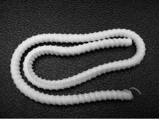
276 BIOMATERIALS: AN OVERVIEW
Figure 7. A photograph of a Carboflo vascular graft made out of expanded polytetrafluoroethylene (ePTFE) impregnated with carbon and marketed by Bard Peripheral Vascular, Inc.
of glycolic and lactic acid can be tailored based on the relative molar ratios of each monomer. Although the local pH of degrading polyesters can cause local inflammation concerns, the degradation byproducts of glycolic and lactic acid can be readily cleared through existing biochemical pathways. As a result, polyester sutures are commonly used within the body in applications where removal of sutures would warrant an invasive procedure (4,5,16).
Polymers in Cardiovascular Applications. Poly(ethylene terephalate) (Dacron) and expanded polytetrafluoroethylene (Teflon) have been used for decades as vascular grafts. An example of a Teflon vascular graft is shown in Fig. 7. Both of these polymers have excellent burst strengths and can be sutured directly to existing vasculature. For applications involving large diameter vascular grafts (> 6 mm), these two materials have worked well. However, neointimal hyperplasia and thrombus formation severely limit the patency of all known polymeric materials used for small diameter vascular grafts (17). Most current strategies to improve vascular graft patency involves chemically modifying the polymers used as vascular grafts to include the anticoagulant heparin, endothelial binding peptide analogues, and growth factors to stimulate endothelialization and minimize proliferation of smooth muscle into the lumen of the graft (15,18).
Polymers for Tissue Engineering. For many in vivo applications, researchers continue to evaluate a variety of polymeric biomaterials. Some more recent additions to the repertoire of biomaterials include naturally derived or recombinantly produced biological polymers. As an example, in the case of articular cartilage repair, it is evident that many types of polymers can be designed, modified, or combined with other materials to create new generations of biomaterials that promote healing and/or restore biological function. For example, synthetic polymers, such as poly(vinyl alcohol) (PVA), PMMA, poly(hydroxyethyl methacrylate), poly(N-isopropylacrylamide), polyethylene, poly(lactic acid), poly(glycolic acid), poly(lactic-co-glycolic
acid), and poly(ethylene glycol) and naturally derived polymers (e.g., alginate, agarose, chitosan, hyaluornic acid, collagen, and fibrin) have been studied extensively with and without biochemical modifications to replace cartilage function or to promote neocartilage formation (15,19,20). These and other polymeric biomaterials have been used in studies related to liver, nerve, cardiovascular, bone, ophthalmic, skin, and pancreatic repair or restoration (15,21).
Hydrogels. As the name implies, hydrogels are polymer networks that contain large amounts of water (up to or > 90% water). As a result, hydrogels generally are hydrophilic materials, although, the presence of hydrophobic domains within the hydrogel backbone can enhance mechanical properties. To avoid dissolution into the aqueous phase, the polymeric component of the hydrogel must contain cross-links. The majority of hydrogel systems use chemical cross-links, such as covalent bonds to create a three-dimensional (3D) network; however, some hydrogels exist that rely on physical interactions to maintain gel integrity.
The high water content of hydrogels provides many benefits. First, of all the materials within materials science, the physical and mechanical properties of hydrogels most closely resemble those of biological tissue. Due to their polymeric content, hydrogels exhibit viscoelastic behavior. The elastic modulus, G’, of many gel compositions reaches 1 MPa, but some hydrogels can be as strong as 20 MPa. These mechanical properties match well with those reported for many tissues. Second, the large presence of water within hydrogels can limit nonspecific interactions within the body, can shield the polymer from leukocytes and can decrease frictional effects at the site of implantation. Third, the relatively low concentration of polymer within the hydrogel can result in materials with higher porosities. Consequently, it is possible not only for cells to migrate within the hydrogel structure, but also for nutrients and waste products to diffuse into and out of the gel structure (15,22,23).
In addition to high water content, hydrogels possess other characteristics that are beneficial for biomedical applications. For example, chemical composition of polymers used in hydrogel formulations is amenable to chemical modification of the backbone and/or side group structures. These polymer derivatives allow the incorporation of various gelation chemistries, degradation rates and biologically active molecules. Although not a complete list, some of the polymers used as biomaterial hydrogels include poly(ethylene glycol), PVA, poly(hydroxyethyl methacrylate) PHEMA, poly(N-isopropylacrylamide), poly(vinyl pyrrolidone), dextran, alginate, chitosan, and collagen. These hydrogels, in addition to many others, are currently being explored as materials for use in cartilage, skin, liver, nerve, muscle, cardiovascular, and bone tissue engineering applications.
Poly(ethylene glycol). One of the most widely studied hydrogel materials is PEG, which contains repeats of the monomer CH2CH2O and exhibits a large radius of hydration due to its high hydrophilicity. As a result, PEG
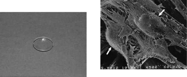
can avoid detection by the body, and often is coupled to pharmaceuticals or other molecules to extend circulation half-life within the body. Of all the materials used in biomedical research, few polymers have better biocompatibility properties than PEG. Also, the chemical structure of PEG is fairly stable within aqueous environments, although hydrolytic degradation can occur. Furthermore, removal of PEG from the body is not a major concern since PEG, with a molecular weight < 20,000 g mol 1, can be cleared readily by the kidneys. Traditionally, PEG hydrogels have been cross-linked through chemical initiators, however, other work has shown that photoinitiators can be used to gel PEG in situ. Recently, more attention has focused on the use of star PEG, which contain a central core out of which proceeds several linear PEG arms. Consequently, these materials offer improved control over mechanical properties and biological interactions since each molecule of polymer contains many more potential sites for cross-linking or for incorporating biologically active molecules. Cell adhesion peptides, polysaccharides, and polysaccharide ligands have all been coupled to various PEG molecules and studied as biomaterials (15,23,24).
Acrylics. One of the greatest success stories involving polymeric biomaterials involves PMMA and PHEMA. Many polymers have not yet been approved by the FDA. However, many polymers of the acrylic family (e.g., PMMA used for bone cement and intraocular lenses) were grandfathered into the Medical Device Amendments of 1976 as approved materials. The PHEMA polymer allows for sufficient gas exchange, and both PHEMA and PMMA have excellent optical properties and a good degree of hydration. As a result, intraocular lenses, hard, and soft contact lenses (see Fig. 8) made in whole or in part from these polymers are commercially available (3,5). Even though contact lenses only touch the eye on one side, the polymers that comprise the contact lenses are still bathed in tears and are therefore subject to protein deposition. This protein deposition can cause eye irritation and lead to contact lens failure if the contacts are not properly cleaned. With
BIOMATERIALS: AN OVERVIEW |
277 |
the development of disposable contact lenses, however, the problem of protein buildup can be minimized since the useful lifespan of each contact lens does not have to extend very long.
Biomimetic Materials. Recently, the field of biomaterials has started to incorporate features found within mechanisms involved in biomolecular assembly and interaction (24). These biomimetic materials show great promise since assembly is directed through biological affinity, recognition and/or interactions. As a result, these materials often have properties more similar to those of natural materials. Biomimetic materials exist as polymer scaffolds and hydrogels, but can also consist of ceramic and metallic materials machined or chemically modified to mimic porous structure of tissue (e.g., bone). Further elucidation of mechanisms responsible for biological self-assembly most likely will lead to improved biomaterials that are capable of interacting very specifically with an environment containing cells, tissue, or ECM molecules. In addition, many researchers are borrowing biological concepts to provide appropriate signals for cellular proliferation or differentiation and to deliver pharmaceuticals in a much more controlled manner. The scanning electron micrograph (SEM) shown in Fig. 9 illustrates a biologically oriented approach of using a biomaterial like chitosan–collagen as a scaffold on which cells can adhere (25).
Drug Delivery. Applications involving biomaterials have evolved from those focused on mostly structural requirements to those combining multiple design considerations including structure, mechanics, degradation, and drug delivery. The latest trend in biomaterials design is to promote healing, repair, or regeneration via the delivery of pharmaceutical agents, drugs, or growth factors. There exist many examples of biomaterials used as delivery vehicles or as drugs (22); however, many of these examples
Figure 8. A photograph of a disposable contact lens made from PHEMA.
Figure 9. A SEM showing chondrocytes (denoted by white arrows) attached to a biomaterial scaffold comprised of chitosan-based hyaluoronic acid hybrid polymer fibers. (Reprinted from Ref. 25 with permission from Elsevier.)

278 BIOMATERIALS: AN OVERVIEW
are just beginning to transition from research materials into commercially available products. One example of a commercially available drug delivery biomaterial is known as Gliadel, made by Guilford Pharmaceuticals. Gliadel wafers consist of a polyanhydride polymer loaded with carmustine, a chemotherapeutic drug. This system is intended as a treatment for malignant gliomas. Following removal of the tumor, the Gliadel wafers are added to the cavity and allowed to degrade and release the carmustine in order to kill remaining tumor cells. In addition to cancer treatments, biomaterials as drug delivery vehicles have been extensively employed in cardiovascular applications. Recently, FDA approval was granted to several types of drug eluting metallic stents. Among these include the Sirolimus-eluting CYPHER stent manufactured by the Cordis Corporation, the TAXUS Express2 Paclitaxel-eluting stent manufactured by the Boston Scientific Corporation. The purpose behind releasing the drugs from the stents is to decrease the occurrence of restenosis, or the renarrowing of vessels treated by the stent. As a result of the drug delivery aspect of the system, the stents are expected to have better long-term viability. Several more examples of drug delivery and biomaterial hybrid systems exist; however, a comprehensive review of biomaterials as drug delivery systems is beyond the scope of this article. It is important to note that more interest and attention have been given to modify biomaterials so that the material is more integrally involved in interacting with and manipulating organ and tissue biology.
FACTORS CONTRIBUTING TO BIOMATERIAL FAILURE
Although there exists a multitude of commercially available and successful metallic, ceramic, and polymeric biomaterials, biomaterials have and will continue to fail. The human body is a very hostile environment for synthetic and natural materials. In some instances, like orthopedic applications, it is much easier to understand why materials can fail since no material can survive cyclical loading indefinitely without showing signs of fatigue or wear. However, for most biomaterial failures, the exact reason for failure is still not well understood. Some factors contributing to the failure of a biomaterial include corrosion, wear, degradation, and biological interactions.
Corrosion
By weight, more than one-half of the human body consists of water. As a result, all implanted biomaterials will encounter an aqueous environment. Moreover, this aqueous environment is also very saline due to the presence of a relatively large concentration of extracellular salts. The aqueous and saline conditions of physiological solutions create favorable conditions for metallic corrosion. Corrosion involves oxidation and reduction reactions between a metal, ions, and species (e.g., dissolved oxygen). In fact, the lowest free energy state of many metals in and oxygenated and hydrated environment is an oxide. Most corrosion reactions are electrochemical. For example, if zinc metal is placed in an acidic environment (e.g., hydrochloric acid),
hydrogen gas will evolve as the zinc become cationic and binds to chloride ions. The actual reaction consists of two half reactions. In the first reaction, zinc metal is oxidized to a Zn2þ state; the second reaction involves the reduction of hydrogen ions to hydrogen gas. During this process, the newly formed metal ions diffuse into solution. Both the oxidation and reduction reactions must occur at the same time to avoid charge buildup within the material. This process occurs at the surface and exposed pore of metals, and, in an attempt to passivate the surface to avoid this process, corrosion resistant oxides have been incorporated into an implant surface (13). Care must be taken, however, to ensure that the protective oxide coating is not damaged during processing, packaging, or surgical procedure.
In addition to oxidative corrosion, bimetallic or galvanic corrosion is a concern with implants composed of more than one type of metal, such as alloys with mixing defects and implants containing parts made from distinct metals. Galvanic corrosion can occur because all metals have a different tendency to corrode. If two distinct metals are in contact with one another through a conductive medium, oxidation of one metal will occur while reduction of the other occurs. In both oxidative corrosion and bimetallic corrosion, bits of metal, metal ions, and oxidative debris can enter the surrounding tissue and even travel to distant body parts. This can result in inflammation and even in metal toxicity.
Wear
In addition to corrosion, metal, as well as other materials can wear as a result of friction. For example, in hip implants, the acetabular cup is in contact with the ball of the metal or ceramic stem. Every time a movement occurs within the joint, rubbing between the ball and cup occurs and small wear particles of metal and polymer are left behind (see Fig. 10). More often than not, the particles are shed from the softer surface (e.g., ultrahigh molecular weight polyethylene); however, metal particles are also produced. The particles range in size from
Figure 10. A photograph of some worn biomaterials. Examples in this photograph include screws, a femoral head replacement, and a polyethylene acetabular cup. (Used with permission from the Department of Materials at Queen Mary University of London.)
nanometers to microns with the smaller particles able to enter the lymph fluid and travel to distant parts of the body. The small particles increase the surface area of the material, and this increased surface area can result in increased corrosion (5,13). Thus, wear can lead to deleterious effects (e.g., corrosion), described above, and inflammation, as will be discussed below.
Degradation
Although not affected by corrosion, certain bioactive ceramics and polymers are susceptible to degradation. In the case of bioactive ceramics, however, this process is relatively slow compared with the potential rate of bone regeneration. For polymers, degradation can occur via hydrolytic or, in some cases, enzymatic mechanisms. The chemical structures of both polyamides and polyesters lend themselves toward enzymatic degradation. For polyesters, acidic or alkaline conditions will lead to a deesterification reaction that will eventually destroy the backbone of the polymer. The degradation rate varies greatly depending on the composition of the polymer. For example, within the body, poly(lactic acid) will degrade over many months to years, but poly(glycolic acid) can degrade over a few days or weeks. The degradation rate of polyamides is slower than that of polyesters, but is still an important design consideration when choosing a polymeric biomaterial for a specific application. For applications (e.g., sutures), degradation of the material is a beneficial property since the sutures only need to remain in place for a few days to weeks until the native tissue heals. For applications needing a material with a longer lifespan, degradation poses a larger problem.
Increasingly, degradable polymers or polymers with degradable cross-links are being studied as biomaterials. This interest in degradable systems stems largely from more current research involving tissue engineering and drug delivery (15,16,22,24,26,27). The philosophy of tissue engineering holds that the polymeric biomaterial acts as a scaffold with or without viable cells or biological molecules to promote tissue ingrowth. As cells proliferate and migrate within these scaffolds and begin to create new tissue, the material can and should degrade to leave, ultimately, regenerated or repaired tissue in its place. One of the engineering design constraints, therefore, is to balance the rate of degradation with that of tissue ingrowth. If the biomaterial degrades too rapidly and the newly formed tissue cannot provide the necessary mechanical support, then the biomaterial will have failed. At the same time, if the biomaterial degrades too slowly, then the process of tissue ingrowth may become inhibited or may not occur at all. To this end, more recent research has attempted to include enzymatically sensitive crosslinks, usually made from synthetic peptide analogues of enzyme substrates, within polymer networks. Instead of relying upon relatively uncontrolled hydrolytic degradation, the polymeric biomaterial would degrade at a rate controlled by migrating cells. Thus, the cells themselves could degrade the material and produce new tissue in a much more controlled and physiologically relevant manner.
BIOMATERIALS: AN OVERVIEW |
279 |
Biological Interactions
Most modern biomaterials are intended to come into direct contact with living tissue and biological fluids. This interaction often makes the biomaterial a target for the protective mechanisms within the body. These protective mechanisms include protein adsorption, hemostasis, inflammation and the foreign body response, and the immune response. Although it has been well established that all types of tissue-contacting biomaterials invoke some degree of biological response, it has only been during the past decade or so when investigations have revealed that all implanted tissue-containing biomaterials invoke an almost identical inflammatory and foreign body response regardless of whether the biomaterial is of metallic, ceramic, polymeric, or composite origin. Although future research in the field of biomaterials aims to better understand and to eventually mitigate the biological interactions that currently result in the failure of many biomaterials, the following biological responses remain of great importance when considering the design and potential applications of any biomaterial. In fact, most current obstacles related to the design of biomaterials involve the interaction of biomaterials with the body and the reaction of the body to biomaterials. As a result, current biomaterial research trends aim to provide an environment that allows the body to invade, remodel, and degrade the implanted material (23,27,28).
Protein Adsorption. As soon as a biomaterial comes into contact with biological fluid (e.g., blood) the material becomes coated with adsorbed proteins. This adsorption is very rapid and is based primarily on noncovalent interactions between various hydrophilic and hydrophobic domains within the adsorbed proteins and the surface of the implanted biomaterial. Initially, the composition of the protein layer depends on the relative concentration of various proteins within the biological fluid. Certain proteins (e.g., albumin) are very abundant in serum and will initially be found abundantly in the adsorbed protein layer. However, over time the adsorbed protein layer will change its composition as proteins with higher affinities for the surface of the material, but lower serum concentrations will displace proteins with lower affinities and higher serum concentrations. This rearrangement and equilibration of the protein layer is known as the Vroman effect. When biomaterials become coated with proteins, surrounding cells no longer see the surface of the material. Instead, they see a layer of serum-soluble proteins. Increasingly, biomaterials design has focused on optimizing surface chemistries and incorporating selective reactive domains that will promote a specific biological response. In reality, these engineered surfaces become masked by a nonspecific protein layer, and it is this protein layer that drives the biological response to an implanted biomaterial. Some successful examples of surface modifications aimed at reducing nonspecific protein adsorption involve the use of nonfouling hydrophilic polymers (e.g., PEG and dextran), the pretreatment of the biomaterial with a specific protein, and the replacement of certain chemically reactive functional groups with others. Time, however, remains the
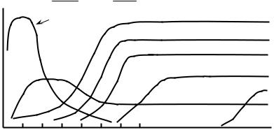
280 BIOMATERIALS: AN OVERVIEW
 Acute Chronic
Neutrophils
Intensity
Granulation tissue 
Macrophages
Neovascularization
Foreign body giant cells
Fibroblasts
Fibrosis
Mononuclear
Leukocytes
Time
Figure 11. A schematic representing the temporal events involved in acute and chronic inflammation as well as the foreign body response. (Adopted from Anderson et al. as found in Ref. 5.)
largest obstacle with any of these surface treatments. Often, surface treatments will only function for a limited time before serum and other extracellular proteins becomes adsorbed. Once adsorbed, proteins can undergo conformational changes that expose cryptic sites, allow autoactivation, or that influence the behavior of other proteins or cells (5,6,18).
Blood Contact. Direct contact with blood is a major concern of all biomaterials regardless of whether or not they were designed for cardiovascular applications. During surgical implantation, blood vessels are broken, which results in an increased probability that the biomaterial will contact blood. Although exact mechanisms remain unclear, serum proteins (e.g., Factor XII, Factor XI, plasma prekallikrein, and high molecular weight kininogen) interact to initiate contact activation of the coagulation cascade through the intrinsic pathway. This calciumand plateletdependent cyclic network involves the activation of thrombin, which ultimately cleaves specific protein domains within fibrinogen and Factor XIII. Activated Factor XIIIa and fibrinogen then react to form a cross-linked fibrin clot. The formation of blood clots as well as the activation of various serum proteins and platelets can lead to local inflammation. Recent approaches have attempted to passivate the blood contact response of implanted biomaterials by incorporating heparin or other antithrombotic agents on the biomaterial surface (5,6,18).
Inflammation and the Foreign Body Response. The human body is well equipped to handle injuries that affect hemostasis. During trauma, proteins within blood can initiate a relatively large biological response lasting days, weeks, and even months. Initially, the area around a trauma site, including the implantation of a biomaterial, becomes inflamed. Inflammation is a normal process involved with healing that is characterized by four major events: swelling, pain, redness, and heat. The vasculature around an injury will become leaky to allow extravasation of various leukocytes (e.g., neutrophils and macrophages). With the presence of cytokines and other growth factors, leukocytes, primarily macrophages, are stimulated to remove bacteria and foreign material. Macrophages also
recruit fibroblasts and other cells to the injury site to aid in healing by forming granulation tissue. Over the course of several days or weeks, this initial granulation tissue is remodeled and replaced with restored, functional tissue or, more commonly, scar tissue (Fig. 11).
In the case of implanted biomaterials, the implantation site is the injury site and will become inflamed. As a result, macrophages will be recruited to the site and attempt to remove the ‘‘foreign’’ biomaterial. Unlike smaller injuries, macrophages are unable to remove biomaterials through phagocytosis. When they become frustrated, macrophages will fuse together to form foreign body giant cells. These foreign body giant cells can secrete superoxides and free radicals, which can damage biomaterials, but these cells usually cannot completely remove the foreign biomaterial. In the event that the body cannot eliminate a foreign object through phagocytosis, activated macrophages and foreign body giant cells remain around the implant and can promote a chronic localized area of inflammation. Remaining fibroblasts and other cells around the biomaterial then will begin to secrete a layer of avascular collagen around the biomaterial to effectively encapsulate it and wall it off from the rest of the body (5,6,18). Although the function of some biomaterials is not affected by this foreign body response, biomaterials ranging from sensors to orthopedic implants to soft tissue replacements are adversely affected by this biological reaction. To date, it is not known how to minimize or eliminate an inflammation or foreign body reaction. However, a great deal of research is attempting to create biomaterials that do not evoke a tremendous inflammatory response or that degrade in a way that allows the restoration or repair of native tissue without the adverse affects of chronic inflammation.
Immune Response. The innate and adaptive immune responses of the body also pose a challenge for biomaterials designed for long-term applications. Increasingly, new biomaterials have attempted to incorporate cellular components in an attempt to create new tissues in vitro or to seed materials with autologous, allogeneic, or xenogenic cells, including stem cells, to promote tissue repair. Unfortunately, the adaptive immune response will actively eliminate allogeneic or xenogenic cell types. As a result,
biomaterials have been designed to act as barriers that limit lymphocyte activation. Often, cells are encapsulated in microspheres made from various polymers or layers of polymers. For example, pancreatic Islets of Langerhans from animal and human donors have been encapsulated within polymers [e.g., polysulfones, poly(N-isopropylacryl- amide)] and alginates, to provide an immunoisolated environment that still retains enough permeability to allow for the diffusion of insulin. One of the major complications of this type of biomaterials design is to balance the creation of volume within the microsphere to accommodate enough Islets to allow for sufficient insulin production with the need to provide appropriate diffusion rates so that the cells within the center of the microsphere remain viable. As more polymeric biomaterials incorporate or consist of peptide and protein motifs, there remains a concern as to whether or not these motifs might elicit an adaptive immune response. Even if protein domains derived from human proteins are incorporated into biomaterials, these domains might not be presented the same way to lymphocytes. As a result, the body may start producing antibodies against these domains, which might also lead to certain forms of autoimmune diseases (29).
Although the adaptive immune system is playing an increasingly important role in the rejection of new types of biomaterials, the innate immune system remains a very large threat to the success of a biomaterial. As mentioned above, proteins bind to biomaterials upon implantation. One of the most abundant proteins within the blood is the complement protein C3. Within the blood, C3 can spontaneously hydrolyze to form an active convertase complex, which can cleave C3 into C3a and C3b. Although C3b is rapidly inactivated within the blood, it can remain active if it binds to a surface (e.g., a biomaterial). As a result, the alternative pathway of the complement system can be activated very rapidly leading to formation of membraneattack complexes but more importantly, the formation of the soluble anaphylotoxins C3a, C4a, and C5a. These anaphylotoxins induce smooth muscle contraction, increase vascular permeability, recruit phagocytic cells, and promote opsonization by phagocytic cells. These phagocytic cells (e.g., macrophages) have receptors recognizing C3b. As a result, macrophages will attempt to engulf the C3b-coated biomaterial. When this fails, the macrophages will form foreign body giant cells, and the body will attempt to encapsulate the biomaterial in a manner similar to that described above for the inflammation and foreign body response (29). Overall, all of the above mentioned biological responses can affect the performance of any biomaterial, and active biomaterials research is striving not only to better understand the mechanisms of inflammation, protein adsorption, hemostasis, and innate and adaptive immune responses, but also to develop strategies to minimize, eliminate, evade, or alter adverse biological responses to materials.
BIOCOMPATIBILITY
Since biomaterials are intended for direct contact with biologically viable tissue, all biomaterials need to possess
BIOMATERIALS: AN OVERVIEW |
281 |
some degree of biocompatibility. In a manner similar to that of the term biomaterials, the term biocompatibility has experienced many changing definitions over the past several decades. Initially, biocompatibility implied that the biomaterial remained inert to its surroundings in order to refrain from being toxic, carcinogenic, or allergenic. As the definition of biomaterials evolved to include biologically derived materials and molecules, the term biocompatibility needed to encompass these changes. In 1987, David Williams suggested that biocompatibility is ‘‘the ability of a material to perform with an appropriate host response in a specific application’’ (30). Although there does not yet exist a universal consensus with regard to the definition of the term biocompatibility, the definition proposed by Williams provides enough generality to serve as an adequate and accurate description of biocompatibility.
Instead of remaining inert, biomaterials are becoming increasingly reliant on biochemical reactions and physiological processes in order to serve a useful function. In some cases (e.g., in the case of bone plates and artificial joints), biomaterials can remain inert and still provide satisfactory performance. In other instances (e.g., drug delivery vehicles), tissue engineering applications, and in vivo organ replacement therapies, biomaterials not only need to actively minimize or adapt to the surrounding biological responses (e.g., inflammation and foreign body responses), but also need to depend on interactions with surrounding tissues and cells in order to provide a useful function (15,22,24–27). In addition, the performance of traditionally inert biomaterials is being enhanced by incorporating chemical or mechanical modifications that interact with biology at the cellular level. For example, the bone-contacting surfaces of metallic femoral stems, for hip replacement, have been modified to contain bioactive ceramic porous networks or hydroxyapatite crystal networks. These ceramic networks allow better osteointegration of the implant with the host tissue and, in some cases, eliminate the need to use bone sealants (e.g., PMMA).
Obviously, if a successful biomaterial needs to show some level of biocompatibility, then there must exist various testing conditions and manufacturing standards to establish safety controls. Organizations [e.g., ASTM International and the International Organization for Standardization (ISO)] do have guidelines and standards for the testing and evaluation of biomaterial biocompatibility. These regulations include tests include the measuring of cytotoxicity, sensitization, skin irritation, intracutaneous reactivity, acute systemic toxicity, genotoxicity, macroscopic and microscopic evaluation of implanted materials and devices, hemocompatibility, subchronic and chronic toxicity, carcinogenicity, the effect of degradation byproducts, and the effect of sterilization (31). For many of these paramenters, the associated standards dictate the size and shape of the material to be tested, appropriate in vitro testing procedures and analysis schemes, and relevant testing and evaluation protocols for in vivo experimentation. Although standards related to the manufacturing and performance of some biomaterials exist, there remains a lack of uniform biocompatibility testing standards for new classes of biomaterials that rely heavily upon cellular and tissue interactions or that contain biologically active

282 BIOMATERIALS: AN OVERVIEW
molecules. New developments in biologically active biomaterials have resulted in not only nonuniform approaches to biocompatibility testing, but also confusions related to the regulatory classification of new types of biomaterials.
FUTURE DIRECTIONS
As more information becomes available regarding biological responses to materials, mechanisms that control embryonic development and early wound healing, and matrix biology, materials will be designed to more adequately address, promote or inhibit biological responses as needed. As a result, the field of biomaterials will not only incorporate principles from materials science and engineering, but also rely increasingly upon design constraints governed by biology (see Fig. 12). Recent trends in biomaterial research show an increased emphasis in designing materials that better match the biological environment with respect to mechanics and biological signals. Materials promote cell attachment using biologically derived signals, degrade through relevant enzymatic degradation and release and store bioactive factors using methods derived from biology. Continued adaptation of materials to more appropriately interact with the living system will result in devices that work with the body to promote tissue regeneration and healing.
Factors to Consider When Designing a New Biomaterial
Required
Mechanical Properties
Required |
ScienceMaterials |
Engineeringand |
Required |
|
|
||
Degradation Rate |
|
|
Porosity |
Moldability |
|
|
Required |
or Shapability |
|
|
Surface Properties |
New Material for
Tissue Regeneration
Inhibit Immune and |
Biology medicineand |
Required |
Growth Factor |
Bioactivity |
|
Foreign Body Reactions |
|
|
|
|
How to Connect |
Requirements |
|
New Tissue to |
|
Existing Vasculature |
|
|
|
|
Number of |
|
Nutrient |
Cell Types to Support |
|
Requirements |
Figure 12. An illustration depicting the various engineering and biological factors that need to be considered in the design of modern biomaterials. Successful, new biomaterials will require the optimization of a variety of parameters and the cooperation of interdisciplinary scientists, engineers, and clinicians. (Reprinted from Ref. 15 with permission from Elsevier.)
BIBLIOGRAPHY
Cited References
1.Black J. The education of the bio-materialist—report of a survey. 1980–1981. J Biomed Mater Res 1982;16(2):159– 167.
2.Galletti PM, Boretos JW. Report on the consensus development conference on clinical-applications of biomaterials, 1–3 november 1983. J Biomed Mater Res 1983;17(3):539–555.
3.Lindstrom RL. The polymethylmetacrylate (pmma) intraocular lenses. In: Steinert RF, et al. editors. Cataract surgery: Technique, Complications, and Management. Philadelphia: Saunders; 2003.
4.Barbucci R, editor. Integrated Biomaterials Science. New York: Kluwer Academic; 2002.
5.Ratner BD, Hoffman AS, Schoen FJ, Lemons JE, editors. Biomaterials Science: An Introduction to Materials in Medicine. San Diego: Academic Press; 1996.
6.Greco RS, editor. Implantation Biology: The Host Response and Biomedical Devices. Boca Raton (FL): CRC Press; 1994.
7.Lysaght MJ, O’Loughlin JA. Demographic scope and economic magnitude of contemporary organ replacement therapies. Asaio J 2000;46(5):515–521.
8.Harris G. Step forward for implants with silicone. New York: The New York Times; 2005.
9.Park JB, Lakes RS. Biomaterials: An introduction. 2nd ed. New York: Plenum Press; 1992.
10.Black J, Hastings G, editors. Handbood of Biomaterial Properties. New York: Chapman & Hall; 1998.
11.Wen CE, et al. Novel titanium foam for bone tissue engineering. J Mater Res 2002;17(10):2633–2639.
12.Graham, et al. The use of SMARTeR stents in patients with binary obstrevition. Cliro Radiol 2004;59:288–291.
13.Smith WF, Foundations of Materials Science and Engineering. 2nd ed. New York: McGraw-Hill, Inc.; 1993.
14.Fernandez E, et al. Calcium phosphate bone cements for clinical applications—part ii: Precipitate formation during setting reactions. J Mater Sci-Mater M 1999;10(3):177–183.
15.Seal BL, Otero TC, Panitch A. Polymeric biomaterials for tissue and organ regeneration. Mater Sci Eng R 2001; 34(4–5):147–230.
16.Langer R. 1994 Whitaker lecture—polymers for drug-deliv- ery and tissue engineering. Ann Biomed Eng 1995;23(2): 101–111.
17.Greenwald SE, Berry CL. Improving vascular grafts: The importance of mechanical and haemodynamic properties. J Pathol 2000;190(3):292–299.
18.Dee KC, Puleo DA, Bizios R. An Introduction to TissueBiomaterial Interactions. Hoboken (NJ): John Wiley & Sons, Inc.; 2002.
19.Temenoff JS, Mikos AG. Review: Tissue engineering for regeneration of articular cartilage. Biomaterials 2000;21 (5):431–440.
20.Temenoff JS, Mikos AG. Injectable biodegradable materials for orthopedic tissue engineering. Biomaterials 2000;21(23): 2405–2412.
21.Palsson BØ, Bhatia SN. Tissue Engineering. Upper Saddle River (NJ): Pearson Prentice Hall; 2004.
22.Peppas NA, Bures P, Leobandung W, Ichikawa H. Hydrogels in pharmaceutical formulations. Eur J Pharm Biopharm 2000;50(1):27–46.
23.Hubbell JA. Biomaterials in tissue engineering. Bio-Technol 1995;13(6):565–576.
24.Sakiyama-Elbert SE, Hubbell JA. Functional biomaterials: Design of novel biomaterials. Ann Rev Mater Res 2001;31: 183–201.
