

188BIOHEAT TRANSFER
74.Arena JG, Bruno GM, Brucks AG, Searle JD, Sherman RA, Meador KJ. Reliability of an ambulatory electromyographic activity device for musculoskeletal pain disorders. Int J Psychophysiol 1994;17:153–157.
75.Arena JG, Bruno GM, Brucks AG, Searle JD, Sherman RA, Meador KJ (unpublished manuscript). The measurement of surface EMG in tension-headache subjects in the natural environment: Ambulatory recordings of data from five consecutive days.
76.Gatchel RJ, Oordt MS. Future trends and opportunities. In: Gatchel RJ, Oordt MS, editors. Clinical Health Psychology and Primary Care: Practical Advice and Clinical Guidance for Successful Collaboration. Washington, DC: American Psychological Association; 2003.
77.Blanchard EB, Andrasik F, Appelbaum KA, Evans DD, Jurish SE, Teders SJ, Rodichok LD, Barron KD. The efficacy and cost-effectiveness of minimal-therapist contact, nondrug treatments of chronic migraine and tension headache. Headache 1985a;25:214–220.
78.Blanchard EB, Appelbaum KA, Nicholson NL, Radnitz CL, Morrill B, Michultka D, Kirsch C, Hillhouse J, Dentinger MP. A controlled evaluation of the addition of cognitive therapy to a home-based biofeedback and relaxation treatment of vascular headache. Headache 1990a;30:371–376.
79.Jurish SE, Blanchard EB, Andrasik F, Teders SJ, Neff DF, Arena JG. Home versus clinic-based treatment of vascular headache. J Consult Clin Psychol 1983;51:743–751.
80.Tobin DL, Holroyd KA, Baker A, Reynolds RVC, Holms JE. Development and clinical trial of a minimal contact, cogni- tive-behavioral treatment for tension headache. Cognit Ther Res 1988;12:325–339.
81.Blanchard EB, McCoy GC, Musso A, Gerardi RJ, Cotch PA, Siracusa K, Andrasik F. A controlled comparison of thermal biofeedback and relaxation training in the treatment of essential hypertension: I. Short-term and long-term outcome. Behav Ther 1986;17:563–579.
82.Devineni T, Blanchard EB. A Randomized Controlled Trial of an Internet-based Treatment for Chronic Headache. Behav Res Ther 2005;43:277–292.
83.Arena JG, Dennis N, Devineni T, McClean R, Meador KJ. A pilot study of the feasibility of a telemedicine delivery system for psychophysiological treatments for vascular headache. Tele Meo J E-Health 2005;10:449–454.
See also BIOELECTRODES; ELECTROENCEPHALOGRAPHY; ELECTROGASTROGRAM; ELECTROMYOGRAPHY.
BIOHEAT TRANSFER
JONATHAN W. VALVANO
The University of Texas
Austin, Texas
INTRODUCTION
Bioheat transfer is the study of the transport of thermal energy in living systems. Because biochemical processes are temperature dependent, heat transfer plays a major role in living systems. Also, because the mass transport of blood through tissue causes a consequent thermal energy transfer, bioheat transfer methods are applicable for diagnostic and therapeutic applications involving either mass or heat transfer. This article presents the characteristics of
Capillary |
|
Arteriole |
Venole |
|
Terminal
Terminal
artery
vein
Supply vessels
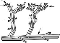
Figure 1. Countercurrent blood vessels have arterial blood flowing in the opposite direction as venous blood.
bioheat transfer that distinguish it from nonliving systems, including the effects of blood perfusion on temperature distribution, coupling with biochemical processes, therapeutic and injury processes, and thermoregulation.
The study of bioheat transfer involves phenomena that are not found in systems that are not alive. For example, blood perfusion is considered a three-dimensional (3D) process as fluid traverses in a volumetric manner through tissues and organs via a complex network of branching vessels. Heat transfer is affected by vessel geometry, local blood flow rates, and thermal capacity of the blood (1). One factor that makes modeling blood perfusion difficult is the complex network of pairs of arteries and veins with countercurrent flow (2), as shown in Fig. 1. Arterial and venous blood temperatures may be different, and it is possible that neither is equal to the local tissue temperature. These temperatures may vary as a function of many transient physiological and physical parameters. The regulation of temperature and blood flow is quite nonlinear and has presented a major challenge to understand and model. Nevertheless, these critical processes must be accounted for in the design of many types of systems that interface with humans and animals.
Many scientists view life from either the macroscopic (systems) or the microscopic (cellular) level, but in reality one must be aware that life processes exist continuously throughout the spectrum. In order to better understand life processes at the molecular level, a significant research effort is underway associated with molecular biology. Because temperature and blood flow are critical factors, bioengineers are collaborating with molecular biologists to understand and manipulate the molecular and biochemical processes that constitute the basis of life. Research has found that the rates of nearly all physiological functions are altered 6–10%/8C (3). Similarly, heat can be added or removed during therapeutic or diagnostic procedures to produce or measure a targeted effect, based on the fact that a change in local temperature will have a large effect on rates of biochemical process rates. Thus, the measurement and control of temperature in living tissues is of great value in both the assessment of normal physiological function and the treatment of pathological states.
The study of the effects of temperature alterations on biochemical rate processes has been divided into three broad categories: hyperthermia (increased temperature), hypothermia (decreased temperature), and cryobiology

(subfreezing temperature). An extensive review of these domains has been published (4), to which the reader is referred for further details and bibliography.
Effects of Blood Perfusion on Heat Transfer
Blood perfusion through the vascular network and the local temperature distribution are interdependent. Many environmental (e.g., heat stress and hypothermia), pathophysiologic (e.g., inflammation and cancer), therapeutic (e.g., heating–cooling pads) situations create a significant temperature difference between the blood and the tissue through which it flows. The temperature difference causes convective heat transport to occur, altering the temperatures of both the blood and the tissue. Perfusion-based heat transfer interaction is critical to a number of physiological processes, such as thermoregulation and inflammation.
The convective heat transfer depends on the rate of perfusion and the vascular anatomy, which vary widely among the different tissues, organs of the body, and pathology. Diller et al. published an extensive compilation of perfusion data for many tissues and organs and for many species (5). Charney reviewed the literature on mathematical modeling of the influence of blood perfusion on bioheat transfer phenomena (6).
The rate of perfusion of blood through different tissues and organs varies over the time course of a normal day’s activities, depending on factors, such as physical activity, physiological stimulus, and environmental conditions. Further, many disease processes are characterized by alterations in blood perfusion, and some therapeutic interventions result in either an increase or decrease in blood flow in a target tissue. For these reasons, it is very useful in a clinical context to know what the absolute level of blood perfusion is within a given tissue. Many thermal techniques have been developed that directly measure heat flux to predict blood perfusion by exploiting the coupling between vascular perfusion and local tissue temperature using inverse mathematical solutions.
In 1948, Pennes (7) published the seminal work describing the mathematical coupling between the mass transfer of blood perfusion and the thermal heat transfer. His work consisted of a series of experiments to measure temperature distribution as a function of radial position in the forearms of nine human subjects. A butt-junction thermocouple was passed completely through the arm via a needle inserted as a temporary track, with the two leads exiting on opposite sides of the arm. The subjects were unanesthetized so as to avoid the effects of anesthesia on blood perfusion. Following a period of normalization, the thermocouple was scanned transversely across the mediolateral axis to measure the temperature as a function of radial position within the interior of the arm. The environment in the experimental suite was kept thermally neutral during experiments. Pennes’ data showed a temperature difference of 3–48 between the skin and the interior of the arm, which he attributed to the effects of metabolic heat generation and heat transfer with arterial blood perfused through the microvasculature.
Pennes proposed a model to describe the effects of metabolism and blood perfusion on the energy balance
BIOHEAT TRANSFER |
189 |
within tissue. These two effects were incorporated into the standard thermal diffusion equation, which is written in its simplified form as
@T
rc t ¼ r krT þ rblcblwðTa TÞ þ Qmet ð1Þ
@
Metabolic heat generation, Qmet, is assumed to be homogeneously distributed throughout the tissue of interest as rate of energy deposition per unit volume. It is assumed that the blood perfusion effect is homogeneous and isotropic, and that thermal equilibration occurs in the microcirculatory capillary bed. In this scenario, blood enters capillaries at the temperature of arterial blood, Ta, where heat exchange occurs to bring the temperature to that of the surrounding tissue, T. There is assumed to be no energy transfer either before or after the blood passes through the capillaries, so that the temperature at which it enters the venous circulation is that of the local tissue. The total energy exchange between blood and tissue is directly proportional to the density, rbl, specific heat, cbl, and perfusion rate, w, of blood through the tissue. The basic principle that couples mass transfer to heat transfer is the change in sensible energy caused by the moving blood. The units of perfusion in equation 1 are volume of blood per volume of tissue per time (s 1). This thermal transport model is analogous to the process of mass transport between blood and tissue, which is confined primarily to the capillary bed.
A major advantage of the Pennes model is that the added term to account for perfusion heat transfer is linear in temperature, which facilitates the solution of Eq. 1. Since the publication of this work, the Pennes model has been adapted by many researchers for the analysis of a variety of bioheat transfer phenomena. These applications vary in physiological complexity from a simple homogeneous volume of tissue to thermal regulation of the entire human body (8,9). As more scientists have evaluated the Pennes model for application in specific physiological systems, it has become increasingly clear that many of the assumptions to the model are not valid. For example, it is now well established that the significant heat transfer due to blood flow occurs in the terminal arterioles (vessels 60–300 mm in diameter) (10–17). Thermal equilibration is essentially complete for vessels <60 mm (precapillaries and capillaries). Therefore, no significant heat transfer occurs in the capillary bed; the exchange of heat occurs in the larger components of the vascular tree. The vascular morphology varies considerably among the various organs of the body, which contributes to the need for specific models for the thermal effects of blood flow (as compared to the Pennes model that incorporates no information concerning vascular geometry). It would appear as a consequence of these physiological realities that the validity of the Pennes model is questionable.
Many investigators have developed alternative models for the exchange of heat between blood and tissue. These models have accounted for the effects of vessel size, countercurrent heat exchange, as well as a combination of partial countercurrent exchange and bleed-off perfusion. All of these models provided a larger degree of rigor in the analysis, but at the compromise of greater complexity and
190 BIOHEAT TRANSFER
reduced generality. These studies also led to an increased appreciation of the necessity for a more explicit understanding of the local vascular morphology as it governs bioheat transfer, which has given rise to experimental studies to measure and characterize the 3D architecture of the vasculature in tissues and organs of interest (18). The quantitative analysis of the effects of blood perfusion on the internal temperature distribution in living tissue remains a topic of active research after one-half of a century of study (19).
THERAPEUTIC APPLICATIONS OF BIOHEAT TRANSFER
The elevation of tissue temperature into the 40–42 8C range provides some relief from pain (analgesia). In addition, wound healing can be enhanced by this modest increase in temperature. Increased temperature does not cause healing by itself, but rather it creates the improved conditions for the natural processes to heal wounds.
Hot or cold packs can be used to create therapeutic heating for injuries near the skin surface. Heating packs are effective for injuries such as sprains, muscle strains, and postoperative swelling. Elevated temperatures cause an increase in blood perfusion, supplying nutrients to the injured tissue. During the first 12–24 h after injury, cold packs will reduce perfusion, thereby reducing vascular pressure and tissue swelling. Afterward, hot packs (up to 45 8C) are applied to increase perfusion and promote healing (20).
When the injury is deeper, the therapy requires a volumetric heater, such as radio frequency (rf) electromagnetic heating, microwave frequency electromagnetic heating, and ultrasonic heating. The Industrial-Medical- Scientific (ISM) frequencies are 6.78, 13.56, 27.12, and 40.68 MHz. The ISM frequencies typically used in medical applications of microwaves are 915 MHz and 2.45 GHz. Medical ultrasonic devices operate in the 500 kHz to 10 MHz range. There are three engineering parameters to consider when designing a therapeutic device. The first parameter is the amount of local volumetric heat generation, the second parameter is the shape of the heating field, and the third parameter is the depth of penetration. Higher frequencies have shorter wavelengths, causing higher absorption and less penetration. The electrical properties of the tissue, which depend on structure and composition, strongly affect the effectiveness of volumetric heating using rf and microwave EM fields. Similarly, acoustic properties of the tissue are important in ultrasonic systems. Often the design of an effective therapeutic device hinges on proper control of the boundary conditions where energy is transferred across the trans- ducer–tissue interface.
There is a systemic effect of local heating, controlled by the hypothalamus, involving both neuronal and hormonal signals. Local heating of organs or peripheral muscles can also cause a spinal cord mediated response. A local release of bradykinins can affect the vascular tone of the terminal arterioles (see Fig. 1), which in turn affect the vascular resistance to blood flow. In general, an increase in local temperature causes an increase in local blood flow,
whereas a decrease in local temperature creates a decrease in local blood flow, but the behavior is highly complex.
Smooth muscles surrounding the 40–200 mm diameter arterioles play a dominate role in controlling local blood flow. Normal capillary pressure is 3.3 kPa (25 Torr). When local tissues are heated, these arterioles dilate causing an increase in capillary pressure and capillary blood flow. Edema occurs when this pressure widens gaps in the capillary wall causing excess fluid to leak from the vascular to intravascular space. High capillary flow promotes healing by removing wastes, delivering nutrients, and supplying oxygen. Leukocytes (white blood cells) control the healing process by first breaking down, then removing damaged and dead tissue.
The volumetric heating created by electromagnetic fields
in |
governed by the electrical conductivity, |
s (S m 1), |
|||
the |
|
imaginary part |
of the electrical |
permittivity, |
|
e |
|
|
1 |
|
|
00 (F |
|
m 1), and the magnitude of the local electric field, |
|||
jEj |
(V m ): |
|
|
||
|
|
|
q000 |
¼ ðs þ ve00ÞjEj2 |
ð2Þ |
where v is the angular frequency of the field in rad s 1. Direct heating from the magnetic fields in medical applications is usually neglected. Magnetic fields can be used to heat tissue, but because of Faraday’s law of induction, the time-varying magnetic field will induce an electric field, and it is this electric field that heats the tissue. A comprehensive review of electromagnetic heating can be found in Roussy and Pearce (21). Tables of electrical and acoustic properties can be found in Diller (5).
THERMOREGULATION
Thermoregulation is an elaborate control system, used by mammals, to maintain internal body temperatures near a physiological set point under a large spectrum of environmental conditions and metabolic rate activities. Even though there have been many years of research, much remains unknown about the human thermoregulatory system. Therefore, active investigation continues. Heat transfer due to conduction, convective heat transfer via the blood flow, local generation of thermal energy, and thermal boundary conditions comprise the major components of thermoregulation. Once these individual mechanisms are understood, they can be combined to create mathematical models to simulate and predict thermoregulatory behavior. The mathematical models are used to design systems to interact thermally with the human body (such as a space suit) without compromising the health and safety of the subject.
The prevailing theory is that the main objective of a human thermoregulation system is to maintain the body core temperature at a constant value consistent with that required for normal physiological functions, regardless of the environmental conditions. An alternative theory, suggested by Chappuis et al. (22) and Webb (23), is that the goal of the human regulation system is to maintain the body’s energy balance. In this theory, tissue temperatures are a result, rather than a cause, of the regulation process.
Nunneley (24,25) showed that temperature and internal energy storage of the human body vary with time of day, metabolic activity, and individuality of the human. To maintain body core temperature, the thermoregulatory system incorporates a number of energy production and dissipation mechanisms, many of them controlled by feedback from other body parameters. Examples of such feedback control are that for sweating, shivering, and varying blood flow.
Ganong (26) showed that the main control center for feedback mechanisms is located in the hypothalamus of the brain, where reflex responses operate to maintain the body temperature within its narrow range. The signals that activate the hypothalamic temperature regulating centers come largely from two sources: the temperature-sensitive cells in the anterior hypothalamus and cutaneous temperature receptors. The cells in the anterior hypothalamus sense the temperature of the body core or, specifically, the temperature of the arterial blood that passes through the head.
The theory of energy regulation is based on the demonstration of the existence of temperature sensors at several levels in the skin enabling the sensing of heat flow within and from the body. Evidence has also shown neurological sensing of thermal gradients, of which changes relate to the thermal regulating responses. Therefore, the hypothesis behind the theory of energy content regulation based on Webb’s experimental observations is ‘‘Heat (energy) regulation achieves heat (energy) balance over a wide range of heat (energy) loads. Heat flow to or from the body is sensed, and physiological responses defend the body heat (energy) content. Heat (energy) content varies over a range that is related to heat (energy) load. Changes in body heat (energy) content drive deep body temperatures’’ (23). Energy regulation involves constantly changing metabolic energy production and the adjustment of heat losses to maintain a system in equilibrium. This mechanism is opposed to temperature regulation where adjustments are required to maintain body temperature.
The thermal energy balance over time within the human body combines the heat added by internal production minus the heat lost by various heat-transfer processes.
DE ¼ M ðW þ Qconv þ Qcond þ Qrad þ Qevap þ QrespÞ ð3Þ
where DE is the rate of energy storage in the body (W), M is the metabolic energy production (W), W is the external
work (W), Qconv is the surface heat loss by convection (W), Qcond is the surface heat loss by conduction (W), Qrad is the surface heat loss by radiation (W), Qevap is the surface heat loss by evaporation (W), and Qresp is the respiratory heat loss (W).
The human body produces energy, exchanges heat with the environment, and loses heat by evaporation of body fluids. Thermal energy is produced by metabolism, a biochemical process occurring in cells has adenosine triphosphate (ATP) is combined with oxygen to produce the various life functions. Fulcher (27) defined the basal metabolic rate as ‘‘the minimal metabolism measured at a temperature of thermal neutrality in a resting homeotherm with normal body temperature several hours after
BIOHEAT TRANSFER |
191 |
a meal and not immediately after hypothermia’’. Energy is also produced at an increased rate due to muscle activity, including physical exercise and shivering, and by food intake. Therefore, the total energy production in the body is determined by the energy needed for basic body processes plus any external work. Since the body operates with <100% efficiency only a fraction of the metabolic rate is applied to work. The remainder shows up as heat. The mechanical efficiency associated with metabolic energy utilization is zero for most activities except when the person is performing external mechanical work, such as in walking upstairs, lifting something to a higher level, or cycling on an exercise machine. When external work is dissipated into heat in the human body, the mechanical efficiency is negative. An example of negative mechanical efficiency is walking downstairs.
Convection, radiation, conduction, and evaporation of sweat at the skin surface allow heat to be dissipated from the body. There is also heat transfer, especially when the environmental air temperature is extremely high or low, through the respiratory tract and lungs. Storage of energy takes place whenever there is an imbalance of production and dissipation mechanisms. In many instances, such as astronauts in space suits or military personnel in chemical defense garments, energy storage is forced due to the lack of appropriate heat exchange with the environment.
The human thermoregulatory system is quite complicated and behaves mathematically in a highly nonlinear manner. It contains multiple sensors, multiple feedback loops, and multiple outputs. The control mechanisms to release excess energy include the production of sweat, and vasodilatation of the blood vessels in the skin. Conversely, to conserve energy there can be shivering of the muscles, and vasoconstriction of blood vessels, which engage in the transportation of heat to the surface of the body.
Heat transfer within the body is due to the internal conductance that governs the flow of heat from the core, through the tissue, to the surface. This component of heat transfer is governed by peripheral blood flow, the core-skin temperature gradient, and the conductivity of the body tissue. Blood flow provides the majority of the peripheral conductance where there is convection between blood and tissue and countercurrent heat exchange between the arteries and the veins. Blood flow is controlled according to metabolic needs of the body as well as the need to maintain the appropriate core temperature. When the core becomes too hot, the blood vessels in the skin dilate to allow increased blood flow to the surface of the skin. Then, the environment cools the blood and the cooler blood returns to the core. Increased blood flow to the skin surface increases extravascular pressure enabling greater sweat production, again adding to the cooling process. In contrast, when the core becomes too cold, blood flow to the skin is constricted to conserve the body’s internal energy.
Sweating is centrally controlled by the hypothalamus. When the body senses an increase temperature the hypothalamus increases nerve impulses to the sweat glands. Shivering, on the other hand, is an involuntary response of the skeletal muscles when passive body cooling exceeds metabolic energy production.
192 BIOHEAT TRANSFER
This section briefly introduced the concepts of thermoregulation. For a quantitative analysis of this topic, see Wissler (8,9).
THERMAL INJURY
Thermal injury is defined as irreversible changes to living tissue caused by temperature. Injury can occur when the tissue temperature exceeds the range between which normal life processes exist. Both high and low temperature can cause irreversible changes to biomolecules, resulting in injury. Common examples are burns and frostbite. Recently, it has been discovered that under some kinds of moderate thermal stress that is subthreshold to injury, cells produce molecules that render temporary protection against levels of many types of stress (thermal, mechanical, chemical, etc.) that would normally cause injury. These protective molecules are called heat shock proteins, and they are the subject of widespread investigation to identify the kinetics of their expression and function. It is an effort to develop applications in which they may be induced either before or even after a traumatic event.
The most commonly encountered type of thermal injury is the burn. Accidental burns are encountered most frequently in domestic and industrial settings as well as many other venues of activity. Most burns result from the propagation of heat inward into tissues as a result of contact at the surface (skin) with a hot solid, liquid, or vapor. One exception is electrical burns in which the tissue temperature is elevated owing to I2R dissipation of electric energy when a voltage is applied. In this case, the primary source of heating is internal since the impedance of muscle is higher than of skin and fat.
It is generally assumed that thermal burns can be modeled as a simple Arrhenius rate process such that
V t |
Þ ¼ |
Z t AeDE=RTðtÞdt |
ð |
4 |
Þ |
ð |
0 |
|
where V is a dimensionless damage parameter (e.g., V ¼ 1 means first degree burn), A is a frequency factor (s 1), DE is the activation energy in J mol 1, R is the universal gas constant (8.314 J mol 1 K 1), and T is the tissue temperature (K). The constants A and DE are tissue parameters, and T(t) is the time history of the tissue temperature (28).
This model was first posed for predicting the severity of a burn as a function of the temperature and time of exposure at the skin surface by Moritz and Henriques (29) shortly after World War II. They also performed experiments to determine threshold conditions for eliciting first and second degree burns in humans and applied this data to determine values for the scaling constant and activation energy in their Arrhenius model. Their experiments were conducted at temperatures between 44 and 708C and exposure times between 1 and 25,000 s. The model parameter values are A ¼ 3.1 1098 s 1 and DE ¼ 6.28 105 kJ mol 1. Over the ensuing 50 years many subsequent investigators have studied this process with mathematical models and experimental investigations (30–40). Although a considerable body of literature has been accrued, there is by no means a consensus on how to
accurately predict the occurrence of thermal injury over the wide range of conditions that cause burns.
SUBZERO EFFECTS
One application of subzero temperatures is the long-term preservation of biologic tissue. Therapeutic devices based on subzero temperatures can be used to destroy cancerous cells or remove necrotic tissue. The rate of biochemical processes is governed by local temperature. Lowering the temperature has the effect of reducing reaction rates, and at sufficiently low temperatures, a state of suspended animation can be achieved. Because of the water component of physiological fluids, temperatures low enough to affect suspended animation normally result in freezing. The freezing of native biomaterials is nearly always lethal to the affected tissue upon thawing. The formation of ice has two damaging effects. The first effect is mechanical as intracellular ice crystals physically damage cell structures. The second and more lethal effect is osmotic. The local concentration of ions, such as Naþ, Kþ, and Cl , are critical for sustaining life, and a high concentration of these ions is produced as liquid water freezes into ice. The effected injury can be used to benefit in cryosurgery for the purpose of destroying a target tissue, such as cancer. Alternatively, the tissue can be modified prior to freezing by the introduction of a chemical cryoprotective agent (CPA) to afford protection from freeze–thaw injury. The CPA either protects against the injurious effects of ice formation or blocks the formation of ice so that a glassy state results, which is called vitrification. Organ transplantation, blood banks, and animal husbandry are three applications that require the successful long-term cryopreservation of biologic tissue. The response of living biomaterials to freezing and thawing is intimately tied to the thermal history during processing, especially at subzero temperatures. Thus, bioheat transfer analysis has played a key role in the design and development of effective cryopreservation techniques. Polge was first to report the successful use of glycerol to freeze fowl sperm >55 years ago (41). Successes were reported in succession for other types of tissues having rather simple cell structures, such as erythrocytes, gametes and various cells obtained from primary cultures (42–44). Most of these cryopreservation techniques were derived via largely empirical methods, and starting in the 1970s it came to be realized that the cryopreservation of more complex systems, such as multicellular tissues and whole organs require a more rigorous scientific understanding of the mechanisms of the governing biophysical processes and cellular response to freezing and thawing. Since that time engineers have made significant contributions to the developing science of cryobiology, not the least of which has been to identify some of the key biophysical problems to be solved (45,46).
MEASUREMENT OF THERMAL CONDUCTIVITY AND THERMAL DIFFUSIVITY
While the other sections in this article presented brief overviews of the various disciplines within the field of
Leads
Glass-coating
Leads
Insulator
Thermistor bead
Tissue
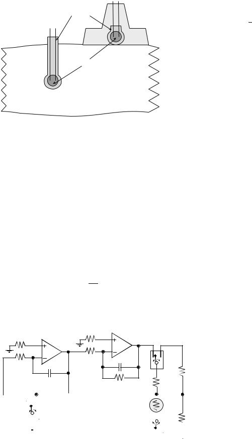
Figure 2. A glass-coated thermistor is placed into or on the surface of the tissue of interest.
bioheat transfer, this section will present in detail a specific measurement technique. In particular, this section presents an instrument used to measure thermal properties in tissue. The section begins with definitions of thermal properties, overviews of the technique, then develops the heat-transfer equations that form the basis of the instrument. Finally, calibration methods and error analyses are presented.
Definitions of Thermal Properties
Thermal conductivity (k) is the ability of a material to transport heat in the steady state. In one dimension, the total heat (Q) transported across a flat surface of area A and thickness Dx is related to the temperature gradient across the surface (DT) and the thermal conductivity of the material.
DT |
ð5Þ |
Q ¼ kA Dx |
Thermal diffusivity (a) is the ability of a material to conduct heat in the transient state. Thermal properties of
BIOHEAT TRANSFER |
193 |
conductivity and diffusivity are related. The quotient of conductivity divided by diffusivity equals density times specific heat.
k |
¼ rc |
ð6Þ |
a |
Diffusivity is often defined in the partial differential equation used to describe transient heat transfer. Assuming homogeneous thermal properties, the Fourier conduction equation in one dimension is
@2T |
1 |
@T |
ð7Þ |
||
|
¼ |
|
|
|
|
@x2 |
a |
|
@t |
||
Measurement Technique
The technique involves inserting a thermistor into the tissue of interest or placing it on the tissue surface, as shown in Fig. 2. Thermometrics P60DA102M and Fenwal 121-102EAJ-Q01 are glass probe thermistors that make excellent transducers (shown on the left of Fig. 2). The diameter of these thermistors is 0.15 cm. The glass-coated spherical probes provide a large bead size and a rugged, stable transducer. The Thermometrics BR55KA102M and Fenwal 112-102EAJ-B01 bead thermistors also provide excellent results (shown on the right of Fig. 2).
If the tissue is living, the properties measured are called effective thermal conductivity, keff, and effective thermal diffusivity, aeff. Effective thermal properties include the contribution to heat transfer due to intrinsic conduction added to the contribution caused by the transport of blood through the tissue.
In the constant temperature heating technique (47– 52), the instrument first measures the baseline tissue temperature, Ts. Then, an electronic feedback circuit applies a variable voltage, V0(t), in order to maintain the average thermistor temperature at a predefined constant, Th. The electrical circuit used to implement the constant temperature heating technique is shown in Fig. 3. Three high quality, gold-plated, electromagnetic relays are used to switch the thermistor (Rs) between ‘‘heat’’ and ‘‘sense’’ mode. Figure 3 shows the position of the three relays in ‘‘heat’’ mode. Initially, the instrument places the circuit in ‘‘sense’’ mode with the three relays in
10 kΩ Integrator |
10 kΩ |
Inverter |
|
|
|
-V0 |
V1 |
||
|
|
+V0 |
||
|
OP27 |
OP27 |
|
|
|
|
|
relay |
|
10 kΩ |
|
20 kΩ |
47 pF |
|
220 pF |
|
|
50 kΩ |
|
|
|
|
||
|
|
|
|
|
|
|
|
20 kΩ |
Rset |
|
|
|
|
|
error
|
|
|
|
|
|
|
|
|
|
|
|
|
|
|
|
|
|
+5.00 V |
|
|
|
|
|
|
|
|
relay |
Thermistor |
|
|
|
Rs |
|
||||
|
|
|
|
|
|
|
|
|
|
|
50 kΩ |
|||||||
|
|
|
|
|
|
|
|
|
|
|
||||||||
|
|
|
|
|
|
|
|
|
|
|
|
|
|
|||||
|
|
|
|
|
|
|
|
|
|
|
|
|
||||||
|
|
|
|
|
|
|
|
|
|
|
|
|||||||
|
|
|
|
|
|
|
|
|
resistors are 1% metal film |
|
|
|
|
|
relay |
|||
|
|
|
|
|
|
|
|
|
|
|
|
|
|
|
||||
|
|
|
|
|
|
|
|
|
|
|
|
|
|
|
|
|||
|
|
|
|
|
|
|
|
|
capacitors are ceramic |
|
|
|
|
|
|
Figure 3. Instrumentation used for the constant |
||
|
|
|
|
|
|
|
|
|
|
|
|
|
|
|
|
|||
|
|
|
|
|
|
|
|
|
relays are gold-contact EM |
|
|
|
|
|
V2 |
|
||
|
|
|
|
|
|
|
|
|
|
|
|
|
|
temperature heating technique. |
||||
|
|
|
|
|
|
|
|
|
|
|
|
|
|
|
|
|||
194 BIOHEAT TRANSFER
the opposite position as shown in Fig. 3. A precision þ5.00 V reference (PMI REF02) supplies voltage to the
four-resistor bridge, formed by the two 50 kV, Rset, and Rs resistors. The voltage difference V2 V1 is fed to a differ-
ential amplifier, passed through a low pass filter, then fed to a 12-bit ADC.
Fundamental Equations
Resistance calibration is performed to determine the relationship between the ADC sample and the unknown Rs. Next, temperature calibration is performed by placing the thermistor adjacent to an accurate temperature monitor and placing the combination in a temperaturecontrolled waterbath. The thermistor resistance varies nonlinearly with its temperature. For small temperature ranges equation 8 can be used for temperature calibration.
Rs ¼ RO eb=ðTsþ273:15Þ |
ð8Þ |
where Ts is the temperature in degrees Celsius, and Rs is the thermistor resistance in ohms.
In heat mode, the integrator–inverter circuit varies the voltage across the thermistor until the thermistor resistance, Rs, matches the fixed resistor, Rset. It takes just a few milliseconds for the electrical control circuit to stabilize. Once stable, Rs is equal to Rset, meaning the volume average thermistor temperature is equal to a constant. The instrument uses a calibration temperature versus resistance curve to determine the heated temperature
Th from the fixed resistor Rset. The power applied to the thermistor, P, is calculated from (V0)2/Rset. The applied
thermistor power includes a steady state and a transient term:
PðtÞ ¼ A þ Bt 1=2 |
ð9Þ |
In order to measure thermal conductivity, thermal diffusivity, and tissue perfusion the relationship between applied thermistor power, P, and resulting thermistor temperature rise, DT(t) ¼ Th Ts, must be known. In the constant temperature method, DT is constant. The thermistor bead is treated as a sphere of radius a embedded in a homogeneous medium. Since all media are considered to have constant parameters with respect to time and space, the initial temperature will be uniform when no power is supplied to the probe.
Tb ¼ Tm ¼ Ts ¼ Ta þ |
Qmet |
at t ¼ 0 |
ð10Þ |
wrblcbl |
|||
Let V be the temperature rise |
above baseline, |
V ¼ |
|
T Ts. Both the thermistor bead temperature rise (Vb)
and the tissue temperature |
rise (Vm) are |
initially |
zero. |
|
|
Vb ¼ Vm ¼ 0 |
at t ¼ 0 |
ð11Þ |
To solve this coupled thermistor–tissue system, equation 7 is written in spherical coordinates and the applied power is deposited into the thermistor, while the perfusion heat sink is added to the tissue, equation 1. Assuming the venous blood temperature equilibrates with the tissue
temperature and that the metabolic heat is uniform in time and space, the Pennes’ bioheat transfer equation in spherical coordinates is given by
|
r c |
|
|
@Vb |
¼ |
k |
|
1 @ |
r2 |
@Vb |
|
|
A þ Bt 1=2 |
|
r < a |
(12) |
||||||||||
|
|
|
|
|
|
|
|
|
|
|
||||||||||||||||
|
b @t |
b r2 @r |
|
@r |
|
|||||||||||||||||||||
|
|
b |
|
|
|
|
þ |
4=3p a3 |
|
|
||||||||||||||||
r |
|
cm |
@Vm |
|
¼ |
km |
1 @ |
r2 |
@Vm |
|
|
wr |
c |
bl |
Vm |
r > a |
(13) |
|||||||||
m |
|
|
|
|
|
|
|
|||||||||||||||||||
|
@t |
|
r2 @r |
|
||||||||||||||||||||||
|
|
|
|
|
|
|
|
@r |
|
|
|
bl |
|
|
|
|
||||||||||
where w is the tissue perfusion (s 1). Perfect thermal contact is assumed between the finite-sized spherical thermistor and the infinite homogeneous perfused tissue. At the interface between the bead and the tissue, continuity of thermal flux and temperature leads to the following boundary conditions:
|
Vb ¼ Vm |
|
at r ¼ a |
(14) |
|
kb |
@Vb |
¼ km |
@Vm |
at r ¼ a |
(15) |
@r |
@r |
||||
The other boundary conditions are necessary at positions r ! 0 and r ! infinity. Since no heat is gained or lost at the center of the thermistor:
@Vb |
¼ 0 as r ! 0 |
ð16Þ |
kb @r |
Because the thermistor power is finite and the tissue is infinite, the tissue temperature rise at infinity goes to zero:
Vm ! 0 as r ! infinity |
ð17Þ |
It is this last initial condition that allows the Laplace transform to be used to solve the coupled partial differential equations. The Laplace transform converts the partial differential equations into ordinary differential equations that are independent of time t. The steady-state solution allows for the determination of thermal conductivity and perfusion (49).
V |
|
r |
A |
|
|
kb |
|
|
1 |
1 |
|
|
r |
|
2 |
18 |
|
||
|
|
kmð1 þ |
Þ þ |
|
|
a |
Þ |
||||||||||||
|
b |
ð Þ ¼ |
4p a k |
b |
2 |
|
ð |
|
|||||||||||
|
|
|
|
|
|
eð1 r=aÞ |
pz |
|
|
|
|
|
|||||||
|
|
|
A |
|
|
|
|
! |
|
|
|
|
|
|
|
|
|
||
|
|
ð Þ ¼ |
m |
|
|
pz |
|
|
|
|
|
|
ð |
|
Þ |
||||
|
|
|
1 þ |
|
|
|
|
|
|
|
|||||||||
Vm |
r |
4p r k |
|
|
|
|
pz |
|
|
|
|
|
|
|
|
19 |
|
||
where z is a dimensionless Pennes’ model perfusion term (wrbl cbl a2/km). The measured thermistor response, DT, is assumed be the simple volume average of the thermistor temperature:
R |
0aVbðrÞ4pr2dr |
|
|
DT ¼ |
|
4=3pa3 |
ð20Þ |
Inserting equation 18 into Eq. 20 yields the relationship used to measure thermal conductivity assuming no perfusion (49).
km ¼ |
1 |
|
|
ð21Þ |
|
|
|
||
4p a DT |
0:2 |
|||
|
A |
kb |
|
|
A similar equation allows the measurement of thermal diffusivity from the transient response, again assuming no
perfusion (49).
|
|
|
|
a |
!2 |
|
am ¼ |
pp |
B |
ð1 |
km |
Þ |
ð22Þ |
|
|
A |
þ 0:2 kb |
|
||
Calibration Equations
The first calibration determines relationship between the ADC sample and the thermistor resistance when in sense mode. For this calibration, precision resistors are connected in place of the thermistor, and the computer-based instrument is used to sample the ADC in sense mode. A simple linear equation works well for converting ADC samples to measured resistance. In this procedure, the device acts like a standard ohmmeter.
The second calibration determines the relationship between thermistor temperature and its resistance. The instrument measures resistance, and a precision thermometer determines true temperature. Equation 23 yields an accurate fit over a wide range of temperature:
T ¼ |
1 |
273:15 |
ð23Þ |
H0 þ H1lnðRÞ þ H3½lnðRÞ&3 |
where T is in degrees Celsius. Temperature resistance data are fit to Eq. 23 using nonlinear regression to determine the calibration coefficients H0, H1, and H3.
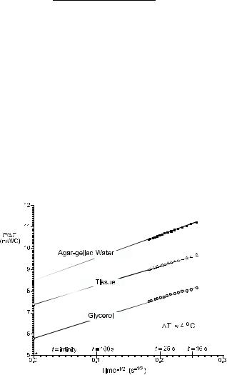
The applied power, P(t), is measured during a 30 s transient while in heat mode. Nonlinear regression is used to calculate the steady-state and transient terms in equation 9. Figure 4 shows some typical responses. The steady-state response (time equals infinity) is a measure of the thermal conductivity. The transient response (slope) indicated the thermal diffusivity.
The third calibration maps measured power to thermal properties while operating in heat mode. Rather than using the actual probe radius (a) and probe thermal conductivity (kb), as shown in Eqs. 21 and 22, the following empirical
Figure 4. Typical P/DT versus t 1/2 data for the constant temperature heating technique. The agar-gelled water and glycerol curves are used for empirical calibration.
|
|
|
BIOHEAT TRANSFER |
|
195 |
||||
equations are used to calculate thermal properties. |
|
|
|
||||||
km ¼ |
|
1 |
|
|
ð24Þ |
||||
|
|
|
|
||||||
ðc1 DT=AÞ þ c2 |
2 |
||||||||
am |
|
c3 |
ð |
25 |
Þ |
||||
B=Að1 þ km=c4Þ |
|||||||||
|
¼ |
|
|
|
|||||
The coefficients c1, c2, c3, and c4 are determined by operating the probe in two materials of known thermal properties. Typically, agar-gelled water and glycerol are used as thermal standards. This empirical calibration is performed at the same temperatures at which the thermal property measurements will be performed.
Error Analysis
It is assumed that the baseline tissue temperature, T0, is constant during the 30 s transient. Patel has shown that if the temperature drift, dT0/dt, is >0.002 8C s 1, then significant errors will occur (52). The electronic feedback circuit forces Th to a constant. Thus, if T0 is constant then DT does not vary during the 30 s transient.
The time of heating can vary from 10 to 60 s. Shorter heating times are better for small tissue samples and for situations where there is baseline tissue temperature drift. Another advantage of shorter heating times is the reduction in the total time required to make one measurement. Longer heating times increase the measurement volume and reduce the effect of imperfect thermistor–tissue coupling. Typically, shorter heating times are used in vivo because it allows more measurements to be taken over the same time period. On the other hand, longer heating times are used in vitro because accuracy is more important than measurement speed.
Thermal probes must be constructed in order to measure thermal properties. The two important factors for the thermal probe are thermal contact and transducer sensitivity. The shape of the probe should be chosen in order to minimize trauma during insertion. Any boundary layer between the thermistor and the tissue of interest will cause a significant measurement error. The second factor is transducer sensitivity that is the slope of the thermistor voltage versus tissue thermal conductivity. Equation 21 shows for a fixed DT km and kb the thermistor power (A) increases linearly with probe size (a). Therefore larger probes are more sensitive to thermal conductivity. For large tissue samples, multiple thermistors can be wired in parallel, so they act electrically and thermally as one large device. There are two advantages to using multiple thermistors. The effective radius, a ¼ c1/4p, is increased from 0.08 cm for a typical single P60DA102M probe to0.5 cm for a configuration of three P60DA102M thermistors. The second advantage is that the three thermistors are close enough to each other that the tissue between the probes will be heated by all three thermistors. This cooperative heating tends to increase the effective measurement volume and reduce the probe/tissue contact error. Good mechanical–thermal contact is critical. The probes are calibrated after they are constructed, so that the thermistor geometry is incorporated into the coefficients
196 BIOHEAT TRANSFER
c1, c2, c3, and c4. The same waterbath, and probe configuration should be used during the calibration and during the tissue measurements.
Calibration is a critical factor when using an empirical technique. For temperatures <0 8C, ice and ethylene glycol are used as thermal standards. For temperatures between 0 and 15 8C, agar-gelled water and ethylene glycol can be used as thermal standards. For temperatures between 15 and 75 8C, agar-gelled water and glycerol were used. To prevent convection, 1 g of agar/100 mL of water should be added. A mixture of water and glycerol can be used to estimate the accuracy of the technique. The mass fraction, m, can be used to determine the true thermal properties of the mixture (53,54). The ability to determine measurement accuracy is critical for the acceptance of new technology. These two equations provide for the capability to create reference materials of known thermal properties, which can be used to experimentally determine measurement accuracy.
km ¼ m kg þ ð1 mÞkw þ 1:4 mðm 1Þðkw kg 2Þ |
ð26Þ |
0:014 mðm 1ÞðT 20 CÞ |
|
am ¼ m ag þ ð1 mÞaw |
ð27Þ |
where T is in degrees Celsius. Self-heat thermistors have also been successfully used to measure the convective heat transfer coefficient on the endocardial surface of the heart (55,56).
ADDITIONAL STUDIES
In this article, the general concepts of bioheat transfer were introduced, and a detailed design and analysis of an instrument that measures thermal properties was presented. Although out of print, the 1985 book Heat Transfer in Medicine and Biology, is a wonderful collection of detailed works that address a wide spectrum of topics in bioheat transfer. The book Optical-Thermal Response of Laser Irradiated Tissue covers the issues involved in high temperature effect such as tissue damage and thermal ablation. Valvano’s chapter titled Temperature Measurements, in Advances In Heat Transfer: Bioengineering Heat Transfer, covers many practical issues involved in measuring temperature in the biomedical setting. An in depth treatment of bioheat transfer topics can be found in the 2005 edition of CRC Handbook of Heat Transfer. This reference has excellent treatments of thermoregulation and low temperature effects.
BIBLIOGRAPHY
Cited References
1.Baish JW, Ayyaswamy PS, Foster KR. Heat transport mechanisms in vascular tissues: a model comparison. J Biomech Eng 1986;108:324–331.
2.Baish JW. Heat transport by countercurrent blood vessels in the presence of an arbitrary temperature gradient. J Biomech Eng 1990;112:207–211.
3.Johnston KA, Bennett AF, editors. Animals and Temperature: Phenotypic and Evolutionary Adaptation. Cambridge: Cambridge University Press; 1996.
4.Diller KR. Modeling of bioheat transfer processes at high and low temperatures. Adv Heat Trans 1992;22:157–357.
5.Diller KR, Valvano JW, Pearce JA. Bioheat Transfer. In: Kneith F, editor. CRC Handbook of Heat Transfer, 2nd ed. 2005.
6.Charney CK. Mathematical models of bioheat transfer. Adv Heat Trans 1992;22:19–155.
7.Pennes HH. Analysis of Tissue and Arterial Blood Temperature in the Resting Human Forearm. J Appl Phys 1948;1:93–102.
8.Wissler E. A review of human thermal models. In: Morrison MB, editor. Environmental Ergonomics. New York: Taylor and Francis; 1988. p 267–285.
9.Wissler EH. Mathematical simulation of human thermal behavior using whole-body models. In: Shitzer A, Eberhart RC, editors. Heat Transfer in Medicine and Biology. Vol. 1. New York: Plenum Press; 1985. p 325–373.
10.Chato JC. Heat transfer to blood vessels. J Biomech Eng 1980;102:110–118.
11.Chen MM, Holmes KR. Microvascular contributions in tissue heat transfer. Annals NY Acad Sci 1980;335:137–150.
12.Weinbaum S, Jiji L, Lemons DE. Theory and Experiment for the Effect of Vascular Temperature on Surface Tissue Heat Transfer—Part 1: Anatomical Foundation and Model Conceptualization. ASME J Biomech Eng 1984;106:246–251.
13.Weinbaum S, Jiji L, Lemons DE. Theory and Experiment for the Effect of Vascular Temperature on Surface Tissue Heat Transfer—Part 2: Model Formulation and Solution. ASME J Biomech Eng 1984;106:331–341.
14.Weinbaum S, Jiji L. A New Simplified Bioheat Equation for the Effect of Blood Flow on Average Tissue Temperature. J of Biomech Eng 1985;107:131–139.
15.Charny CK, Weinbaum S, Levin RL. An Evaluation of the Weinbaum-Jiji Bioheat Equation for Normal and Hyperthermic Conditions. ASME J Biomech Eng 1990;112:80–87.
16.Xu LX, Chen MM, Holmes KR, Arkin H. The Evaluation of the Pennes, the Chen-Holmes, the Weinbaum-Jiji Bioheat Transfer Models in the Pig Kidney Cortex. ASME WAM Proc HDT 1991;189:15–21.
17.Arkin H, Xu LX, Holmes KR. Recent Developments in Modeling Heat Transfer in Blood Perfused Tissues. IEEE Trans Biomed Eng 1994;41(2):97–107.
18.Wissler EH. Pennes’ 1948 paper revisited. J Appl Physiol 1998;85:35–41.
19.Pennes HH. Analysis of tissue and arterial blood temperatures in the resting forearm. J Appl Physiol 1948;1:93–122 (republished for fiftieth anniversary issue of J Appl Physiol 1998;85:5–34).
20.Scully RM, Barnes MR. Physical Therapy. Philadelphia: J.B. Lippincott Co.; 1989.
21.Roussy G, Pearce JA. Foundations And Industrial Applications Of Microwaves Physical And Chemical Processes. New York: John Wiley & Sons, Inc.; 1995.
22.Chappuis P et al. Heat storage regulation in exercise during thermal transients. J Appl Physiol 1976;40:384–392.
23.Webb P. The physiology of heat regulation. Am J Physiol 1995;268:R838–R850.
24.Nunneley SA. Water cooled garments: a review. Space Life Sci 1970;2:335–360.
25.Nunneley SA. Physiological response of women to thermal stress: A review. Med Sci Sports 1978;10:250–255.
26.Ganong WF. Review of Medical Physiology. 16th ed. Norwalk (CT): Appleton and Lange; 1993.
27.Fulcher CWG. Control of a liquid cooling garment for extravehicular astronauts by cutaneous and external auditory meatus temperatures, Ph.D. dissertation, Houston (TX): University of Houston; 1970.