

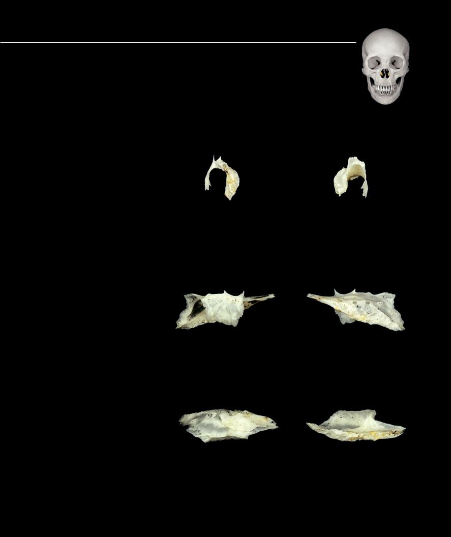
Cranial Bones – Inferior Nasal Concha
This is a small, delicate bone that projects from the lateral wall of the nasal cavity. It is scroll-like in appearance as it arches inferiorly and laterally from the nasal cavity’s lateral wall. The medial surface of the bone is convex and furrowed by many longitudinal grooves that transport blood vessels beneath the thick nasal mucosa that covers this surface. The lateral surface of the bone is concave and forms most of the superior and medial boundary of the inferior nasal meatus. The inferior border of the bone has a rough, spongy appearance. Superiorly the bone forms an articular border with four bones.
1 |
Lacrimal process |
|
|
2 |
Maxillary process |
|
|
3 |
Ethmoidal process |
|
|
4 |
Lateral surface |
|
5 |
5 |
Medial surface |
4 |
Left inferior nasal concha
Anterior view, lateral at left
3 2 1
4
Left inferior nasal concha
Lateral view, anterior at right
|
5 |
|
|
3 |
4 |
2 |
1 |
|
|||
|
|
|
Left inferior nasal concha
Superior view, anterior at right
5
4
Left inferior nasal concha
Posterior view, lateral at right
1 2 3
5
Left inferior nasal concha
Medial view, anterior at left
5
Left inferior nasal concha
Inferior view, anterior at right
69

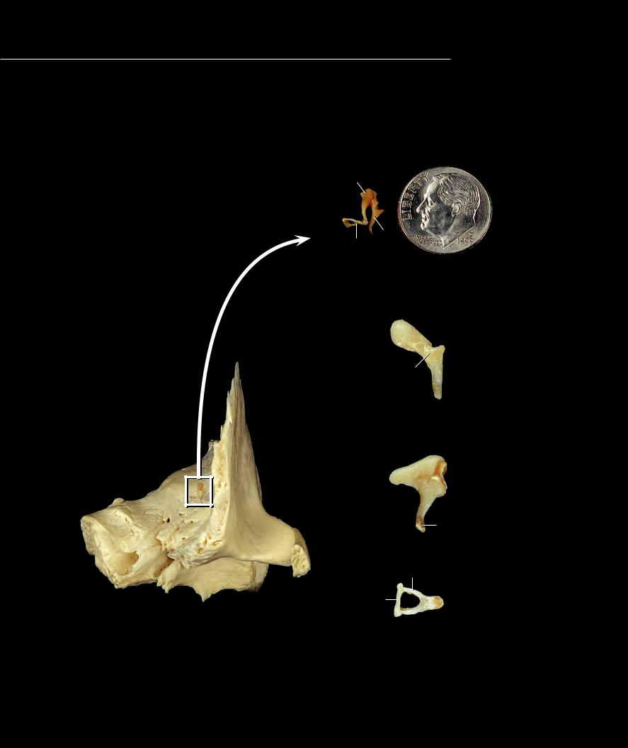
The lacrimal bone derives its name from the Latin word meaning tear because the bone houses
the “tear duct.” This small, delicate, quadrate-shaped bone has a vertical axis that is slightly longer than its horizontal axis. It is extremely thin. When it is held up to a light source, the light easily penetrates the bone. The bone sits in the anterior part of the medial wall of the orbit. The orbital surface is smooth and fl at in its posterior half where it contributes to the medial wall of the orbit. Anteriorly this surface has a longitudinal groove that ends posteriorly in a longitudinal crest that is hook-shaped inferiorly. This groove supports the nasolacrimal duct. Covered with mucous membrane, the slightly rough, medial surface of the bone contributes to the nasal cavity. The lacrimal bone articulates with four bones.
1 Posterior lacrimal crest
2 Lacrimal groove
3 Lacrimal hamulus
1
2
Left lacrimal bone |
Left lacrimal bone |
Anterior view, lateral at right |
Posterior view, lateral at left |
1 |
|
|
|
|
|
|
|
3 |
3 |
||
|
|
|
|
Left lacrimal bone |
Left lacrimal bone |
||
Lateral view, anterior at left |
Medial view, anterior at right |
||
3 |
2 |
3 |
2 |
Left lacrimal bone |
Left lacrimal bone |
|
Inferior view, lateral at left |
||
Superior view, lateral at right |
||
|
70
The auditory ossicles are the smallest bones of the human skeleton. These
three small bones occupy the middle ear cavity, where they transmit and amplify the sound waves from the tympanic membrane to the inner ear. From lateral to medial the bones are the malleus, the incus, and the stapes, or in layman’s terms the hammer, the anvil, and the stirrup, because of their striking resemblance to these structures.
1 Malleus
2 Incus
3 Stapes
4 Handle of malleus
5 Head of malleus
6 Neck of malleus
7 Lateral process
8 Anterior process
9 Body of incus
10Long limb
11Lenticular process
12Short limb
13Head of stapes
14Anterior limb
15Posterior limb
16Footplate
Auditory ossicles in situ within temporal bone
Anterior view, left temporal bone
2
1
3
Left auditory ossicles
Anterior view, lateral at left
5
6
7
8
4
Left malleus
Anterior view, lateral at left
12 9
10
11
Left incus
Lateral view, anterior at left
15
16 |
|
|
|
13 |
|||
|
|||
|
14 |
||
Left stapes
Superior view, lateral at left
71
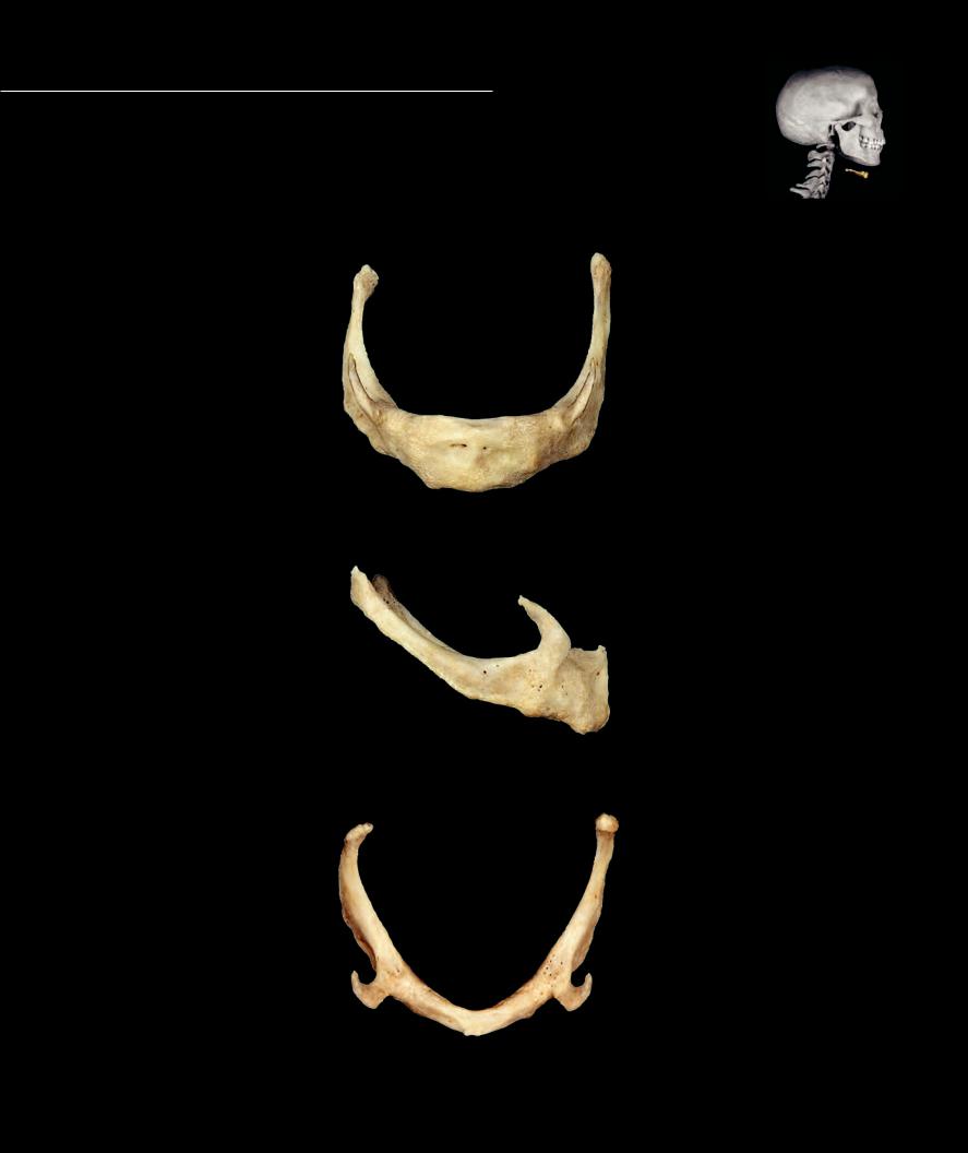
Suspended from the styloid processes of the temporal bones by the stylohyoid ligaments, the U-shaped hyoid bone
occupies the ventrosuperior neck just inferior to the mandible. It serves as a skeletal attachment site for muscles associated with the tongue, larynx, and pharynx. It consists of fi ve elements — a body and bilateral lesser and greater cornua. The body is the rectangular ventral element that sits in the transverse plane. Projecting posterolaterally from the body are the paired, long, slender greater cornua. At the junction of the greater cornua and the body are smaller superior projections, the lesser cornua.
1 Body
2 Lesser horn
3 Greater horn
72
3
2
1
Hyoid bone
Anterior view
2
3
1
Hyoid bone
Lateral view, anterior at right
3
2
1
Hyoid bone
Superior view, anterior at bottom