

- •Preface
- •Content
- •Tissues
- •Nerve Tissue
- •Skin - Epidermis
- •Skin - Dermis
- •Skin - Glands
- •Subcutaneous Layer
- •Skeleton
- •Axial Skeleton
- •Cranium
- •Cranial Bones – Inferior Nasal Concha
- •Vertebral Column
- •Sacrum and Coccyx
- •Ribs
- •Sternum
- •Clavicle
- •Scapula
- •Humerus
- •Ulna
- •Radius
- •Metacarpals and Phalanges
- •Pelvis - Male
- •Femur
- •Tibia
- •Fibula
- •Tarsal Bones - Cuboid and Navicular
- •Phalanges
- •Patella
- •Skeletal Muscles
- •Transversospinales Muscles
- •Cervical Hypaxial Muscles
- •Thoracic and Abdominal Hypaxial Muscles
- •Shoulder Muscles - Rotator Cuff
- •Shoulder Muscles - Prime Movers
- •Anterior Brachial Muscles
- •Posterior Brachial Muscles
- •Posterior Thigh Muscles
- •Thigh Muscles
- •Lateral Leg Muscles
- •Posterior Leg Muscles
- •Spinal Nerves
- •Dorsal Rami
- •Intercostal Nerves
- •Cutaneous Nerves
- •Autonomic Nerves
- •Spinal Cord
- •Brain
- •Cerebrum
- •Cerebellum
- •Meninges
- •Hypothalamus
- •Pituitary Gland
- •Pineal Gland
- •Thymus
- •Pancreas
- •Ovaries
- •Testes
- •Blood
- •Heart
- •Lymphatics
- •Larynx
- •Lungs
- •Cast of Trachea and Bronchial Tree
- •Esophagus
- •Stomach
- •Pancreas
- •Large Intestine
- •Mesenteries
- •Omenta
- •Female Reproductive Organs
- •Ovary
- •Vagina
- •Ductus Deferens and Spermatic Cord
- •Penis
- •Index
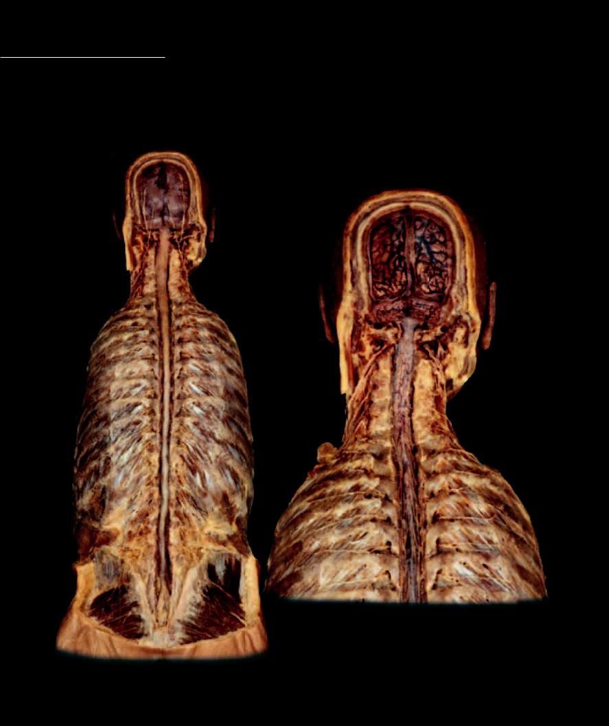
Meninges Within the cranium and vertebral column, the meninges form a protective encasement for the tissue of the brain and spinal cord. There are three meningeal membranes, the tough outer
connective tissue pachymenix, the dura mater, and the epithelial inner leptomeninges, the arachnoid mater and pia mater. Between the leptomeningeal layers there is a fl uid compartment called the subarachnoid space. Cerebrospinal fl uid, secreted from the choroid plexuses of the ventricles, exits the ventricles to fi ll this compartment. The cerebrospinal fl uid forms a hydraulic shock absorber and suspension system for the brain and spinal cord. In addition to protecting the central nervous system, the meninges support many of the blood vessels that are associated with the brain. Within the cranium the subdivisions of the dura mater split to form large venous channels, the dural venous sinuses, which drain all the tissues of the cranial vault, and these splits also form strong, fibrous septa that separate different parts of the brain.
1
4
2
3
5
Dura removed to expose leptomeninges
Posterior view
Dissection of cranial and spinal dura mater
Posterior view
246
1 Cranial dura mater
2 Spinal dura mater
3 Dural venous sinus
4 Cranial leptomeninges - arachnoid is superficial to and covering pia mater 5 Spinal leptomeninges - arachnoid is superficial to and covering pia mater 6 Middle meningeal artery and branches in dura mater
7 Superficial middle cerebral vein and tributaries in subarachnoid space
1
6
6
6
4
7
2
3
Dural sac (above), Leptomeninges (below)
Lateral views
247
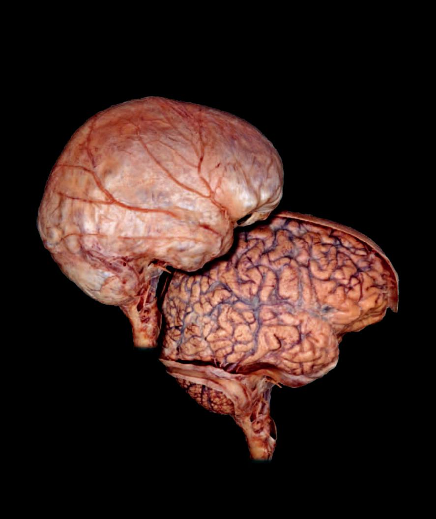
Meninges
1 |
Falx cerebri |
6 |
Lateral ventricle |
11 |
Cerebellum |
2 |
Tentorium cerebelli (cut) |
7 |
Septum pellucidum |
12 |
Corpus callosum |
3 |
Superior sagittal sinus |
8 |
Third ventricle |
13 |
Choroid plexus |
4 |
Straight sinus |
9 |
Fourth ventricle |
14 |
Optic chiasm |
5 |
Transverse sinus |
10 |
Cerebrum |
15 |
Trigeminal nerve |
1
12
7
8
3
15
11 4
1
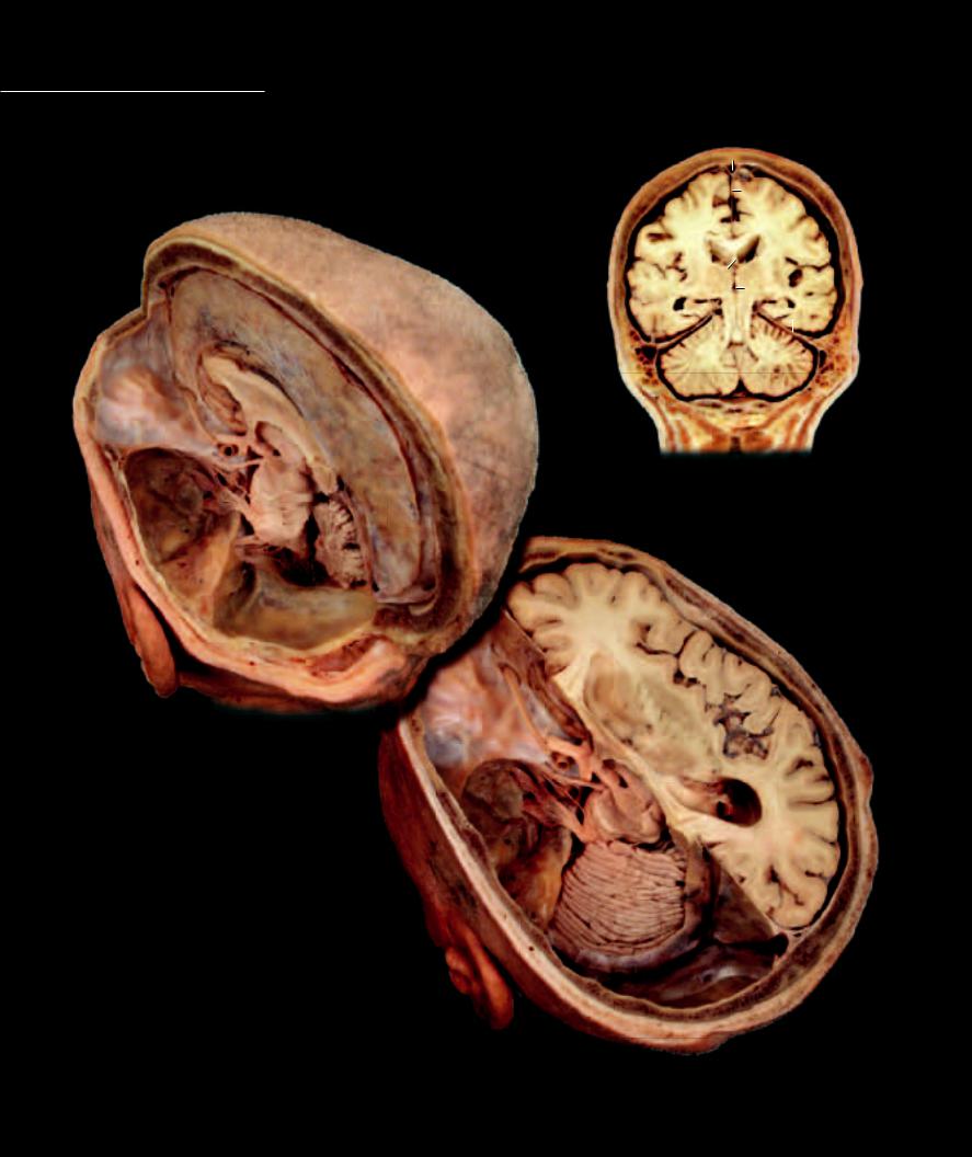
Dissection of cranium |
14 |
Superoposterior view
15
3
1
10
12
6
7
8
2
9
5
11
Head frontal section revealing dural septa
Anterior view
10
6
7
13
6
10
4
11
1
2 |
3 |
5
Dissection of cranium
Superoposterior view
248
15 Endocrine System
Like the nervous system, the endocrine system is a control system within the body. The nervous system administers its control over the body tissues via long wirelike cells that originate form complex circuits in the central nervous system. This circuitry receives sensory input, processes this input,
and generates regulatory output. Endocrine control works in a much different fashion. The endocrine system consists of a number of different glands that function like radio transmitting stations. Just as different radio stations send radio signals of different wavelengths into the air, endocrine glands distribute different types of
small molecules called hormones throughout the body via the circulatory system. These small molecules travel through the blood stream and are detected by effector organs in different parts of the body, much like radio waves are detected by radios in different parts of a city. Effector organs have receptor sites that are specific to specific hormones. This results in a “lock and key” function at the effector cell. When the hormone binds to the receptor site, it initiates a regulatory effect on the cell.
Because the hormones are distributed by the circulatory system, the speed of endocrine regulation is slower than that of nervous regulation, many minutes compared to milliseconds. Also, because of the distribution of the hormones via the circulatory system, endocrine effects can be experienced anywhere there are cells with the appropriate receptor site. In comparison to the nervous system, endocrine distribution is potentially very widespread. Because the hormone can lock into
the receptor site and not be degraded instantly, the duration can be longer lasting than that initiated by a single nervous impulse.
Find more information about the endocrine system in
R E A L A N AT O M Y
249