Добавил:
kiopkiopkiop18@yandex.ru Вовсе не секретарь, но почту проверяю Опубликованный материал нарушает ваши авторские права? Сообщите нам.
Вуз: Предмет: Файл:
Скачиваний:
8
Добавлен:
03.02.2024
Размер:
76.04 Mб
Скачать
Intercostal Nerves

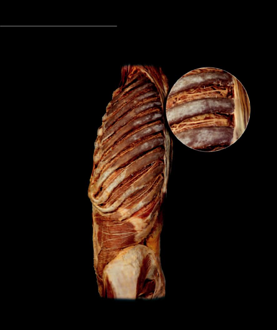
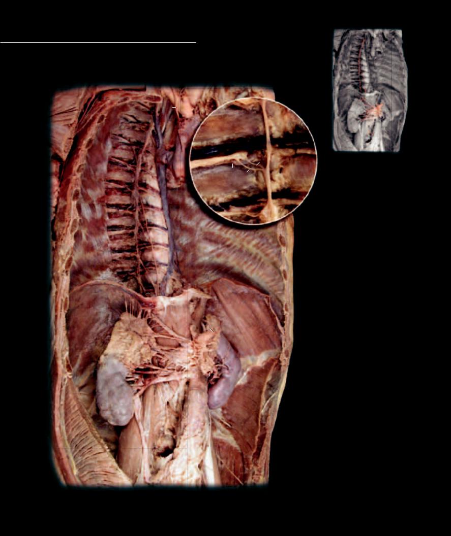
Unlike the ventral rami in the cervical, lumbar, and sacral regions, which form plexuses, most of the thoracic ventral rami remain segmental like their dorsal counterparts. These thoracic ventral rami, called the inter-

costal and subcostal nerves, emerge from the spinal nerve trunk and enter the intercostal space just inferior to each of the twelve ribs. Each of these segmental nerves has a similar structural design. The main trunk of the nerve runs through the intercostal space, with the segmental arteries and veins, between the middle and internal muscle layers of the body wall. Accompanying the main branch is a smaller collateral branch, which emerges from the main branch near the angle of the rib, and runs inferior to the main branch through the intercostal space. The main branch also gives rise to lateral and anterior cutaneous branches that supply the skin, or dermatome, of each segment.

Intercostal Nerves

6


1

Main trunk


2

Collateral branch

1

Other Nerves and Structures


2

3

Subcostal nerve


4

Iliohypogastric nerve


5

Posterior intercostal vein


6

Posterior intercostal artery


7

Innermost intercostal muscle


8

Transversus abdominis muscle


9

Gluteus medius muscle


10Piriformis muscle

11Iliocostalis muscles

12Rib 12

6

Dissection of intercostal space

Lateral view

5

7

7

12

11

3

4

8

9

10

Dissection of intercostal nerves

Lateral view

220

Cutaneous Nerves Many small nerves, named cutaneous nerves, branch from the spinal and cranial nerves and course through and between muscles to emerge

into the integumentary covering of the body. These detailed dissections reveal all the cutaneous nerves of the body.


1


Greater occipital nerve


2


Transverse cervical nerves


3


Supraclavicular nerves


4


Medial cutaneous branches (dorsal rami)


5


Lateral cutaneous branches (dorsal rami)


6


Anterior cutaneous branches (ventral rami)

2


7


Lateral cutaneous branches (ventral rami)


3


8


9


6


10


11


7


13


12


17


14


16


8


Superior lateral brachial cutaneous nerves


9


Posterior brachial cutaneous nerves


10


Inferior lateral brachial cutaneous nerves


11


Posterior antebrachial cutaneous nerve


12


Lateral antebrachial cutaneous nerve


13


Medial antebrachial cutaneous nerve


14


Femoral branch of genitofemoral nerve


15


Lateral cutaneous branch of subcostal nerve


16


Anterior cutaneous branch of femoral nerve


17


Lateral femoral cutaneous nerve


21

18


Superior cluneal nerves


19


Inferior cluneal nerve


20 Posterior femoral cutaneous nerve


21


Saphenous nerve


22 Lateral sural cutaneous nerve

1

4

5

15

18

19

20

22

Dissections exposing cutaneous nerves

Anterior view to left, Posterior view to right

221

Autonomic Nerves

In contrast to the somatic branches of the spinal nerve, the visceral branches leave the body wall to form nerve pathways that

enter the body cavities. Within the cavities these nerves form the autonomic nerve pathways, sympathetic and parasympathetic, to the viscera. The autonomic nerves relay input signals from the wall of the tubular gut and other viscera, while carrying output signals to smooth muscle,

10

1

20


19


32


17


21

22


18

4


2


19

18


17


32

33

5

6


31


30

24


27


7


8


28

26

9


25


23


28

37

35

29

34

36


1

3

2

Autonomic Nerves

1 Sympathetic trunk nerve

2 Sympathetic trunk ganglion

3 White communicating ramus

4 Gray communicating ramus

5 Greater splanchnic nerve

6 Lesser splanchnic nerve

7 Coeliac ganglion

8 Superior mesenteric ganglion

9 Aorticorenal ganglion

10Vagus nerve

11Recurrent laryngeal nerve

12Anterior vagal trunk

13Posterior vagal trunk

14Inferior cardiac plexus

15Pulmonary plexus

16Esophageal plexus

Other Structures

17Intercostal nerve

18Posterior intercostal artery

19Posterior intercostal vein

20Right superior intercostal vein

21Azygous vein

22Superior vena cava

23Aorta

24Celiac trunk

25Superior mesenteric artery

26Renal artery

27Suprarenal gland

28Kidney

29Ureter

30Diaphragm

30 Esophageal hiatus

32Subcostal muscle

33Innermost intercostal muscle

34Internal oblique muscle

35Transversus abdominis muscle

36Psoas major muscle

37Psoas minor muscle

Deep dissection of sympathetic nerves, callout of communicating rami

Anterolateral view

222

cardiac muscle, and glands. Some of the autonomic nerves even rejoin the somatic pathways to supply the blood vessels and glands of the body wall. The sympathetic pathways are primarily associated with vascular smooth muscle control, and the parasympathetic pathways are principally responsible for the regulation and control of gut tube smooth muscle and glands. The sympathetic nerves are depicted on the opposite page, while the vagus nerve, which carries 75% of the parasympathetic output, is shown below as it follows the derivatives of the gut tube.


53


10


38


10


54


45


47

46


11

39


48


49


44


23

11


55


50

50


41


15


51


51


43


13


16


12

52

30

Thoracic dissection revealing vagus nerve

Anterior view

38Thyroid gland

39Trachea

40Principal bronchus

41Lobar bronchus

42Segmental bronchus

43Esophagus

44Lung

45Right common carotid artery

46Left common carotid artery

47Right subclavian artery

48Left subclavian artery

49Brachiocephalic artery

50Pulmonary arteries

51Pulmonary veins

52Inferior vena cava

53Cricothyroid muscle

54Anterior scalene muscle

55Ligamentum arteriosum


45


38


10


10


54


47


46


11


48


44


39


23


41


11


40


14


41


42


51


43


51


13


30


52


12


Deeper thoracic dissection revealing vagus nerve

Anterior view

223

Cranial Nerves

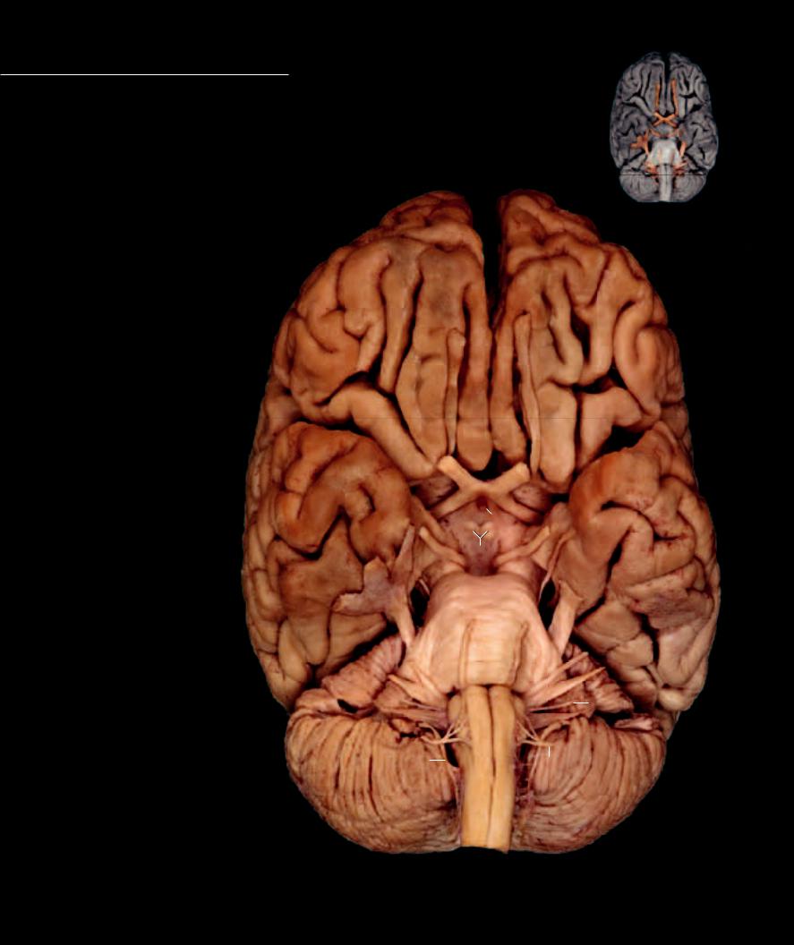
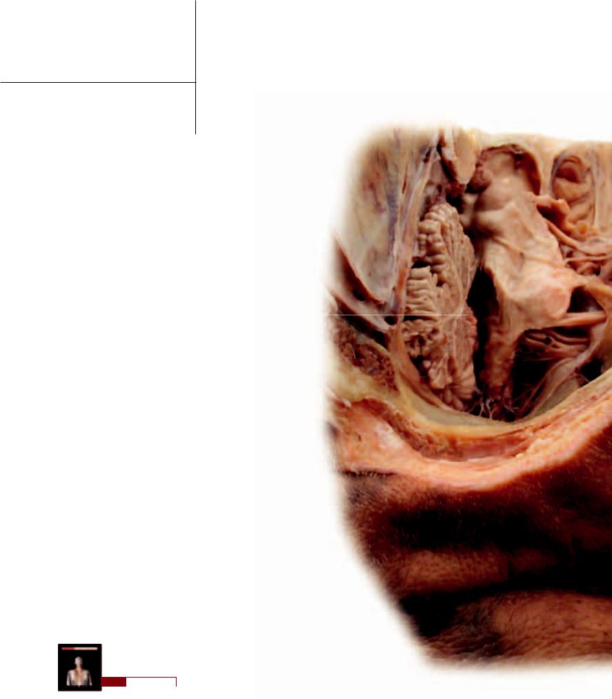
Cranial nerves segregate into three distinct groups based on associations they form during development. In number there are twelve cranial nerves, which originate in pairs

from a rostral to caudal sequence from the brain. The fi rst category, the special sensory cranial nerves, are afferent pathways established between the the brain and the special sensory structures of the nose, eye, and ear. The second category, the ventral or somitic motor cranial nerves, are homologous with the ventral roots of the spinal nerves. They originate from the brainstem as efferent pathways to somitic skeletal muscles within the head. The fi nal category, comprising the largest of the

Special Sensory Nerves 1 Olfactory nerve

2 Optic nerve

3 Vestibulocochlear nerve

Somitic Motor Nerves


4

Occulomotor nerve


27


5

Trochlear nerve


6

Abducens nerve


7

Hypoglossal nerve


Pharyngeal Arch Nerves


8

Trigeminal nerve


9

Trigeminal ganglion


17


10

Opthalmic branch


11

Maxillary branch


12

Mandibular branch


13

Facial nerve


14

Glossopharyngeal nerve


1


15

Vagus nerve


16

Accessory nerve


Other Structures


17

Olfactory bulb


18

Optic chiasm


2


19

Optic tract


18


20

Infundibulum


21

Mammillary bodies

28


19


22

Cerebral peduncle


20


23

Pons


24

Cerebellum


10


4


25

Medulla oblongata


21


26

Spinal cord


5


11


22


27

Frontal lobe


28

Temporal lobe


9


29

Insular lobe

12

23


30

Parietal lobe


8


31

Occipital lobe


32

Right lateral ventricle


33

Choroid plexus


6


34

Falx cerebri


13


35

Falx cerebelli


36

Straight sinus


3


37

Superior sagittal sinus


14


38

Corpora quadrigemina


25


39

Pineal gland


15


40Third ventricle

41Fourth ventricle

42

Geniculate ganglion

24

16


7

43

Anterior cerebral artery


44Internal carotid artery

45Levator palpebrae superioris muscle

46Superior rectus muscle

47Lateral rectus muscle

48

Superior oblique muscle

26

49

Nasociliary nerve


50

Long ciliary nerve


51

Ciliary ganglion

Base of brain with cranial nerves

52

Eye

Inferior view

224

cranial nerves, are those cranial nerves associated with the pharyngeal arches. The dorsal or pharyngeal arch cranial nerves are developmentally similar to the dorsal roots of the spinal nerves. These fi ve dorsal cranial nerves form the general sensory afferent pathways from the peripheral tissues of the head. However, because these nerve pathways coursed through the specialized arches forming the pharyngeal wall of the foregut, they established parasympathetic efferent pathways to the glandular tissue of the gut wall, along with motor efferent pathways to the skeletal muscles derived from the pharyngeal arch tissues.


27


34


29

28


30


49


45


17

43


33


48


32


40


46

1


18


52


2


39


47


4


44


31


38

51

50


5


11

10

6


34


36

98

12


41

37

13


42


3


24


15


14


7


35


16


Intracranial dissection of cranial nerves

Posterolateral view

225

Cranial Nerves

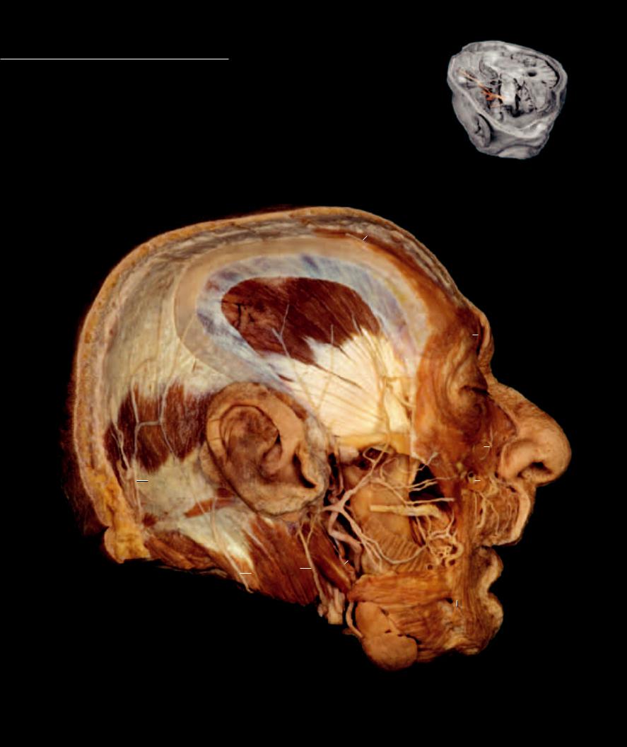
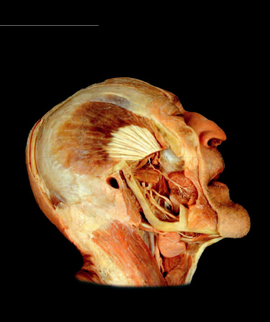
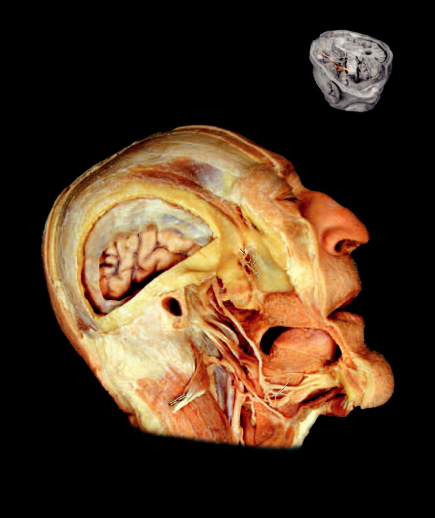
Cranial nerves V and VII, the trigeminal and facial nerves respectively, have the most extensive distribution to the tissues of the head. This page

and the three pages that follow depict the peripheral distribution of many of the branches of the trigeminal and facial nerves.

Trigeminal Nerve

9

Superior posterior lateral nasal branch

16

Buccal branches

1

Auriculotemporal nerve

10

Inferior posterior lateral nasal branch

17

Mandibular branches

2

Supraorbital nerve

11

Pharyngeal branch

18

Cervical branch

3

Infraorbital nerve

12

Lesser palatine nerve


4

Mental nerve

13

Greater palatine nerve

Other Nerves and Structures

5

Maxillary branch


19

Greater occipital nerve

6

Nerve of the pterygoid canal

Facial Nerve

20

Lesser occipital nerve

7

Pterygopalatine ganglion

14

Temporal branches

21

Great auricular nerve

8

Nasopalatine nerve (cut)

15

Zygomatic branches

22

Auricularis posterior muscle

2

24

25

26

27

2


28


1


23


14

3


19


15

29


3


22


34


31


35

16


33

18

32


21


17


20


30

4

36

Dissection of head exposing branches of the facial nerve

Lateral view

226

23

Occipital belly of epicranius muscle

32

Masseter muscle

41

Septum pellucidum

50

Pituitary gland

24

Galia aponeurotica

33

Posterior digastricus muscle

42

Thalamus

51

Torus tubarius

25

Frontal belly of epicranius muscle

34

Parotid duct

43

Midbrain

52

Maxillary sinus

26

Temporal fascia

35

External carotid artery

44

Pons

53

Middle nasal concha

27

Temporalis muscle

36

Submandibular gland

45

Cerebellum

54

Inferior nasal concha

28

Orbicularis oculi muscle

37

Frontal sinus

46

Fourth ventricle

55

Hard palate

29

Zygomaticus major muscle

38

Cerebrum

47

Choroid plexus

56

Soft palate

30

Risorius muscle

39

Falx cerebri

48

Medulla oblongata

57

Uvula

31

Buccinator muscle

40

Corpus callosum

49

Spinal cord

58

Tongue

38


40


41

39


42


37


43


50

46


5


44

45


8


6

47


7


53

9


52


11


10


48


54


51


13


12


55

56


49


57


58

Parasagittal section and dissection of head exposing branches of the trigeminal and facial nerve

Medial view

227

Cranial Nerves

1

Nerve to temporalis muscle

7

Inferior alveolar nerve

13

Superior laryngeal nerve

2

Buccal nerve

8

Nerve to mylohyoid muscle


3

Middle superior alveolar nerve

9

Pterygopalatine ganglion

Other Structures

4

Posterior superior alveolar nerve

10

Infraorbital nerve

14

Orbicularis oculi muscle

5

Lingual nerve

11

Hypoglossal nerve

15

Temporal fascia

6

Chorda tympani nerve

12

Submandibular ganglion

16

Temporalis muscle

15

16

14

3

1

4

17

19

35

2

18

5

7

8

20

8

21

31

32

22

30 23

24

Dissection of head exposing branches of the trigeminal nerve

Lateral view

228

17

Lateral pterygoid muscle

24

Omohyoid muscle

31

Internal jugular vein

18

Medial pterygoid muscle

25

Styloglossus muscle

32

Common carotid artery

19

Buccinator muscle

26

Stylohyoid muscle

33

Dura mater

20

Posterior digastricus muscle

27

Geniohyoid muscle

34

Cerebrum

21

Anterior digastricus muscle

28

Mylohyoid muscle

35

External acoustic meatus

22

Sternocleidomastoid muscle

29

Superior pharyngeal constrictor

36

Tongue

23

Thyrohyoid muscle

30

Inferior pharyngeal constrictor


15

16

14

33

34

10

9 3

4

6

5


35


29


19


36


26

25


20


12


31

11


27


13

28


32


21


22


23


30


24


Dissection of head with mandible removed

Lateral view

229

Sensory Receptors

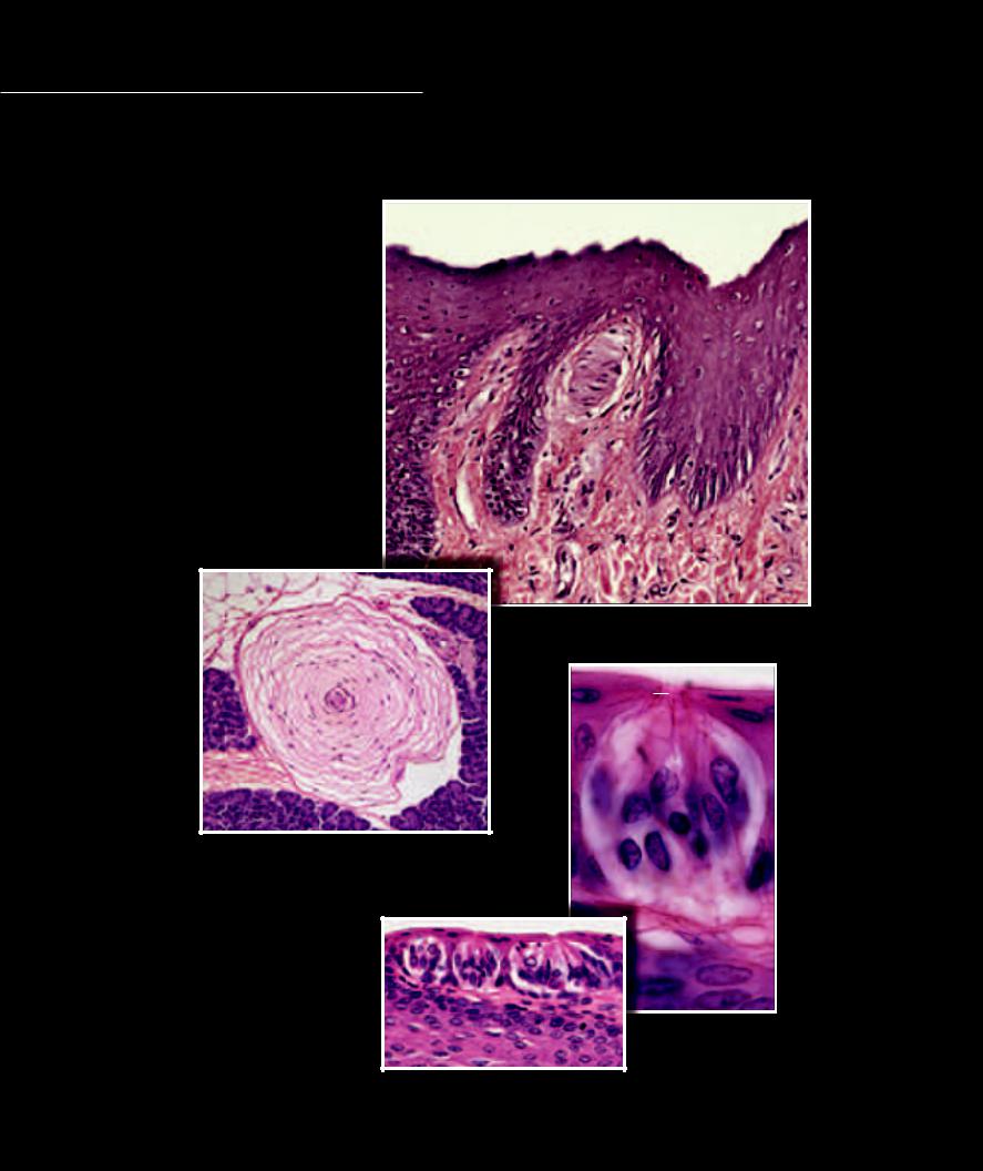
Sensory receptors are the transducers of the nervous system; that is, they convert the different types of energy we experience such as mechanical energy (touch, pressure, sound waves, etc.), thermal

energy (heat), chemical energy (taste, smell), and electromagnetic energy (light) into the electrical energy of the nervous impulse. They do this by facilitating the depolarization of the peripheral terminals of the sensory neurons. This initiates the nervous impulse along the sensory neuron, and this input is carried by the sensory neuron to the processing centers of the brain and spinal cord, which will be the topic of the next chapter.

1

Epidermis


2

Corpuscle of touch (Meissner’s)

1


3

Dermis


4

Dermal papilla


5

Neuron


6

Lamellated corpuscle


7

Taste bud

4

2

8

Taste pore


9

Gustatory hair


10Gustatory receptor cell

11Supporting cell

12Basal cell

3


Photomicrograph of corpuscle of touch


200x


8

5

6

9


10

Photomicrograph of lamellated corpuscle

11


100x


12


7


Photomicrographs of taste bud

200x (left), 700x (right)

230

14 Central Nervous System

While the neuronal circuitry of the central nervous system is awe inspiring to say the least, the basic concepts behind this complex integration and control center have a simple design. At its simplest, the fundamental design of the central nervous system involves two features: gray matter and white matter.

The gray matter centers represent the synaptic integration and control circuits; that is, these centers contain numerous highly dendritic interneurons along with the cell bodies of efferent neurons and axon terminals of incoming afferent neurons, all forming a myriad of synaptic circuits. In these gray centers input is integrated, compared, sensed, and stored to give rise to coordinated, controlled output. The white matter, on the other hand, represents conduction tracts between the synaptic gray centers. These white tracts consist mainly of the myelinated axons of interneurons relaying signals from one gray center to another.

A second simple concept to keep in mind is that the complexity of the central nervous system increases from a caudal to cranial direction. There is logic to this pattern because in the spinal cord the gray centers primarily function as integration networks that regulate input and output for their specific spinal nerve levels. In other words, they are segmental control centers. Input entering a spinal nerve level initiates reflexive output back to the peripheral tissues at that same spinal level. Connecting these segmental gray centers via interneuronal tracts leads to greater association between neighboring levels, therefore improving integration and control. If one segmental gray center can relay information received from its center to neighboring centers, then there can be a greater spread of control generated in response to local segmental input. Now take this a step further by relaying information via white tracts from each of the segmental control centers to higher centers. These higher centers receive in-

put from all the lower segmental centers, integrating the input to gain a full body perspective, while generating the necessary output signals to exert coordinated full body control. Because of this added circuitry the cranial or brain end of the central nervous system increases in size. This additive accumulation of interconnected gray centers accounts for the structure of the brain and its amazing functional properties.

Because much of the central nervous system circuitry is of a more microscopic nature and beyond the scope of this book. In this chapter we attempt to depict the basic gross anatomy of the central nervous system and its protective coverings.

Find more information about the central nervous system in

R E A L A N AT O M Y

231