

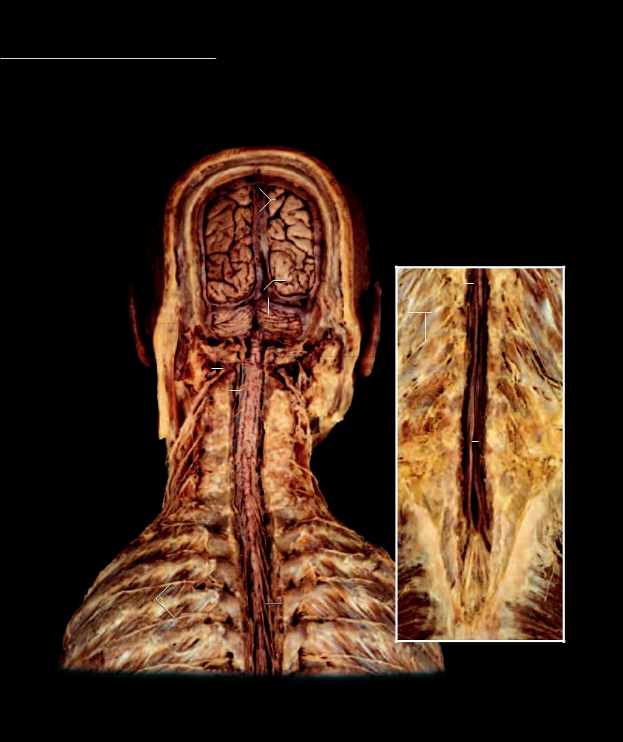
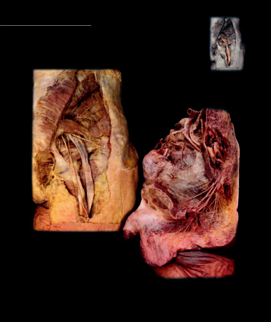
With slight variation, the basic pattern of the spinal nerve repeats itself thirty-one times along the entire length of the spinal cord. With the exception of the fi rst spinal nerve, each spinal nerve level emerges from within the vertebral column to pass
peripherally between successive vertebrae. Because of the developmental differences in the growth rate of the vertebral column and associated spinal cord, the lower roots of the spinal nerves are dragged downward by the lengthening vertebral column. With each succeeding spinal nerve level the roots become longer and more oblique in their course, eventually extending beyond the end of the spinal cord as the vertically oriented cauda equina.
Spinal Nerves |
|
|
|
|
|
|
|
|
Other Structures |
12 |
Dura mater |
|||||||
1 |
Spinal nerve |
|
|
|
|
|
|
|
|
8 |
Cerebrum |
13 |
Superior sagittal sinus |
|||||
2 |
Cervical dorsal rootlets |
|
|
|
|
|
|
|
|
9 |
Cerebellum |
14 |
Transverse sinus |
|||||
3 |
Thoracic dorsal rootlets |
|
|
|
|
|
|
|
|
10 |
Medulla oblongata |
15 |
Opening of straight sinus |
|||||
4 |
Lumbosacral dorsal rootlets |
|
|
|
|
|
|
|
|
11 |
Spinal cord |
16 |
Confluence of sinuses |
|||||
5 |
Dorsal rami |
12 |
|
|
|
|
|
|
|
|
|
|
|
|
|
|||
6 |
Cauda equina |
|
|
|
|
|
|
|
|
|
|
|
|
|
||||
|
|
|
|
|
|
|
|
|
|
|
|
|
|
|
|
|
||
7 |
Filum terminale |
|
|
|
|
|
|
|
13 |
|
|
|
|
|
|
|
|
|
|
|
8 |
|
|
|
|
|
|
|
|
|
|
|
|
|
|||
|
|
|
|
|
|
|
|
|
|
|
|
|
|
|
|
|||
|
|
|
|
|
|
15 |
|
|
|
|
4 |
|
|
|
|
|||
|
|
|
|
|
|
|
|
|
|
|
|
|
|
|||||
|
|
|
|
|
|
|
|
|
|
|
|
|
|
|
|
|||
|
|
|
|
|
|
|
14 |
|
|
|
|
|
|
|
|
|
||
|
|
|
|
|
|
|
|
|
|
|
|
|
|
5 |
|
|
|
|
|
|
9 |
16 |
|
|
|
|
|
|
|
|
|
|
|||||
|
|
|
|
|
|
|
|
|
|
|
|
|
|
|
||||
|
|
1 |
|
|
|
10 |
|
|
|
|
|
|
|
|
|
|
|
|
|
|
|
|
|
|
|
|
|
|
|
|
|
|
|
|
|
||
|
|
|
|
|
|
|
|
|
|
|
|
|
|
|
|
|
|
|
|
|
|
|
|
|
|
|
|
|
|
|
|
|
|
|
|
|
|
|
|
2 |
|
|
|
|
|
|
|
|
|
|
|
6 |
||||
|
|
|
|
|
|
|
|
|
|
|
|
|
||||||
|
|
|
|
|
|
|
|
|
|
|
|
|
|
|
|
|||
|
|
|
|
|
|
11 |
|
|
|
|
|
|
|
|
|
|
7 |
|
|
|
|
|
|
|
|
|
|
|
|
|
|
|
|
|
|||
5 
3
Dissection exposing cauda equina
Posterior view
Dissection revealing spinal cord and brain
Posterior view
214
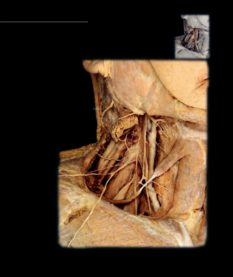
The dorsal rami of the spinal nerves arise at all spinal levels and pursue a posterior course into the muscles, connective tissue, and skin of the back. They innervate all
the epaxial muscles comprising the extensors of the vertebral column. The cutaneous distribution of the dorsal rami spans from the top of the head, down the posterior trunk, to the superior half of the gluteal region. With the exception of levels C1, S4, S5, and the coccygeal, the dorsal rami split into lateral and medial branches as they course posteriorly into the back.
Dorsal Rami |
|
Other Structures |
11 |
Semispinalis cervicis muscle |
|
|
|
||||||||||
1 |
Greater occipital nerve |
6 |
Rectus capitis posterior major muscle |
12 |
Intertransversarii thoracic muscle |
|
|
|
|||||||||
2 |
Least occipital nerve |
7 |
Rectus capitis posterior minor muscle |
13 |
Levatores costarum muscles |
|
|
|
|||||||||
3 |
Dorsal ramus |
|
8 |
Obliquus superioris muscle |
14 |
External intercostal muscle |
|
|
|
||||||||
4 |
Medial branch |
|
9 |
Obliquus inferioris muscle |
15 |
External oblique muscle |
|
|
|
||||||||
5 |
Lateral branch |
|
10 |
Posterior digastricus muscle |
16 |
Internal oblique muscle |
|
|
|
||||||||
|
|
|
|
|
|
|
|
|
|
|
|
|
7 |
8 |
|||
|
|
|
|
|
|
|
|
|
|
|
|
|
|
|
|||
|
3 |
|
|
|
|
|
|
|
|
|
|
|
6 |
|
|
1 |
|
|
|
|
|
|
|
|
|
|
|
|
|
||||||
|
|
|
|
|
|
|
|
|
|
|
|
|
|||||
|
|
|
|
|
|
|
|
|
|
|
|
|
|
|
9 |
||
|
|
|
|
|
|
|
|
4 |
|
|
|
|
10 |
||||
|
13 |
|
|
|
|
|
5 |
|
|
|
2 |
||||||
|
|
|
|
|
|
|
|
||||||||||
|
|
|
|
|
|
|
|
|
|
|
|
|
|
||||
|
3 |
|
|
|
|
12 |
|
|
|
|
|
|
|
|
3 |
|
|
|
|
|
|
|
|
|
|
|
|
|
|
|
|
||||
|
|
|
|
|
|
|
|
|
|
|
|
|
|
|
|
||
14 |
13 |
|
|
|
|
|
|
|
|
|
|
|
|
|
|||
|
|
|
|
|
|
|
|
|
|
|
|
|
|||||
|
|
|
|
|
|
|
|
|
|
|
|
|
|
|
|
|
|
|
|
|
|
|
|
|
|
|
|
|
|
|
11 |
|
|
|
|
|
|
|
|
|
|
|
|
3 |
|
|
|
|
|
|
|
||
14
14
3 |
|
13 |
Dissection of cervical dorsal rami |
||
|
|
|
|
||
|
|
|
|
|
|
|
|
|
|
|
Posterior view |
|
3 |
|
|
|
|
|
|
|
|
|
|
Deep dissection exposing dorsal rami
Posterior view
15 16
Erector spinae muscle removed to expose dorsal rami
Posterior view
215
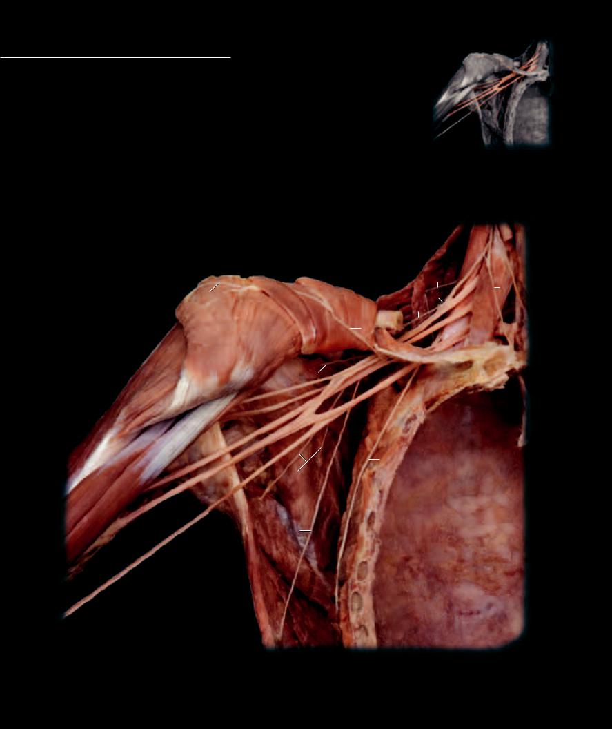
The brachial plexus arises from the last four cervical ventral rami and the fi rst thoracic ventral ramus. The four cervical ventral rami pass later-
ally between the middle and internal layers of the lateral cervical body wall, the middle and anterior scalene muscles, respectively. As they emerge through the scalenes, they connection with with one another as well as with the ascending branch of the fi rst thoracic ventral ramus. This is the beginning of the nerve plexus that will innervate almost all the muscles and associated skin of the upper limb.
Brachial Plexus Nerves |
13 |
Long thoracic nerve |
24 |
Levator scapulae muscle |
|
|
|
|
|
|
|||||
1 |
Dorsal scapular nerve |
14 |
Medial pectoral nerve |
25 |
Subclavius muscle |
|
|
|
|
|
|
||||
2 |
Suprascapular nerve |
15 |
Superior trunk |
26 |
Pectoralis minor muscle |
|
|
|
|
|
|
||||
3 |
Nerve to the subclavius muscle |
16 |
Middle trunk |
27 |
Pectoralis major muscle |
|
|
|
|
|
|
||||
4 |
Lateral pectoral nerve |
17 |
Inferior trunk |
28 |
Deltoid muscle |
|
|
|
|
|
|
||||
5 |
Upper subscapular nerve |
18 |
Lateral cord |
29 |
Biceps brachii muscle |
|
|
|
|
|
|
||||
6 |
Musculocutaneous nerve |
19 |
Posterior cord |
30 |
Subscapularis muscle |
|
|
|
|
|
|
||||
7 |
Axillary nerve |
20 |
Medial cord |
31 |
Teres major muscle |
|
|
|
|
|
|
||||
8 |
Radial nerve |
|
|
32 |
Latissimus dorsi muscle |
|
|
|
|
|
|
||||
9 |
Median nerve |
Other Nerves and Strucures |
33 |
Serratus anterior muscle |
|
|
|
|
|
|
|||||
10 |
Ulnar nerve |
21 |
Phrenic nerve |
34 |
Clavicle |
|
|
|
|
|
|
||||
11 |
Lower subscapular nerve |
22 |
Anterior scalene muscle |
|
|
|
|
|
|
|
|
23 |
|
|
|
12 |
Thoracodorsal nerve |
23 |
Middle scalene muscle |
|
|
|
|
|
|
|
|
|
|
||
|
|
|
|
|
|
|
|
24 |
|
|
|||||
|
|
|
|
|
|
|
|
|
|
|
|
|
|
||
|
|
|
|
|
|
|
|
|
|
|
|
1 |
|
|
|
|
|
4 |
|
|
|
|
|
|
|
|
|
3 |
|
|
21 |
|
|
|
|
|
|
|
|
|
|
|
|
|
|||
|
|
|
|
|
|
|
|
|
|
|
|
|
|||
|
|
|
|
|
|
|
|
|
|
|
|
|
|
||
|
|
|
|
|
|
|
|
|
|
|
|
|
|
|
|
|
|
|
|
|
26 |
|
|
|
2 |
22 |
|
|
|||
|
|
|
|
|
|
|
|
|
|
15 |
|
|
|||
|
|
|
|
|
34 |
|
|
|
|
||||||
|
|
|
|
|
|
|
16 |
|
|
||||||
|
|
|
27 |
|
|
|
14 |
|
|
|
|
|
|||
|
|
|
|
|
|
|
|
|
|
|
|
|
|||
|
|
|
|
|
|
|
|
|
|
|
|
|
|
|
|
|
|
|
|
|
25 |
|
|
17 |
|
|
|||||
|
28 |
|
|
5 |
18 19 |
|
|
|
|
|
|
|
|||
|
|
|
|
|
|
|
|
|
|
|
|
|
|
||
|
|
|
|
|
|
|
|
|
|
|
|
|
|
|
|
|
|
|
6 |
|
20 |
|
|
|
|
|
|
|
|||
|
|
|
|
|
|
|
|
|
|
|
|
|
|
|
|
|
|
|
7 |
|
33 |
|
|
|
|
|
|
|
|||
|
|
|
|
|
|
|
|
|
|
|
|
||||
|
|
|
8 |
|
|
|
|
|
|
|
|
|
|
|
|
|
|
|
9 |
|
|
|
|
|
|
|
|
|
|
|
|
|
|
|
10 |
|
|
|
13 |
|
|
|
|
|
|
||
|
|
|
|
|
|
|
|
|
|
|
|||||
|
|
|
11 |
|
|
|
|
|
|
|
|
|
|
|
|
|
29 |
|
|
|
|
|
|
|
|
|
|
|
|
|
|
30
12
31
32
Dissection of brachial plexus
Anterior view
217
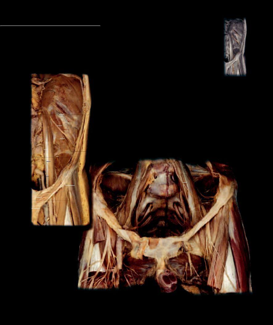
The lumbar plexus arises from the ventral rami of the first four lumbar spinal nerves. The plexus emerges laterally through the intervertebral foramina to pass anterolateral between the two
heads of the psoas major muscle. The more superior branches of the plexus enter the abdominal body wall to innervate the abdominal muscles. The lower nerves of the plexus course into the lower limb as the lateral femoral cutaneous, femoral, and obturator nerves. The lumbar plexus is a transitory plexus that begins as a series of body wall nerves and eventually transitions into limb innervation. The first ventral ramus of the plexus is basically a segmental nerve that follows the basic segmental nerve pattern in the ventral body wall. The second lumbar ventral ramus forms segmental branches in the body wall and other branches that contribute to limb innervation. The third and fourth ventral rami contribute solely to innervation of the lower limb anatomy.
|
|
|
|
|
|
|
|
|
|
|
|
Lumbar Plexus Nerves |
Other Structures |
||
|
|
|
|
|
|
|
|
|
|
|
|
1 |
Subcostal nerve |
20 |
Diaphragm |
|
|
|
|
|
|
|
|
|
|
|
|
2 |
Iliohypogastric nerve |
21 |
Psoas major muscle |
|
|
|
20 |
|
|
|
3 |
Ilioinguinal nerve |
22 |
Psoas minor muscle |
|||||
|
|
|
|
|
|
|
|
|
|
|
|
4 |
Genitofemoral nerve |
23 |
Quadratus lumborum muscle |
|
|
|
|
|
|
|
|
|
|
|
|
5 |
Genital branch of genitofemoral nerve |
24 |
Iliacus muscle |
|
|
|
|
|
|
|
|
|
|
|
|
6 |
Femoral branch of genitofemoral nerve |
25 |
Obturator externus muscle |
|
|
|
|
|
|
|
|
|
|
|
|
7 |
Lateral femoral cutaneous nerve |
26 |
Sartorius muscle |
|
|
|
|
|
|
|
|
|
|
|
|
8 |
Femoral nerve |
27 |
Tensor fasciae latae muscle |
|
|
|
|
|
|
|
|
|
|
|
|
9 |
Obturator nerve |
28 |
Gluteus maximus muscle |
|
|
|
1 |
|
|
|
|
|
|
|
10 |
Lumbosacral trunk |
29 |
Gluteus medius muscle |
|
|
|
|
|
|
|
|
|
|
|
Sacral Plexus Nerves |
30 |
Gluteus minimis muscle |
|||
|
|
|
|
|
|
|
|
|
|||||||
|
|
|
|
|
|
|
|
|
|
|
|
||||
|
|
|
|
|
|
|
|
|
|
|
|
31 |
Piriformis muscle |
||
|
|
|
|
|
|
|
|
|
|
|
|
11 |
Superior gluteal nerve |
||
|
|
|
|
|
|
|
|
2 |
|
32 |
Superior gemellus muscle |
||||
|
|
|
|
|
|
|
|
|
12 |
Inferior gluteal nerve |
|||||
22 |
|
|
|
|
|
|
33 |
Obturator internus muscle |
|||||||
|
|
|
|
|
|
|
|
|
|
13 |
Posterior femoral cutaneous nerve |
||||
|
|
|
|
|
|
|
|
|
|
34 |
Inferior gemellus muscle |
||||
|
|
|
|
|
|
|
|
|
|
|
|
14 |
Nerve to the obturator internus muscle |
||
|
|
|
|
|
|
|
|
|
|
|
|
35 |
Sacrotuberous ligament |
||
|
|
|
3 |
|
|
|
|
|
|
15 |
Pudendal nerve |
||||
|
|
|
|
|
|
|
|
|
36 |
Penis |
|||||
|
|
|
23 |
|
|
|
|
16 |
Perforating cutaneous nerve |
||||||
|
|
|
|
|
|
|
|
|
|||||||
|
|
|
|
|
|
|
17 |
Inferior cluneal nerve |
|
|
|||||
|
|
4 |
|
|
|
|
|
|
|
|
|
|
|
||
|
|
|
|
|
|
|
|
|
|
|
18 |
Sciatic nerve |
|
|
|
|
|
|
|
|
|
|
|
|
|
|
|
||||
21 |
|
|
7 |
|
|
|
19 |
Upper bands of sacral plexus |
|
|
|||||
|
|
|
|
|
|
|
|
|
|
|
|
||||
524
6 |
|
|
|
|
|
8 |
|
|
|
|
|
|
|
7 |
24 |
|
4 |
|
|
|
|||
|
|
|
|
||
|
|
|
 7
7
3 |
|
|
|
|
|
|
|
8 |
21 |
|
|
||||
|
|
|
|
||||
|
|
|
|
|
|
|
|
2 |
|
|
|
|
19 |
|
10 |
|
|
|
|
|
|||
|
|
|
|
|
|
||
|
|
26 |
|
|
|||
|
|
19 |
|
|
|||
|
|
|
|
|
31 |
9 |
|
|
|
|
|
|
|
||
|
|
|
|
|
|
8 |
|
|
|
|
|
|
|
|
|
Abdominal dissection of lumbar plexus |
|
|
|
||||
|
|
|
|
Anteriorview |
8 |
|
26 |
|
|
|
|
|
|
||
|
|
|
|
|
|
|
|
27
9
25
36
Pelvic dissection exposing lumbar and sacral plexus
Anterior view
218
The sacral plexus forms from the ventral rami of the last two lumbar and the fi rst four sacral spinal nerves. The fourth and fi fth lumbar spinal nerves form a descending communication,
the lumbosacral trunk, that joins with the upper sacral spinal nerves as they exit the anterior foramina of the sacrum. On the anterior surface of the sacrum the large roots of the plexus are noticeable before they exit through the greater sciatic notch on their course into the pelvic wall and lower limb. This plexus forms the total nerve supply to the pelvic body wall, and, along with the limb branches from the lumbar plexus, is the nerve supply for the lower limb.
|
|
|
|
|
|
|
|
|
29 |
|
|
||
|
|
|
|
|
|
|
|
|
30 |
|
|
||
|
|
|
|
|
|
|
|
|
11 |
|
|
||
|
|
|
28 |
|
|
|
|
|
|
|
|
|
|
|
|
|
|
|
|
|
|
|
|
|
|
||
|
|
|
|
|
|
|
|
|
|
|
|
||
|
|
|
31 |
|
|
|
|
|
29 |
||||
|
|
|
|
|
|
|
|
|
|
|
|
||
16 |
|
|
32 |
|
|
|
12 |
||||||
|
|
|
|
|
|
|
|||||||
|
|
|
|
|
|
|
|
|
|||||
|
|
|
14 |
|
|
|
|
|
|
|
|
||
|
|
|
33 |
|
|
|
|
|
|
18 |
|||
|
|
|
35 |
|
|
|
|
34 |
|||||
|
|
|
|
|
|
|
|
|
|||||
15 |
|
|
|
|
|
|
|
|
|
13 |
|
|
|
|
|
|
|
|
|
|
|
|
|
||||
|
|
|
17 |
|
|
|
18 |
15 |
|||||
|
|
|
|
|
|
|
|
|
|||||
36
Dissection of sacral plexus nerves
Posterior view
Dissection of pudendal nerves and vessels
Lateral view
219