

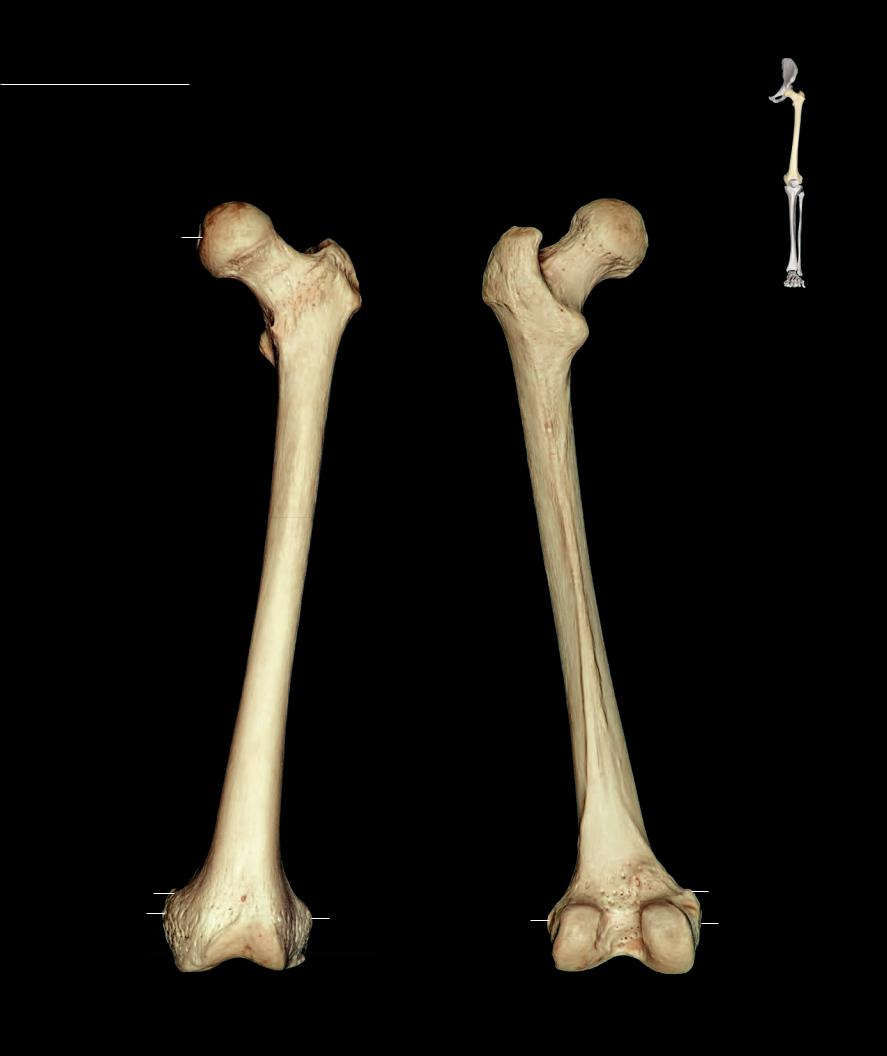
Femur The femur is the longest bone of the body. The strong shaft forms a long cylindrical tube with a slight forward bow. The strong wall of the shaft is thickest near the narrow center of the bone
where the medullary cavity is also the most spacious. As the shaft becomes progressively wider toward each end, the compact wall of bone becomes thinner and the medullary cavity accumulates spongy bone. The proximal end consists of a short cantilevered neck capped by a smooth, round articular head. Projections of bone, the trochanters, form at the base of the cantilevered neck. The distal end consists of two large, knuckle-like processes separated by an intermediate groove. The femur articulates with three bones: the os coxae, patella, and tibia.
1
2
4
3
7
6
10
18 |
|
|
|
|
|
|
|
|
|
|
|
|
|
||
17 |
|
|
16 |
19 |
|
20 |
|
|
|
|
|||||
|
|
|
|
||||
|
|
|
|
|
|||
|
|
|
|
|
|
|
|
|
|
|
|
|
22 |
|
|
1
4
3
9
8
6
13
12
11
15
14
20 |
|
|
|
|
|
|
18 |
|
|
|
|
|
|
||||
|
|
|
|
|
17 |
|||
|
|
|
|
|
|
|||
|
|
|||||||
19 |
23 |
16 |
|
|
|
|
||
|
|
|
|
|
|
|
|
|
Left femur |
Left femur |
108 |
Anterior view, lateral to rigjt |
Posterior view, lateral to left |
|
|
1 |
Head |
9 |
Quadrate tubercle |
17 |
Medial epicondyle |
2 |
Fovea for ligament of head |
10 |
Shaft or body |
18 |
Adductor tubercle |
3 |
Neck |
11 |
Linea apsera |
19 |
Lateral condyle |
4 |
Greater trochanter |
12 |
Pectineal or spiral line |
20 |
Lateral epicondyle |
5 |
Trochanteric fossa |
13 |
Gluteal tuberosity |
21 |
Groove for popliteus |
6 |
Lesser trochanter |
14 |
Medial supracondylar line |
22 |
Patellar surface |
7 |
Intertrochanteric line |
15 |
Lateral supracondylar line |
23 |
Intercondylar fossa |
8 |
Intertrochanteric crest |
16 |
Medial condyle |
|
|
4 |
2 |
|
4
5
3
6
10
11
10
11
|
18 |
20 |
17 |
|
21 |
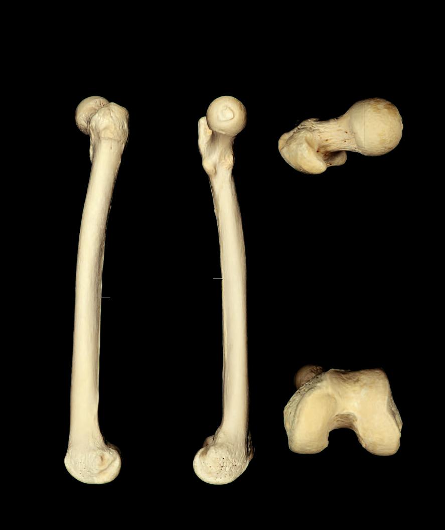
Left femur |
Left femur |
Lateral view, anterior to left |
Medial view, anterior to right |
1
|
3 |
4 |
5 |
|
6 |
Left femur
Superior view, lateral to left
1
22
16 |
19 |
|
23 |
Left femur
Inferior view, lateral to right
109
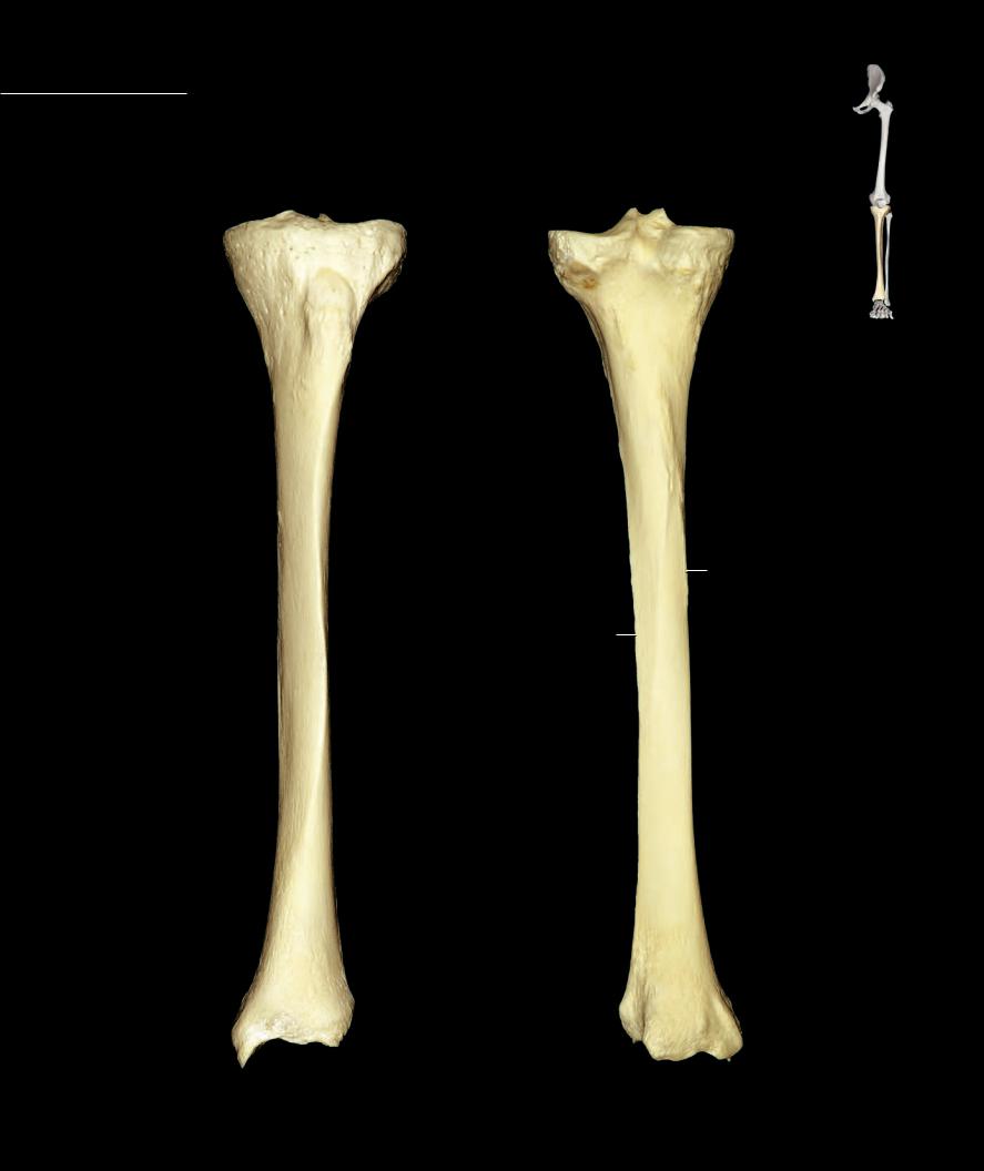
Tibia The tibia is the large, medial bone of the leg skeleton. It is the second longest bone of the body, only exceeded in length by the femur. Its strong shaft, consisting of thick walls of compact bone, is
triangular in cross-section. The shaft expands proximally into a fl uted extremity of spongy bone with a fl at plateau-like superior surface largely covered with articular cartilage. The smaller distal end is more knoblike with a pronounced medial projection, the malleolus. The shaft has a strong anterior crest with sloping surfaces to either side. The bone is easily palpable throughout its length. The tibia articulates with three bones — the femur, fi bula, and talus.
|
|
|
7 |
2 |
3 |
3 |
2 |
|
|
4 |
|
|
11 |
|
|
12
14
15
10 |
10 |
13
|
19 |
17 |
|
16 |
16 |
||
|
Left tibia |
Left tibia |
|
Posterior view, lateral to left |
||
Anterior view, lateral to right |
||
|
110

1 Superior articular surface
2 Medial condyle
3 Lateral condyle
4 Fibular articular facet
5 Anterior intercondylar area
6 Posterior intercondylar area
7 Intercondylar eminence
7
3
4
11
10
14
13
19
Left tibia
Lateral view, anterior to left
8 |
Medial intercondylar tubercle |
15 |
Posterior border |
9 |
Lateral intercondylar tubercle |
16 |
Medial malleolus |
10 |
Shaft or body |
17 |
Malleolar groove |
11 |
Tibial tuberosity |
18 |
Malleolar articular facet |
12 |
Soleal line |
19 |
Fibular notch |
13 |
Interosseous border |
20 |
Inferior articular surface |
14 |
Anterior border |
|
|
7
2
11
10
15
17 |
16 |
Left tibia
Medial view, anterior to right
11 |
|
|
|
|
5 |
|
|
||
|
|
|
||
|
8 |
|
|
|
1 |
7 |
9 |
1 |
|
|
|
|||
|
|
|
|
|
|
6 |
|
|
|
Left tibia
Superior view, lateral to left
19
18
Left tibia
Close-up of lateral view
16 |
20 |
|
Left tibia
Inferior view, lateral to right
111