- •Preface
- •Content
- •Tissues
- •Nerve Tissue
- •Skin - Epidermis
- •Skin - Dermis
- •Skin - Glands
- •Subcutaneous Layer
- •Skeleton
- •Axial Skeleton
- •Cranium
- •Cranial Bones – Inferior Nasal Concha
- •Vertebral Column
- •Sacrum and Coccyx
- •Ribs
- •Sternum
- •Clavicle
- •Scapula
- •Humerus
- •Ulna
- •Radius
- •Metacarpals and Phalanges
- •Pelvis - Male
- •Femur
- •Tibia
- •Fibula
- •Tarsal Bones - Cuboid and Navicular
- •Phalanges
- •Patella
- •Skeletal Muscles
- •Transversospinales Muscles
- •Cervical Hypaxial Muscles
- •Thoracic and Abdominal Hypaxial Muscles
- •Shoulder Muscles - Rotator Cuff
- •Shoulder Muscles - Prime Movers
- •Anterior Brachial Muscles
- •Posterior Brachial Muscles
- •Posterior Thigh Muscles
- •Thigh Muscles
- •Lateral Leg Muscles
- •Posterior Leg Muscles
- •Spinal Nerves
- •Dorsal Rami
- •Intercostal Nerves
- •Cutaneous Nerves
- •Autonomic Nerves
- •Spinal Cord
- •Brain
- •Cerebrum
- •Cerebellum
- •Meninges
- •Hypothalamus
- •Pituitary Gland
- •Pineal Gland
- •Thymus
- •Pancreas
- •Ovaries
- •Testes
- •Blood
- •Heart
- •Lymphatics
- •Larynx
- •Lungs
- •Cast of Trachea and Bronchial Tree
- •Esophagus
- •Stomach
- •Pancreas
- •Large Intestine
- •Mesenteries
- •Omenta
- •Female Reproductive Organs
- •Ovary
- •Vagina
- •Ductus Deferens and Spermatic Cord
- •Penis
- •Index
Female Reproductive Organs
The female genital organs consist of the internal genitalia and the external genitalia. The ovary, uterine tube, uterus, and vagina form the internal genitalia. These organs are responsible for production of the female gamete, the oocyte, and for nourishing, protecting, and delivering the new life that results from fertilization of the oocyte by the sperm. The external genitalia consist of the erectile tissues, glands, and folds of skin that proctect the entry into the female internal genitalia. These organs are the clitoris, vestibular glands, and labia majora and minora.
3
1
4 2
5
16 |
|
|
|
|
15 |
|
|
6 |
|
|
|
|
18 |
||
7 |
20 |
||
|
|||
|
|
|
19 |
|
|
17
8 |
11 |
|
9
10
Sagittal section of female pelvis
Medial view
322
1 |
Ovary |
9 |
Labia minora |
17 |
Rectum |
2 |
Uterine tube |
10 |
Labia majora |
18 |
Bladder |
3 |
Fimbriae |
11 |
Mons pubis |
19 |
Urethra |
4 |
Fundus of uterus |
12 |
Broad ligament |
20 |
Pubic symphysis |
5 |
Body of uterus |
13 |
Round ligament of uterus |
21 |
Cecum |
6 |
Cervix of uterus |
14 |
Ovarian ligament |
22 |
Sigmoid colon |
7 |
Vagina |
15 |
Vesicouterine pouch |
23 |
Ileum |
8 |
Clitoris |
16 |
Rectouterine pouch |
24 |
Mesentery |
23
24
21
24
17
22
3
12
14
4 |
1 |
|
13
2
Dissection of female abdominoplevic cavity
Superoanterior view
323
Ovary The ovaries are the site of oocyte, “egg,” production in the female. These solid organs are approximately the size of an unshelled almond and project into the lower abdominal cavity at the boundary of the pelvis where they are
covered and supported by folds of the peritoneum. During embryonic life, millions of oogonia, potential oocytes, surrounded by nursing follicular cells begin their development. Of these millions of cells only about 500 are ever ovulated during the female’s reproductive life. The follicular cells not only nurse the ooytes, but also are the endocrine cells of the ovary that produce the estrogens and progesterone.
3
2
1 |
Ovary |
|
|
|
|
|
2 |
Tunica albuginea |
|
|
|
|
|
3 |
Primordial follicle |
|
|
|
|
|
4 |
Granulosa cells |
|
|
|
|
|
5 |
Theca folliculi |
|
|
|
|
|
6 |
Zona pellucida |
|
|
|
|
|
7 |
Primary oocyte |
|
|
|
|
|
8 |
Seconary follicle |
|
|
|
|
|
9 |
Follicular antrum |
|
|
5 |
|
|
10 |
Corona radiata |
|
|
|
||
|
|
|
|
|
||
11 |
Corpus luteum |
|
|
8 |
|
|
12 |
Infundibulum of uterine tube |
|
|
10 |
|
|
13 |
Ampulla of uterine tube |
8 |
7 |
|
|
|
14 |
Isthmus of uterine tube |
|
|
|
|
6 |
|
|
|
||||
15 |
Fimbriae of uterine tube |
|
|
7 |
|
|
16Round ligament of uterus
17Ovarian ligament
18 Uterus |
4 |
|
|
|
9 |
|
4 |
|
11 |
11
Photomicrograph of ovary
50x
1
|
|
|
12 |
18 |
17 |
|
15 |
|
13 |
||
|
|
||
|
|
|
|
|
14 |
|
|
|
16 |
|
|
Ovary in situ
Anterior view
324
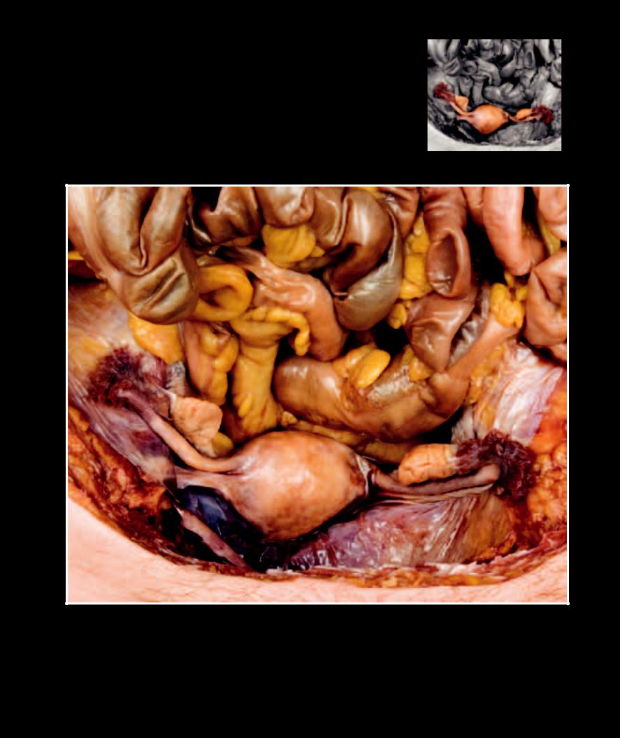
The uterine tubes, also called the oviducts or fallopian tubes, are suspended in the perito-
neal fold, the broad ligament, along with the ovaries. In addition to transporting the oocyte toward the uterus, they are the site of fertilization of the oocyte by the sperm. The uterus is the thick smooth muscle organ that functions as the internal nest of mammalian animals. Note the vascular and glandular changes exhibited by the uterine endometrium as it progresses through the menstrual cycle.
1 |
Uterine tube |
10 |
Peg cells |
Myometrium: |
|
|||
2 |
Fimbriae |
11 |
Ciliated columnar cells |
16 |
Inner longitudinal muscle |
14 |
||
3 |
Mesosalpinx |
12 |
Lamina propria |
17 |
Middle circular muscle |
|
||
4 |
Fundus of uterus |
13 |
Perimetrium |
18 |
Outer longitudinal muscle |
|
||
5 |
Body of uterus |
Endometrium: |
|
|
14 |
|||
6 |
Cervix of uterus |
14 |
Stratum functionalis |
|
|
|||
7 |
Vagina |
15 |
Stratum basalis |
|
|
15 |
||
8 |
Mucosa of uterine tube |
|
|
|
|
|||
|
|
|
|
|
||||
9 |
Muscularis of uterine tube |
|
|
|
|
|
||
|
8 |
|
|
|
|
|
10 |
|
|
|
|
|
9 |
|
|
|
|
|
|
|
|
|
|
11 |
|
|
|
|
|
|
|
|
|
16 |
|
|
|
|
|
|
|
|
12 |
|
|
|
|
|
|
|
|
|
|
|
8 |
|
|
|
8 |
|
|
15 |
|
|
|
|
|
|
|||
|
|
|
|
|
|
|
||
|
|
|
|
|
|
|
|
|
|
9 |
|
|
|
|
|
|
|
|
|
|
|
|
Photomicrograph of tunica mucosa |
|
||
|
|
|
|
|
|
|
of uterine tube |
16 |
|
|
|
|
|
|
|
400x |
|
Photomicrograph of uterine tube |
|
17 |
|
|
|
25x |
|
|
2 |
4 |
|
1 |
|
|
|
|
|
3 |
17 |
|
|
5
13 |
|
|
6 |
|
|
7 |
18 |
|
|
||
|
18 |
|
|
1313 |
|
|
1313 |
|
Female internal genitalia |
Photomicrograph of uterine wall, |
|
2nd week of menstrual cycle left, |
||
Anterior view |
3rd week of menstrual cycle right
16x (left), 20x (right)
325