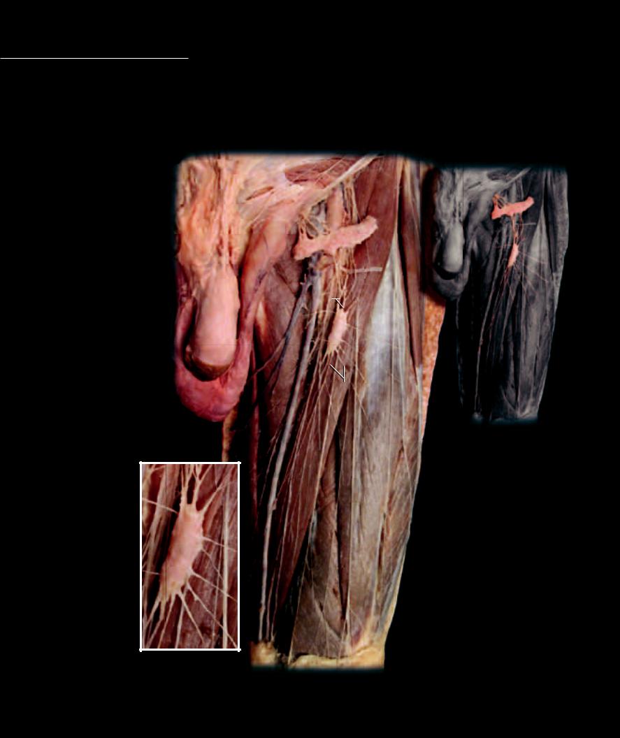
Lymphatics Even under normal circumstances, slightly more fl uid is fi ltered out of the capillaries into the interstitial fl uid than is reabsorbed from the interstitial fl uid back into the plasma. On
average, the net fi ltration pressure starts at 11 mm Hg at the beginning of the capillary, whereas the net reabsorption pressure only reaches 9 mm Hg by the vessel’s end. Because of this pressure differential more fluid is filtered out of the first half of the capillary than is reabsorbed in its last half. If this extra filtered fluid were not drained away, the consequence of this unbalanced exchange would be accumulation of excess interstitial fluid, or edema. To circumvent this potentially disastrous problem, a system of accessory drainage vessels, the lymphatic vessels, evolved in vertebrate animals. This lymphatic system of vessels consists of an extensive network of one-way tubes that provide an accessory route through which fluid is returned from the interstitial fluid to the blood to keep the cardiac output and return equal.
|
|
|
11 |
|
1 |
Superficial inguinal lymph node |
9 |
|
6 |
|
|
2 |
Afferent lymphatic vessels |
5 |
|
|
|
3 |
Efferent lymphatic vessels |
1 |
|
4 |
Great saphenous vein |
|
|
|
|
|
5 |
Femoral vein |
|
|
|
|
6 |
Femoral artery |
7 |
|
|
|
7 |
Spermatic cord |
|
|
|
|
|
|
|
8 |
Penis |
|
|
|
|
9 |
Sartorius muscle |
|
|
|
|
10 |
Rectus femoris muscle |
3 |
|
|
|
|
|
|
11 |
Femoral nerve |
|
|
|
|
|
|
8 |
|
|
|
|
|
1 |
|
4
2
10
3 3
3
1
2
Dissection of lymphatic vessels and nodes in the thigh
Anterior view
17 Respiratory System
The respiratory system consists of a network of passageways that begin at the openings into the nose and mouth and terminate in about 600 million microscopic air spaces within the substance of the lungs. The passageways are typically divided into upper respiratory passageways and lower respiratory passageways. The upper respiratory tract consists of the nose, the nasal cavity and associatied sinuses, and the pharynx. While the mouth is typically included in the digestive system, it can also serve as a passageway for air entering the respiratory system. The lower respiratory
tract consists of the larynx, trachea, and the bronchial and alveolar tubes that form a large, branching network of passageways within the lungs. This branching bronchial tree within each lung begins as a large, finger-sized tube called the main or principal bronchus
and terminates in the lungs as the microscopic air sacs called alveoli.
Like other systems that form an environmental exchange surface with the cardiovascular system, the respiratory system forms an extensive surface area in contact with the capillaries. It is estimated that the surface area of the small dead-end air sacs in the lungs is about the size of a tennis court. This extensive interface is essential for
the exchange of oxygen and carbon dioxide between the inhaled air and the blood. If body cells are deprived of oxygen, they cannot function and they die as a result. So the acquisition of oxygen through the respiratory passageways and its subsequent exchange with the capillary blood is an important function of the respiratory system.
In addition to gas exchange, the portion of the respiratory passageways referred to as the larynx is responsible for generating the sound waves that we manipulate into voice. Internal folds in the lining of the larynx, the vocal folds, vibrate as air passes upward from the lungs to produce the vibrations. For this reason the larynx is often referred to as the voice box.
Find more information about the respiratory system in
R E A L A N AT O M Y
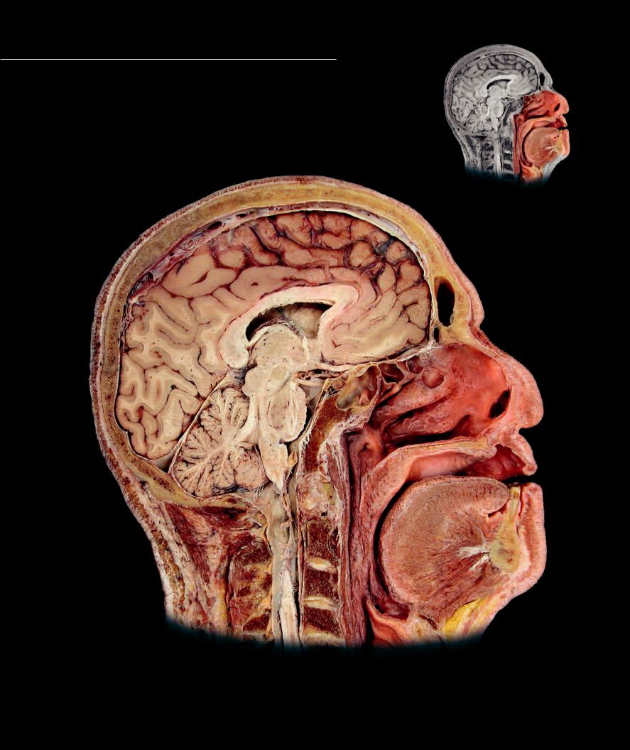
Upper Respiratory Tract
The upper respiratory tract consists of the initial series of passage-
ways that carry the inspired air through the head. The various sections of the head seen on this and the facing page show the passageways of the upper respiratory tract, which include the nose and nasal vestibule, the nasal cavity, the paranasal sinuses, nasopharynx, oropharynx, laryngopharynx, and even the oral cavity. The nasal cavity functions in fi ltering, warming, and humidifying the inspired air, while also detecting chemical odorants.
18
17
20
6
21
|
|
7 |
|
|
|
8 |
2 |
3 |
1 |
|
|
|
|
|
|
19 |
22 |
|
|
4 |
|
|
|
24 |
5 |
|
|
9 |
23 |
|
|
|
|
|
|
|
|
25 |
|
|
10 |
|
12 |
11 |
|
|
|
|
|
|
|
|
16 |
Sagittal section of head
Medial view
290
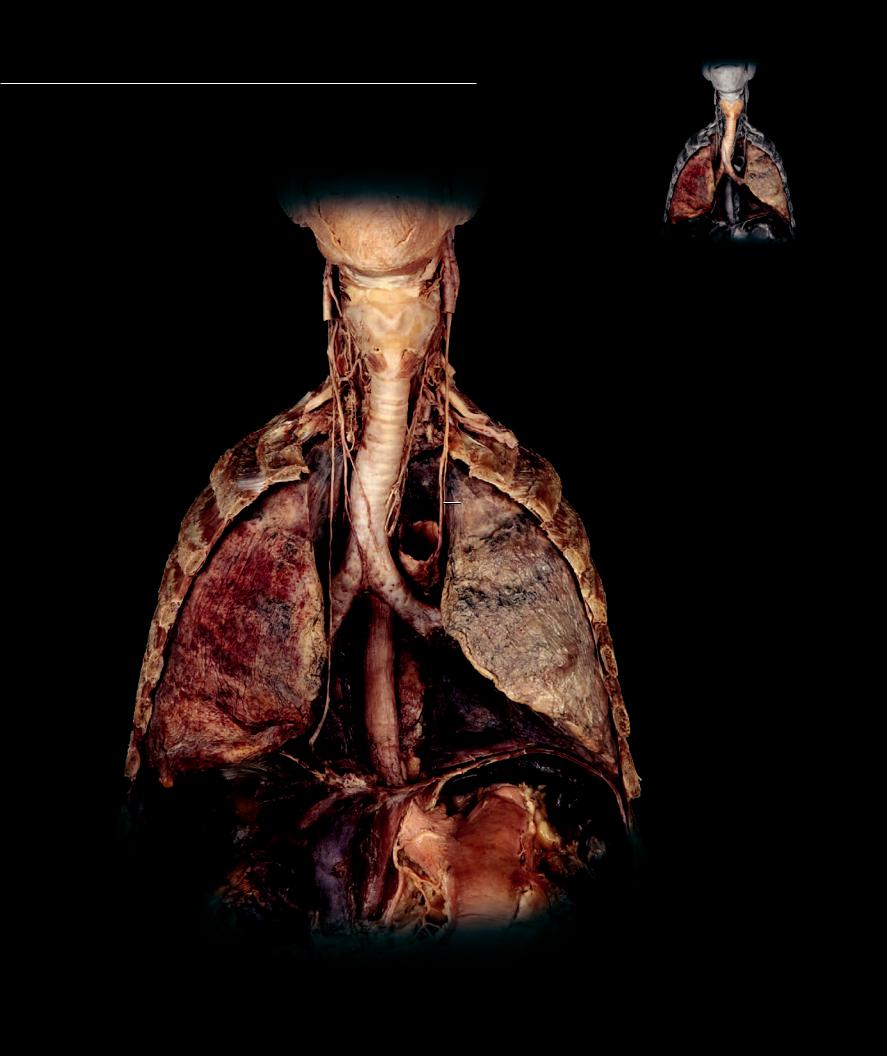
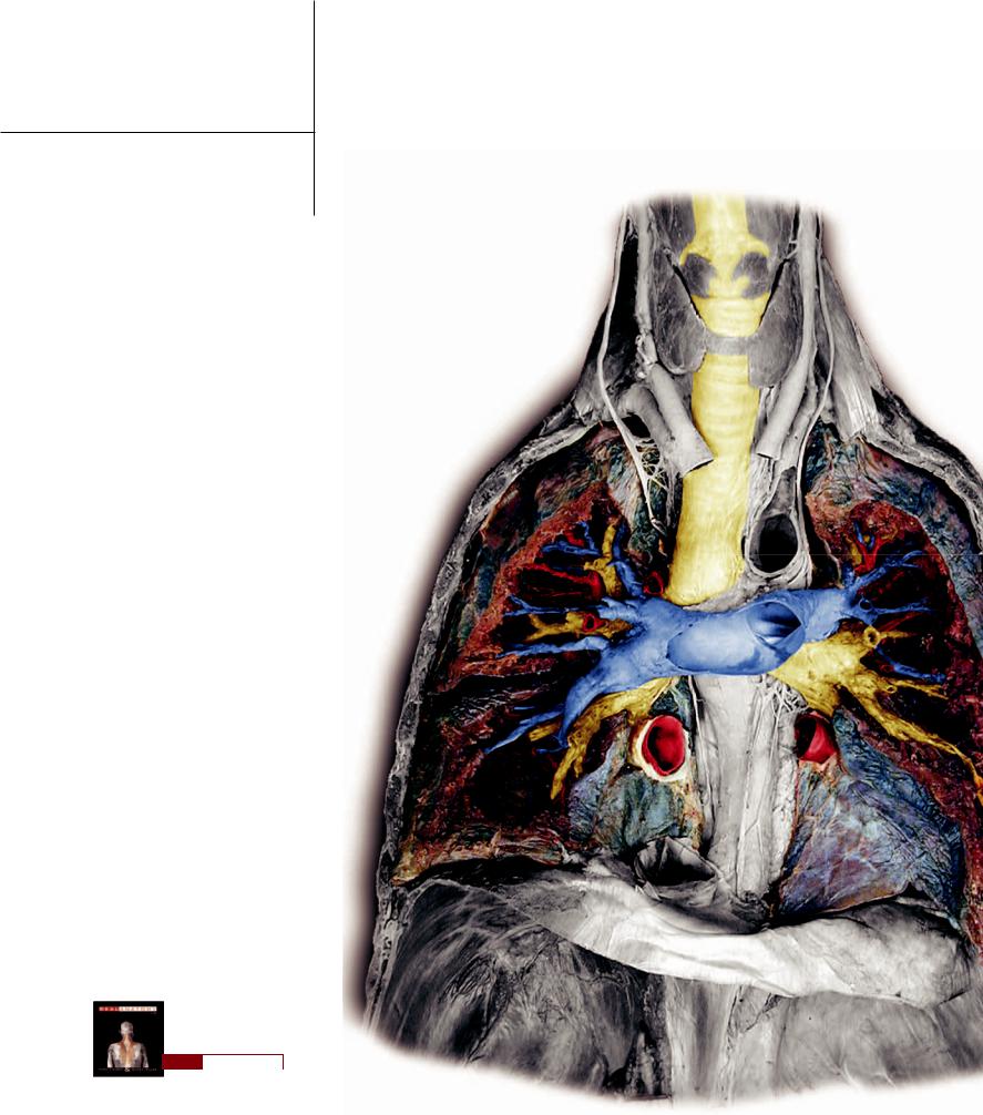
Lower Respiratory Tract
The lower respiratory tract arises as an outgrowth of the tubular gut during embryonic development. This anterior outgrowth of the gut tube begins at the
larynx (voice box), which is the upper expanded portion of the lower respiratory tract. It continues from the neck into the thorax as the trachea (windpipe), and forms a large branching network of tubes that enter the lungs, the bronchial tree. The pages that follow show the tubular organs and histology of the lower respiratory tract.
1
2
3
20
11
21
20
12
13
19
12
16
18
Lower respiratory tract and lungs in situ
Anterior view
292
1 |
Thyroid cartilage of larynx |
10 |
Subclavian artery |
18 |
Stomach |
2 |
Cricoid cartilage of larynx |
11 |
Vagus nerve |
19 |
Phrenic nerve |
3 |
Trachea |
12 |
Esophagus |
20 |
Rib |
4 |
Main (primary) bronchus |
13 |
Aorta |
21 |
Intercostal muscle |
5 |
Right lung |
14 |
Pulmonary artery |
22 |
Anterior scalene muscle |
6 |
Left lung |
15 |
Pulmonary vein |
23 |
Thyrohyoid muscle |
7 |
Bronchial tree |
16 |
Inferior vena cava |
24 |
Cricothyroid muscle |
8 |
Thyroid gland |
17 |
Diaphragm |
25 |
Spleen |
9 |
Common carotid artery |
|
|
|
|
|
|
|
|
|
23 |
1 |
|
|
|
|
|
|
|
24 |
|
|
|
|
|
|
|
2 |
|
|
|
|
11 |
|
|
|
11 |
|
|
|
|
|
|
|
|
|
8 |
|
|
|
|
|
|
|
|
|
|
|
|
|
22 |
|
|
|
9 |
|
9 |
|
|
|
|
|
|
|
|
10 |
3 |
|
|
|
|
|
|
|
|
|
|
|
|
|
|
|
10 |
|
|
|
|
|
|
|
13 |
|
|
5 |
7 |
|
|
7 |
|
|
|
7 |
|
|
14 |
|
|
|
|
|
|
6 |
|
|
|
|
|
|
|
7 |
|
20 |
|
|
|
|
12 |
7 |
|
|
|
15 |
|
20 |
|
|
|
7 |
|
|
|
|
11
16
17
17
Dissection of lower respiratory tract and lungs in situ
Anterior view