


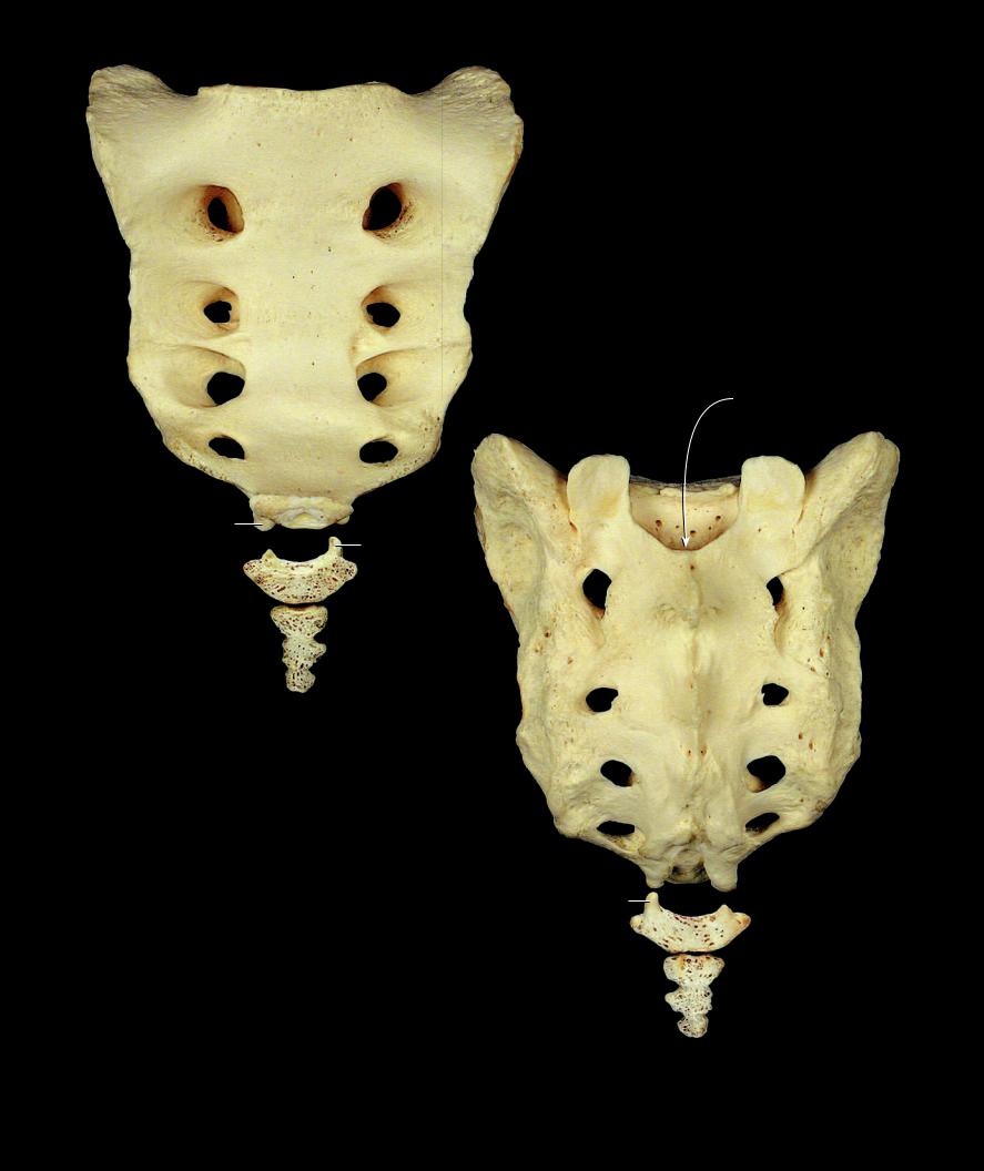
The sacrum is a large triangular-shaped mass that forms from the fusion of fi ve vertebrocostal segments. The base of the triangle is superior and tapers to a fl at-
tened apex inferiorly. It is concave anteriorly and convex posteriorly. The lateral margins of the triangle are widest superiorly where the bone articulates with the two ilia. Forming the large basal portion of the vertebral column, the bone wedges between the two os coxae to form the posterior element of the pelvic skeleton. Its ventral surface, smoother than the rough dorsal surface, forms the posterior wall of the pelvis. Within this triangular mass of bone is a hollow sacral canal. This canal opens through foramina onto the ventral and dorsal surfaces of the bone. It forms a large oval surface superiorly that articulates with the fi fth lumbar vertebra and a smaller oval facet at its apex for articulation with the coccyx.
The coccyx is the terminal end of the vertebral column. It is a triangular bone that forms from the fusion of three to fi ve vertebral segments, most commonly from four fused vertebrae. The superior surface of the fi rst segment’s body forms an oval articular surface with the inferior surface of the fifth sacral segment.
|
|
|
2 |
1 |
Promontory |
|
|
2 |
Ala or wing |
|
|
3 |
Superior articular process |
|
|
4 |
Auricular surface |
|
|
5 |
Sacral tuberosity |
|
1 |
6 |
Pelvic surface |
|
|
|
|
||
7 |
Transverse ridges |
|
|
8 |
Anterior sacral foramina |
|
|
9 |
Posterior sacral foramina |
|
|
10 |
Median sacral crest |
10 |
4 |
11Intermediate sacral crest
12Lateral sacral crest
13Sacral cornu
14Sacral canal
15Sacral hiatus
16Apex
17Coccygeal cornu
10 |
12 |
5 |
11
16
13
17
Sacrum and coccyx
Lateral view, anterior at right
78
2
1
7
6
8
7
8
14
7
2
3
16
13
4
17
5
10 11
9 |
12 |
Sacrum and coccyx
Anterior view, superior at top
9
15
13
17
Sacrum and coccyx
Posterior view, superior at top
79
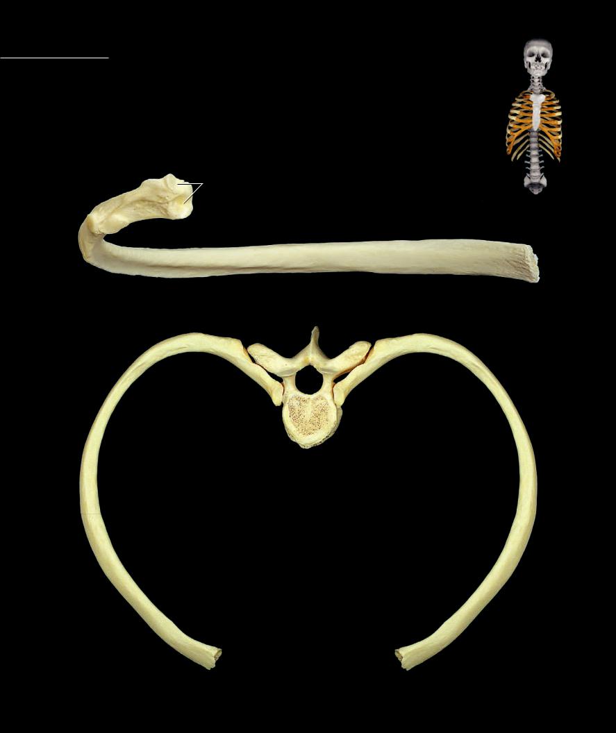
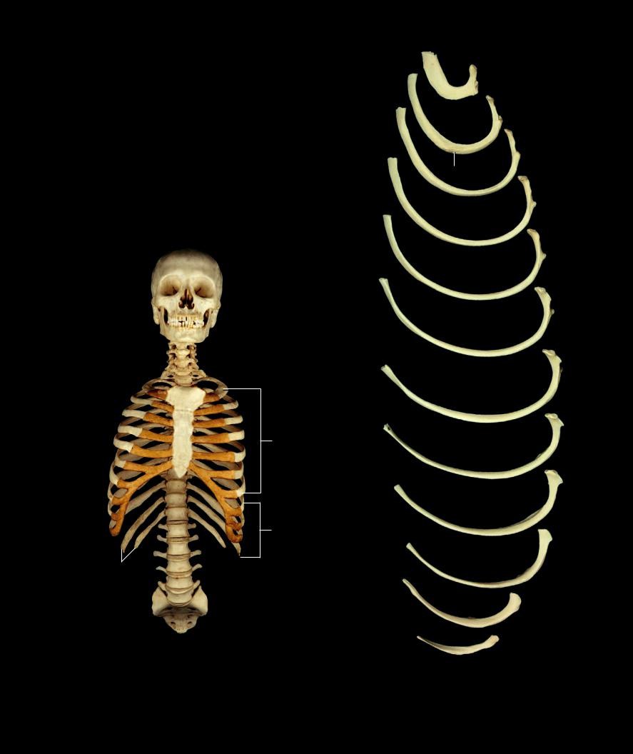
Ribs There are twelve paired ribs, a pair for each of the twelve thoracic vertebrae. The ribs unite the thoracic vertebrae to the sternum via costal cartilages to form the thoracic skeleton, a fl exible, bony wall that protects thoracic viscera and facilitates respiratory function. Although only the
twelve thoracic ribs are named ribs, there are in reality ribs at every vertebral level. The cervical, lumbar, sacral, and coccygeal ribs fuse to their corresponding vertebrae to contribute to the formation of the transverse process. The ribs can be divided into two groups — true ribs and false ribs. The last two false ribs are called fl oating ribs. True ribs, ribs one through seven, are those that have their costal cartilages attached directly to the sternum. False ribs, ribs eight through twelve, have costal cartilages that do not attach directly to the sternum. The costal cartilage of each of the fi rst three false ribs attaches to the cartilage of the rib superior to it. The last two false ribs do not attach to other ribs and are therefore called fl oating ribs.
2
3
1
4
6
7
8 |
5 |
910
Left sixth rib
Posterior view, superior at top
8
6
4
1
5
Ribs and thoracic vertebra
Superior view, posterior at top
80
1 Head
2 Articular facets of head
3 Crest of head
4 Neck
5 Body or shaft
6 Tubercle
7 Articular facet of tubercle
8 Angle
9 Costal groove
10Crest of body
11Scalene tubercle (first rib)
12Tuberosity of serratus anterior (second rib)
13Costal cartilage
14True ribs [I-VII]
15False ribs [VII-XII]
16Floating ribs [XI-XII]
13
14
13
15
16
Rib cage
Anterior view
11
12
Left ribs 1 through 12
Superior view, first rib at top, posterior to right
81