

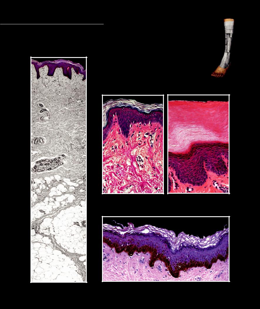
Skin - Epidermis The stratifi ed squamous epithelial epidermis is the superfi cial layer of the skin. This cellular layer and its derivatives — hairs,
nails, and glands — is the most recognizable part of our anatomy. It can range in thickness from a .10 mm (0.0039 in) on the eyelids to 1.5 mm (0.059 in) on the palms and soles. Keratinocytes are the primary cells of the epidermis. They proliferate from the stratum basale and differentiate as they push toward the surface, where they eventually form dead cells fi lled with the protein keratin. Also present in the basal layer are melanocytes that produce the brown pigment melanin to protect the skin from the ultraviolet radiation from the sun.
1 |
Stratum basale |
4 |
Stratum lucidum |
2 |
Stratum spinosum |
5 |
Stratum corneum |
3 |
Stratum granulosum |
6 |
Connective tissue of dermis |
5
3 |
5 |
2
1
|
4 |
6 |
|
|
3 |
|
2 |
|
1 |
|
6 |
Epidermis of skin of a Caucasian |
Epidermis of skin of a Caucasian |
Section of thin skin, 200x |
Section of thick palmar skin, 200x |
|
5 |
|
3 |
|
2 |
|
1 |
|
6 |
Epidermis of integument |
Epidermis of skin of a black |
100x |
Section of thin skin, 200x |
20
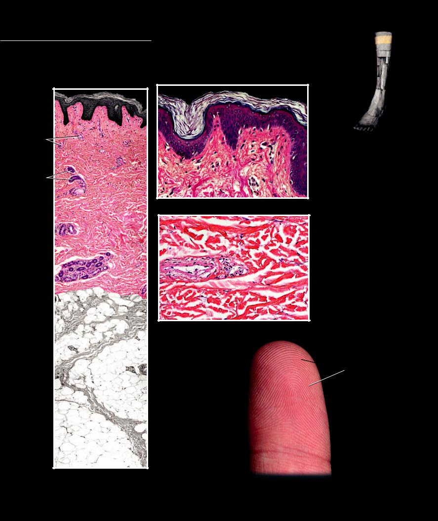
Skin - Dermis The connective tissue dermis sits deep to the epidermis where it forms the strong binding layer of the skin. The zone of interface
between the dermis and epidermis is an intricate peg and socket-like arrangement between the two layers. The dermal pegs are called dermal papillae. This arrangement has multiple functions. It assures that the two layers are strongly united, it increases the surface area to improve the blood supply to the avascular epidermis, and it increases the contact surface for sensory receptors. On the palms and soles the arrangement of the dermal papillae creates the friction ridges we call fingerprints.
3 |
|
|
|
|
|
3 |
|
3 |
|
|
|
|
|
1 |
|
|
|
2 |
|
3 |
|
|
|
|
|
2 |
|
|
|
5 |
|
2 |
|
|
|
|
|
|
|
|
|
|
|
2 |
|
|
|
|
|
|
|
1 |
Epidermis |
6 |
|
|
|
2 |
Loose connective tissue of |
|
|
|
|
stratum papillare |
|
|
|
|
|
|
|
|
|
|
|
3 |
Dermal papilla of the stratum |
|
|
Loose connective tissue of stratum papillare |
|
papillare |
|
|
4 |
4 |
Dense connective tissue of |
||
|
Section of dermis, 200x |
|
|
stratum reticulare |
|
|
|
|
|
||
|
|
|
|
5 |
Blood vessel in dermis |
|
|
|
|
6 |
Sweat glands in dermis |
6 |
|
7 |
8 |
7 |
Longitudinal collagen fibers |
|
|
|
8 |
Transverse collagen fibers |
|
|
|
8 |
|
||
|
|
|
9 |
Friction ridges formed by dermal |
|
|
|
|
|
||
|
|
|
|
|
papillae |
|
|
|
|
10 |
Flexion crease line |
6 |
|
5 |
|
|
|
|
|
|
|
|
|
|
|
|
7 |
|
|
|
|
7 |
|
|
|
Dense irregular connective tissue of stratum reticulare
Section of dermis, 200x
 9
9
|
10 |
Dermis of integument |
Friction ridges (fingerprints) of right index finger |
100x |
|
|
Anterior view |
21
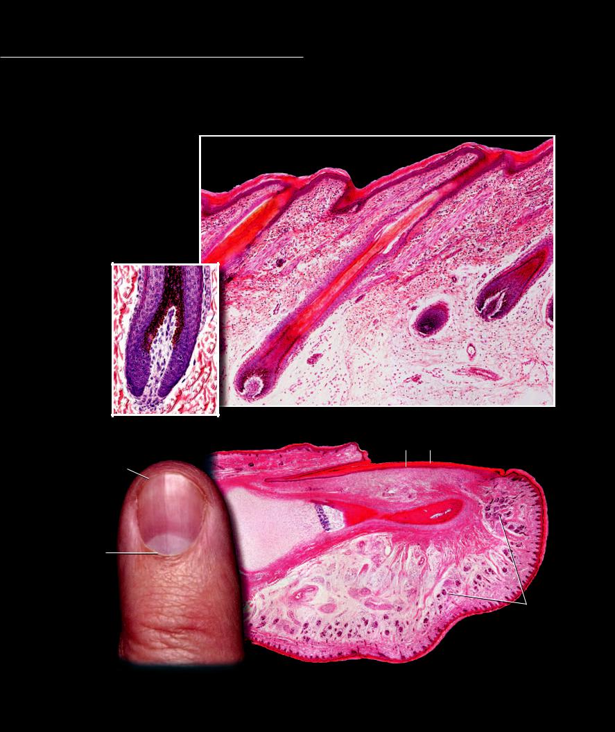
During embryonic and fetal development, the epithelial cells of the epidermis push down (invaginate) into the connective tissue dermis. This developmental process creates a hair follicle, a baglike extension of the epidermis that projects into the dermis and is
responsible for producing the hair. The hair is a column of dead keratinocytes that arise from the basal keratinocytes at the bottom of the hair follicle.Asebaceous gland, also derived from the epidermal epithelium, empties into the hair follicle, and a small band of dermal smooth muscle, the arrector pili muscle, attaches to the base of the follicle. When the muscle shortens it produces “goose bumps” on the surface of the skin and causes the hair to “stand up.” Nails also arise from invaginations that produce the shallow nail fold and root. A plate of strongly keratinized tissue emerges from the nail root to cover the dorsal ends of the fingers and toes.
1 |
Epidermis |
|
2 |
Dermis |
|
3 |
Follicle wall |
|
4 |
Hair |
|
5 |
Papilla |
1 |
6 |
Root of nail |
|
7 |
Nail |
|
8 |
Nail bed |
2 |
9 |
Lunula |
|
10Eponychium (cuticle)
11Hyponychium
12Eccrine sweat glands
13Cartilage
14Bone
|
3 |
|
|
|
|
3 |
|
|
|
|
4 |
|
|
|
5 |
|
|
|
|
5 |
|
|
|
|
Hair bulb |
Hair follicle |
|
|
|
Section of skin, 100x |
|
|
||
Section of skin, 400x |
8 |
7 |
||
|
||||
2 |
10 |
|
|
|
|
|
|
||
11 |
|
|
11 |
|
|
|
|||
6 |
|
|
||
|
|
|
|
|
13 |
14 |
1 |
||
|
|
|||
9 |
|
|
|
|
10
12
2
Fingernail of an adult |
Finger of a child |
|
Longitudinal section, 50x |
||
Dorsal view |
||
|
22
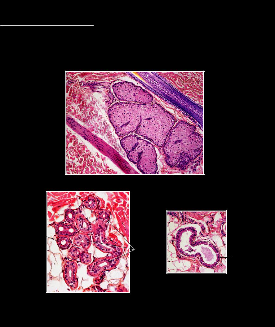
Skin - Glands Like hairs, glands arise as invaginations of the epidermis into the dermis during embryonic and fetal life. The three prominent glands of the skin are the sebaceous
gland, the eccrine sweat gland, and the apocrine sweat gland. The sebaceous and apocrine sweat glands typically empty into a hair follicle, whereas the eccrine sweat gland empties onto the surface of the epidermis.
1 |
Sebaceous secretory cells |
5 |
Hair |
2 |
Eccrine secretory cell |
6 |
Hair follicle |
3 |
Eccrine duct cell |
7 |
Arrector pili muscle |
4 |
Apocrine secretory cell |
|
|
6
5
1
1
7
1
1
Sebaceous gland
Section of dermis, 200x
3 
2
4
Apocrine sweat gland
Section of thin skin, 200x
Eccrine sweat gland
Section of dermis, 200x
23