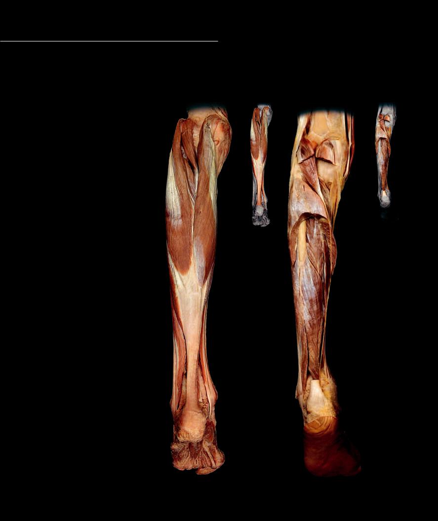
Posterior Leg Muscles
The posterior compartment of the leg comprises the large muscle mass on the back of the leg that is often referred to as the calf. This compartment has two distinct muscle
groups – a large superfi cial group and a smaller deep group. The superfi cial group, the gastrocnemius, the soleus, and the plantaris, each insert on the calcaneus. The gastrocnemius and soleus combine to form the large tendocalcaneus, or Achilles tendon. The smaller, deep group consists of four muscles, three of which form a pulley-like arrangement around the medial malleolus. These are the fl exor hallucis longus, fl exor digitorum longus, and tibialis anterior. The fourth muscle in the group is the deeply situated popliteus that occupies the floor of the popliteal fossa.
Posterior Leg Muscles
1 Tibialis posterior
2 Flexor digitorum longus
3 Flexor hallucis longus
4 Popliteus
5 Plantaris
6 Soleus
7 Gastrocnemius
Other Muscles and Structures 8 Fibularis brevis
9 Fibularis longus (tendon)
10Flexor digitorum brevis
11Abductor hallucis
12Flexor hallucis brevis
13Abductor digiti minimi
14Calcaneal tendon
15Fibula
5
6
7
83
2
14
9
11
13 10 12
Superficial muscles of the crus |
Deep muscles of the crus |
Posterior view |
Posterior view |
|
207
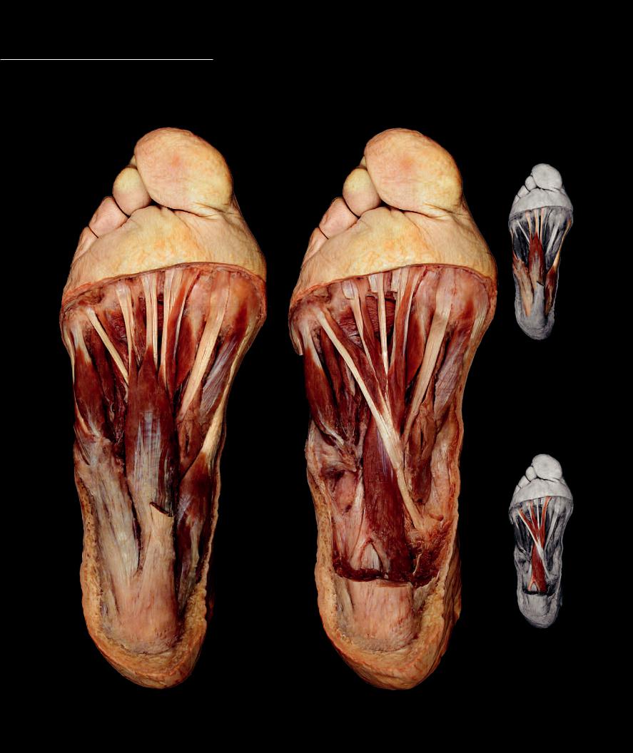
Foot Muscles
Situated on the dorsal surface of the foot are two short digital extensor muscles, the extensor hallucis brevis and extensor digitorum brevis. These thin muscle sheets help the long digital extensors of the anterior compartment extend the digits. Like the
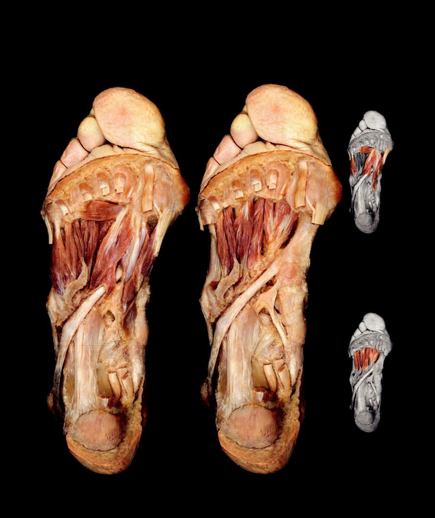
anterior compartment muscles, they are innervated by the deep fi bular nerve. The plantar muscles of the foot are much more substantial than the thin dorsal muscles of the foot. These muscles sit beneath the thick subcutaneous fat pad on the bottom of the foot. From superficial to deep, the plantar muscles form four layers.
7
9
8
2
6
3
1
Dissection of foot, plantar aponeurosis removed
Plantar view
208
7
5
5
8
4
12
Layer two
Dissection of foot, first muscle layer removed
Plantar view

13 Peripheral Nervous System
Look around your city or town and notice the telephone wires that run from telephone pole to telephone pole along the city streets, eventually reaching the homes and places of business throughout the city. They might
not always be visible because in some cities they run underground. Regardless of where they occur, these wires criss-cross throughout the city distributing electrical current from phone to phone in our homes and places of school, work, and entertainment. These wires are not complex
structures; they are simply metal wires that can conduct an electric charge from one phone to another. These telephone wires in our cities and homes are typically insulated from one another and protectively wrapped to prevent damage. Their pathways through the city are not complex; they simply follow logical routes to different parts of the city. The wires are bundled in common groups that follow shared pathways to similar locations. As these wires course through the city they relay to telephone centers operated by the telephone
companies. At these centers the wires enter control rooms where they form complex circuits. This complex circuitry allows the electrical messages
to be processed and directed to the proper phones.
Like the telephone wires of our cities and homes, the nerves of the peripheral nervous system are really rather simple structures.
They consist of long, insulated axons bundled together in protective collagenous wrappings. These axons pass in bundled groups that follow logical routes to the different regions of the body where they communicate with receptor (sensory receptors) or effector strucutres (muscles or glands). Like telephone wires, these neuronal wires conduct electrical messages to and from the central processing center (brain and spinal cord). This chapter will depict the basic design of the structures called nerves and demonstrate the pathways of the nerves throughout the body.
Find more information about the peripheral nervous system in
R E A L A N AT O M Y
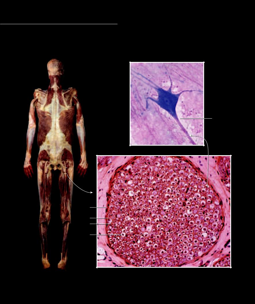
Structure of a Nerve
Nerves are bundles of axons running between the central nervous system and the peripheral tissues of the body. While all nerves have a similar basic structure, they vary in the types and numbers of
neurons bundled within. The basic design of a nerve consists of neurons wrapped by neurolemmocytes to form the nerve fiber. The fibers are protectively wrapped and nourished by a vascular loose connective tissue, the endoneurium. Many endoneurial wrapped fibers are surrounded by a collagenous perineurium to form the fasciculus of the nerve, and all the fasciculi are wrapped in a collagenous sheath, the epineurium, to form the nerve.
1
Dissection of sciatic nerve
8
7
5
6
Photomicrograph of multipolar neuron
400x
Photomicrograph of nerve cross-section
200x
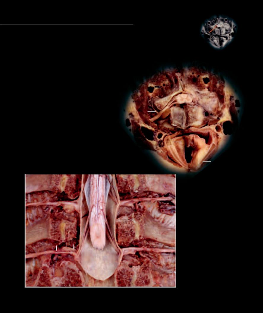
Spinal Nerve Structure
The spinal nerves arise from the spinal cord as a series of small neuronal bundles called rootlets — ventral (motor)rootlets and dorsal (sensory) rootlets. Each series
of ventral rootlets converges to form larger ventral roots. Likewise each series of dorsal rootlets converges to form larger dorsal roots. The dorsal and ventral roots project laterally and converge to form the spinal nerve trunk. A ganglion, the dorsal root ganglion, is present on the dorsal root just prior to the spinal nerve trunk. Branching from the trunk are two large branches and a variable series of smaller branches. Each branch follows a specific course to different peripheral regions. The two largest branches, the ventral ramus and dorsal ramus, are somatic branches that run in the musculoskeletal wall of the body. Smaller visceral branches, the meningeal nerve, the white and gray communicating rami, and the parasympathetic splanchnic nerves form the autonomic pathways to smooth muscle and glandular tissue.
Structure of a Nerve |
Other Structures |
|
|
|
|
|
|
1 |
Sciatic nerve |
17 |
Spinal cord |
|
|
|
|
|
|
2 |
Epineurium |
18 |
Cervical vertebra |
|
|
|
|
|
|
3 |
Perineurium |
19 |
Vertebral artery |
|
|
|
17 |
4 |
Endoneurium |
20 |
Common carotid artery |
|
|
|
|
|
|
10 |
|
5 |
Myelin sheath |
21 |
Internal jugular vein |
|
|
|
9 |
|
6 |
Axon |
22 |
Laryngopharynx |
|
|
|
|
|
|
7 |
Cell body |
23 |
Larynx |
16 |
|
|
19 |
|
|
|
|
|
|
8 |
Dendrite |
24 |
Thyroid cartilage |
14 |
|
|
|
|
|
18 |
|
|
|
25 |
Cricoid cartilage |
15 |
|
|
|
|
|
|
|
|
Spinal Nerve Structures |
26 |
Vocalis muscle |
|
|
|
|
|
|
9 |
Ventral rootlets |
|
|
|
|
|
|
|
|
10 |
Dorsal rootlets |
|
|
21 |
|
|
|
|
11 |
Dorsal root |
|
|
|
|
|
|
|
|
20 |
22 |
12 |
Dorsal root ganglion |
|
|
|
|
|
25 |
|
|
13 |
Ventral root |
|
|
|
|
|
|
|
|
|
|
|
|
|
|
14 |
Spinal nerve trunk |
|
|
|
|
|
|
|
|
15 |
Ventral ramus |
|
|
|
|
|
23 |
16 |
Dorsal ramus |
|
|
|
|
|
26 |
|
|
|
|
|
|
|
|
|
24
Dissection of cervical spinal cord
Superior view
13
16
Dissection of spinal cord, thoracic vertebral bodies removed
Anterior view
213