


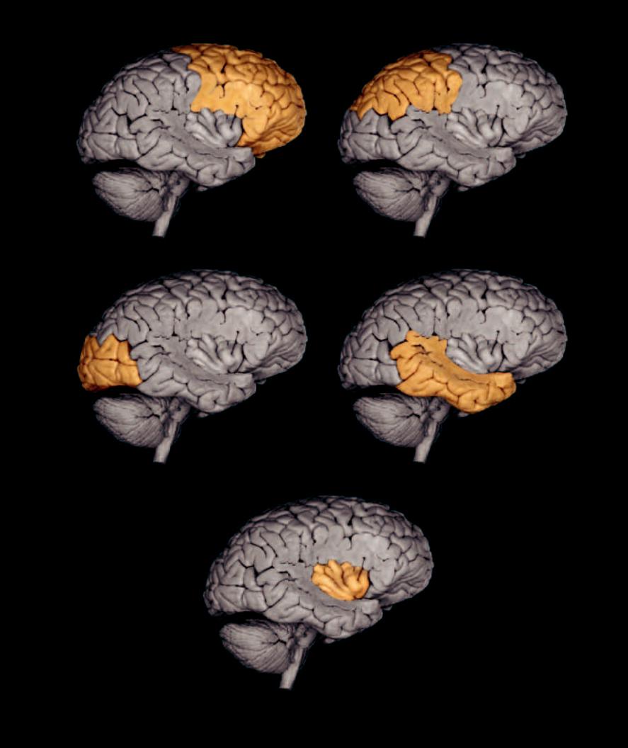

The diencephalon, rostral to the midbrain and almost completely surrounded by the cerebral hemispheres, consists of four
major parts — the thalamus, subthalamus, epithalamus, and hypothalamus. Projecting from the hypothalamus is the hypophysis, or pituitary gland. The brainstem consists of the medulla oblongata, pons, and midbrain. The medulla resembles the spinal cord in many ways. Like the cord it gives rise to many nerve roots; however, these are the roots of cranial nerves rather than spinal nerves. The pons is the bridge between the two cerebellar hemispheres. The ventral portion of the pons forms a large synaptic relay station consisting of scattered gray centers called the pontine nuclei. The dorsal portion of the pons is more like the other regions of the brainstem, the medulla and midbrain. The midbrain sits just above the pons and is obscured by the large, overlapping cerebral hemispheres. It contains nuclei for cranial nerves III and IV, as well as ascending and descending fi ber tracts from the cerebrum.
1 |
Infundibulum |
15 |
Medial eminence |
|
29 |
Superior medullary vellum |
|
|
37 |
|
Abducens nerve |
||||
2 |
Anterior perforated substance |
16 |
Facial colliculus |
|
30 |
Flocculus of cerebellum |
|
|
38 |
|
Trigeminal nerve |
||||
3 |
Tuber cinereum |
17 |
Locus ceruleus |
|
31 |
Caudate nucleus |
|
|
39 |
|
Facial nerve |
||||
4 |
Mammillary body |
18 |
Trigeminal tubercle |
|
32 |
Optic tract |
|
|
40 |
|
Vestibulocochlear nerve |
||||
5 |
Posterior perforated substance |
19 |
Hypoglossal tubercle |
33 |
Optic chiasm |
|
|
41 |
|
Glossopharyngeal nerve |
|||||
6 |
Pulvinar of thalamus |
20 |
Vestibular area |
|
34 |
Optic nerve |
|
|
42 |
|
Vagus nerve |
||||
7 |
Pineal gland |
21 |
Sulcus limitans |
|
35 |
Oculomotor nerve |
|
|
43 |
|
Accessory nerve |
||||
8 |
Superior colliculus |
22 |
Lateral recess |
|
36 |
Trochlear nerve |
|
|
44 |
|
Hypoglossal nerve |
||||
9 |
Inferior colliculus |
23 |
Obex |
|
|
|
|
|
|
|
|
|
|
|
|
10 |
Medial geniculate ganglion |
24 |
Olive |
|
|
|
|
|
|
|
|
|
|
|
|
11 |
Pons |
25 |
Pyramid |
|
|
|
|
|
|
|
|
|
|
|
|
12 |
Superior cerebellar peduncle |
26 |
Third ventricle |
|
|
|
|
|
|
|
|
31 |
|||
13 |
Middle cerebellar peduncle |
27 |
Fourth ventricle |
|
|
|
|
|
|
|
|
|
|
|
|
14 |
Inferior cerebellar peduncle |
28 |
Cerebral crus |
|
|
|
|
|
|
|
|
|
|
|
|
|
|
|
|
34 |
|
|
|
|
|
|
|
|
|
|
|
|
|
|
33 |
2 |
|
|
|
|
|
|
26 |
|
|
|
|
|
|
|
|
|
|
|
|
|
|
|
|
|
|||
|
|
|
|
|
|
|
|
|
|
|
|
|
|
|
|
|
|
1 |
|
32 |
|
|
|
|
|
|
|
|
6 |
||
|
|
3 |
|
|
|
|
|
|
|
7 |
|
|
|
||
|
|
|
|
|
|
|
|
|
|
|
|
|
|
||
|
|
|
4 |
|
|
|
|
|
8 |
|
|
|
|
|
|
|
|
|
|
|
|
|
10 |
|
|
|
|
|
|
||
|
|
|
|
35 |
|
|
|
|
|
|
|
|
|
|
|
|
|
|
5 |
|
|
|
|
|
|
|
|
28 |
|||
|
|
|
|
36 |
|
|
|
|
|
|
|
||||
|
|
|
|
|
|
|
|
|
|
|
|
||||
|
|
|
|
28 |
|
|
|
|
|
|
|
|
|
|
|
|
|
|
|
|
|
|
|
9 |
|
|
|
|
|
||
|
|
|
|
|
|
|
|
|
|
|
29 |
|
|||
|
|
|
11 |
|
|
|
|
|
|
|
|
|
|
|
|
|
|
|
|
|
38 |
|
|
|
|
|
|
|
|
|
|
|
|
|
|
|
|
|
12 |
17 |
15 |
|
|||||
|
|
|
|
|
|
|
|
|
|
|
|
||||
|
|
|
|
37 |
13 |
|
|
|
|
|
|
27 |
|
|
|
|
30 |
|
|
|
|
13 |
|
|
|
|
|
|
|
||
|
|
|
|
|
|
|
|
|
|
16 |
|
||||
|
|
|
|
|
|
|
|
|
|
|
|
|
|||
|
|
|
|
|
39 |
|
|
|
|
|
|
|
|
||
|
|
|
|
|
|
|
|
|
|
|
|
|
|
|
|
|
|
|
|
|
40 |
|
14 |
|
|
|
|
|
20 |
||
|
|
|
|
|
|
21 |
|
|
|
22 |
|||||
|
|
|
|
|
|
|
|
|
|
||||||
|
|
|
|
|
|
|
|
|
|
|
|
|
|||
|
24 |
|
25 |
|
41 |
|
|
|
|
|
|
|
18 |
|
|
|
|
|
|
|
|
|
|
|
|
|
|
|
|
|
|
42
19
44
43
23
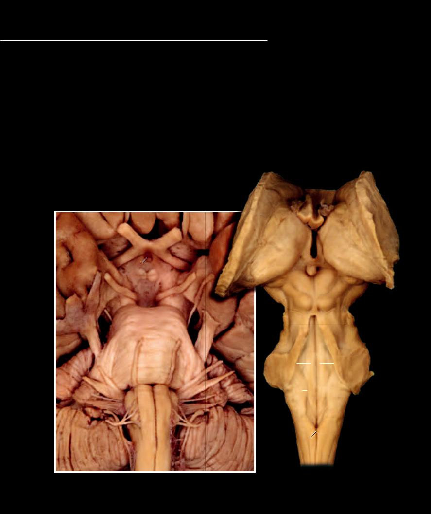
Brainstem |
Brainstem |
Ventral view |
Posterior view |
241
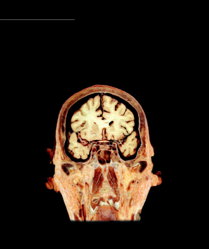
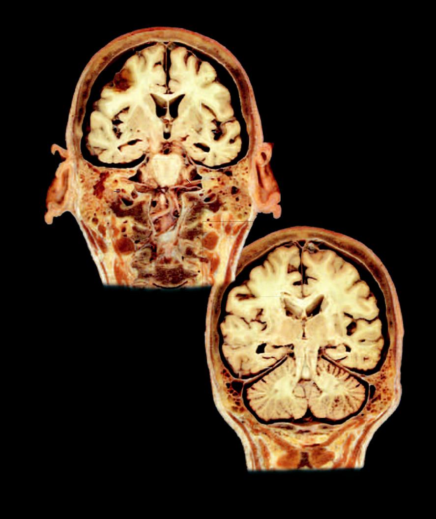
The brain sections on this and the following page depict aspects of brain anatomy that are not evident on the external views of the brain, and the association of the brain with surrounding structures of the head. Each section is approximately 2
centimeters thick and is an anterior view of three sections in succession. The fi rst section begins at the anterior aspect of the ear and the last section is just posterior to the ear.
1 |
Frontal lobe |
14 |
Flocculus |
27 |
Globus pallidus |
40 |
Sigmoid sinus |
2 |
Parietal lobe |
15 |
Superior vermis |
28 |
Medial thalamic nucleus |
41 |
Internal jugular vein |
3 |
Temporal lobe |
16 |
Superior cerebellar peduncle |
29 |
Lateral thalamic nucleus |
42 |
Tympanic cavity |
4 |
Insular lobe |
17 |
Cerebral peduncle |
30 |
Dentate gyrus |
43 |
Cochlea |
5 |
Lateral ventricle |
18 |
Pituitary gland |
31 |
Circular gyrus |
44 |
Sphenoid sinus |
6 |
Third ventricle |
19 |
Pons |
32 |
Optic chiasm |
45 |
Mastoid air cells |
7 |
Cerebral aqueduct |
20 |
Olive |
33 |
Facial nerve |
46 |
Mandibular condyle |
8 |
Fourth ventricle |
21 |
Corpus callosum |
34 |
Vestibulocochlear nerve |
47 |
Occipital condyle |
9 |
Septum pellucidum |
22 |
Caudate nucleus |
35 |
Vertebral artery |
48 |
Atlas |
10 |
Falx cerebri |
23 |
Internal capsule |
36 |
Middle cerebral artery |
49 |
Axis |
11 |
Tentorium cerebelli |
24 |
Putamen |
37 |
Internal carotid artery |
50 |
Lateral pterygoid muscle |
12 |
Anterior lobe of cerebellum |
25 |
External capsule |
38 |
Anterior cerebral artery |
51 |
Medial pterygoid muscle |
13 |
Posterior lobe of cerebellum |
26 |
Body of fornix |
39 |
Superior sagittal sinus |
52 |
Sternocleidomastoid muscle |
 10
10
1 |
|
|
|
|
25 |
5 |
|
|
22 |
||
|
|
|
|
24 |
|
|
38 |
|
|
||
|
|
|
23 |
36 |
|
|
32 |
|
|
|
37 |
3 |
18 |
|
|
|
37 |
|
44 |
46
50
51
Frontal section of head at anterior aspect of auricle
Anterior view
242
 10
10 34
34 10
10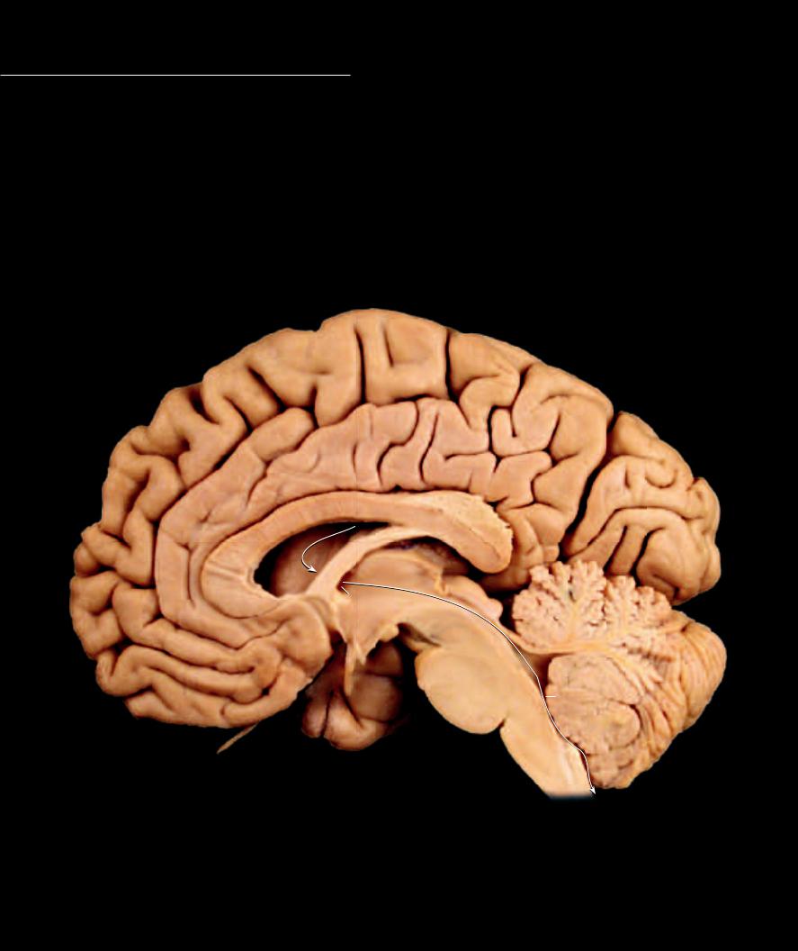
Developmentally the entire central nervous system forms from the hollow neural tube. As development proceeds and the wall of the neural tube becomes increasingly thicker, the hollow lumen of the tube
undergoes changes in relative size and shape throughout different regions of the changing central nervous system. As a result of this developmental history, there remains a hollow interconnected center throughout the entire central nervous system. This hollow core forms the ventricular system. Beginning within the cerebral hemispheres are the large paired lateral ventricles. Each lateral ventricle has a C-shape like its corresponding hemisphere. The lateral ventricles communicate via the interventricular foramina with a midline cavity, the third ventricle. The third ventricle sits within the core of the diencephalon where the right and left thalamus form its lateral walls. From the third ventricle a narrow channel, the aqueduct of the midbrain or cerebral aqueduct, passes through the core of the midbrain. This narrow channel expands in the region of the pons and cerebellum to form the fourth ventricle. The fourth ventricle tapers through the medulla to enter the spinal cord as the central canal. Within the four ventricles of the brain convoluted aggregations of capillaries, called a choroid plexus, project into the cavity of the ventricle. These capillary projections are the principal site for the production of cerebrospinal fluid.
13
16 |
|
8 |
|
14 |
|
|
|
1 |
|
|
|
11 |
|
19 |
|
9 |
|
|
|
|
|
18 |
|
|
|
1 |
|
|
|
|
|
|
|
|
12 |
|
|
3 |
21 |
|
2 |
4 |
||
|
|
|||
|
|
|
||
|
|
|
|
22 |
20
17
25
|
|
5 |
|
|
15 |
23 |
|
|
6 |
|
|
|
||
|
|
24 |
|
|
|
|
|
|
Sagittal section of braining revealing the ventricular system
Medial view, arrows show path of cerebrospinal fluid
244