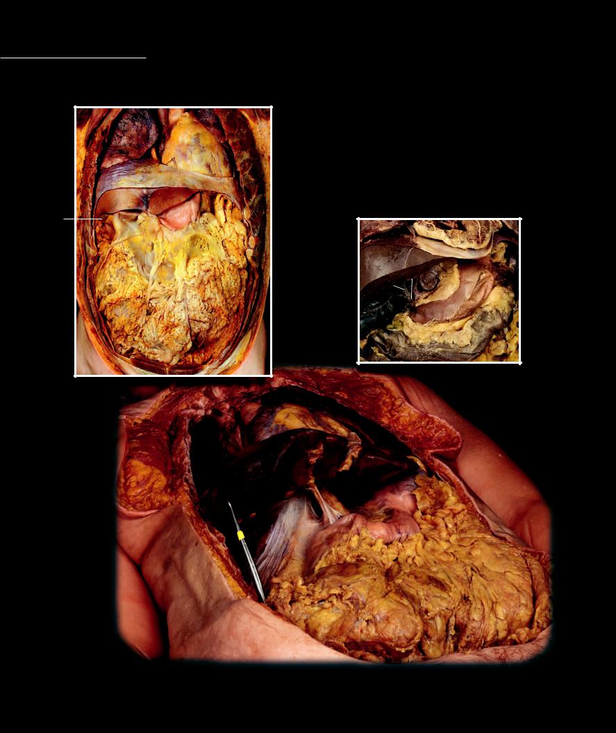
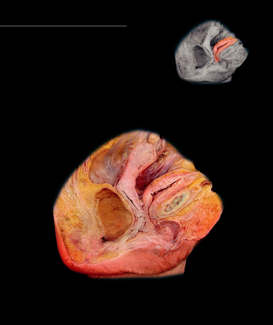
Omenta Omenta are mesenteric structures that unite two digestive organs. These refl ections of the peritoneal membrane course from one abdominal digestive organ to another abdominal digestive organ,
rather than from organ to body wall. There are two omenta in the abdominal cavity. The greater omentum is a peritoneal refl ection between the greater curvature of the stomach and the transverse colon. The lesser omentum is a peritoneal reflection between the lesser curvature of the stomach and the liver.
17
18
20
86
1
Anterior body wall removed exposing body cavity
Anterior view
18
12
19
8
5
1 |
Greater omentum |
|
12 |
Fossa for removed gallbladder |
2 |
Lesser omentum |
|
13 |
Gallbladder |
3 |
Hepatogastric ligament of lesser omentum |
14 |
Common hepatic bile duct |
4 |
Hepatoduodenal ligament of lesser omentum |
15 |
Common bile duct |
5 |
Hepatorenal part of coronary ligament |
|
16 |
Caudate lobe of liver |
6 |
Falciform ligament |
|
17 |
Lung |
7 |
Transverse mesocolon |
|
18 |
Heart |
8 |
Liver |
|
19 |
Breast |
9 |
Stomach |
|
20 |
Diaphragm |
10 |
Duodenum |
|
21 |
Epiploic foramen |
11 |
Transverse colon |
|
22 |
Spleen |
|
|
|
|
18 |
|
20 |
|
|
|
|
8 |
|
|
22 |
|
15 |
16 |
|
|
|
14 |
|
|
|
|
2 |
|
|
|
|
9 |
|
|
|
|
|
13 |
|
7 |
|
|
|
|
|
|
|
|
11 |
|
Dissection of abdominal cavity with anterior aspect of liver removed
Antero-inferior view
19
6
1
Superficial dissection of abdominal cavity with liver elevated
Antero-inferior view
19 Urinary System
Like the respiratory and digestive systems, the urinary system is an environmental exchange system. Like all the exchange systems of the body, the urinary system forms an immense interface with the cardiovascular system for the single purpose of regulating the homeostatic balance of the water environment (extracellular matrix) that surrounds every cell in the body. To make this exchange possible a
large network of microscopic urinary tubes form an intimate interface with an equally large network of cardiovascular capillaries. The urinary system consists of two
blood processing centers called the kidneys, two transport tubes called the ureters, a storage organ called the bladder, and a drain called the urethra. The kidneys continually produce urine, which is then moved via the
ureters to the storage organ, the bladder. When it is convenient to remove the urine from the body, contractions in the wall of the bladder expell the urine through the urethra.
In order to survive, every body cell requires a water environment that is similar to the composition of the oceans in which cellular life first arose. The kidneys help maintain this intercellular water environment by filtering the blood and regulating its contents so the blood can help maintain the correct composition of the extracellular fluid that bathes every cell. By adjusting the amount of water in the plasma and the various plasma constituents, which are either conserved for the body or eliminated in the urine, the kidneys are able to maintain water and electrolyte balance within the very narrow range compatible with life, despite wide variations in intake and losses of these constituents through other avenues.
Find more infomation about the urinary system in
R E A L A N AT O M Y
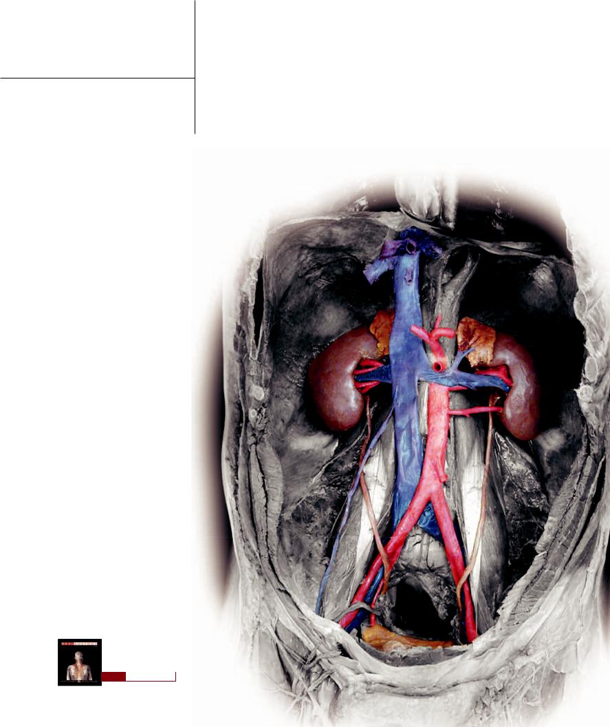
Urinary Organs
The organs of the urinary system include the paired kidneys, paired ureters, bladder, and urethra. The urinary organs occupy the
retroperitoneal and subperitoneal spaces in the abdominopelvic cavity, where they are surrounded by a large amount of adipose tissue and some areolar connective tissue. The dissection images on this and the facing page depict the organs of the urinary system and their relations to other organs in the abdominopelvic cavity.
1 |
Kidney |
7 |
Adrenal gland |
13 |
Liver |
2 |
Renal pelvis |
8 |
Aorta |
14 |
Lumbar vertebra |
3 |
Ureter |
9 |
Inferior vena cava |
15 |
Hilum |
4 |
Bladder |
10 |
Diaphragm |
16 |
Perirenal fat |
5 |
Renal vein |
11 |
Common iliac artery |
17 |
Intestines |
6 |
Renal artery |
12 |
Psoas major muscle |
18 |
Mesenteric fat |
10
|
|
|
|
|
10 |
|
7 |
|
|
7 |
|
|
|
|
|
|
|
6 |
9 |
|
6 |
|
1 |
5 |
5 |
1 |
|
|
|
8 |
2 |
|
|
|
|
|
|
|
|
|
|
3 |
|
|
3 |
|
|
|
|
|
12 |
11 |
|
|
|
|
|
11 |
12 |
|
4
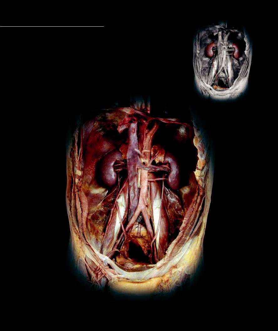
Dissection of the retroperitoneal space of the abdominal cavity
Anterior view
314
Dissection of abdomen showing perirenal fat
Anterior view
18
9
5
16
1
Level of section
Transverse section of abdomen through kidneys
Inferior view
17
17
17
17
6
8
16
Transverse section of abdomen at level of first lumbar vertebra
Inferior view
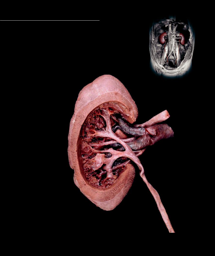
Kidneys and Ureters
The paired kidneys are the processing organs of the urinary system that filter the blood
for the purpose of regulating the water and electrolyte balance of the tissue fl uid, while removing unwanted waste products from the body. They occupy the retroperitoneal space of the abdominal cavity immediately anterior to the 12th ribs. The ureters descend from the kidneys lateral to the lumbar vertebrae, cross anterior to the psoas musculature and the common iliac vessels, and enter the pelvis to join the bladder.
1 |
Hilum |
6 |
Renal artery |
2 |
Renal pelvis |
7 |
Segmental artery |
3 |
Ureter |
8 |
Segmental vein |
4 |
Renal capsule |
9 |
Major calyx |
5 |
Renal vein |
10 |
Minor calyx |
4
6
8
3
Dissection into medulla of left kidney
Posterior view
316
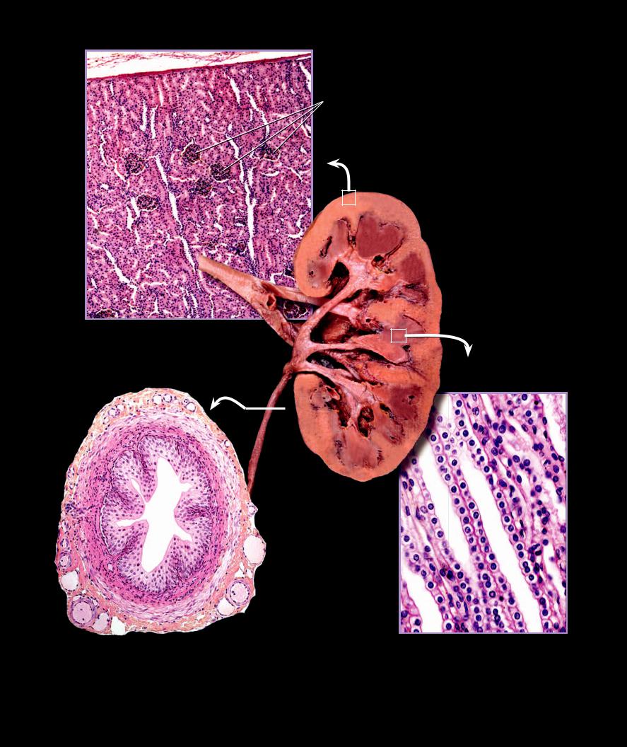
11 Renal cortex
12 Renal pyramid
13 Renal column
1514 Collecting tubule
15 Glomerulus surrounded by urinary tubules
16 Transitional epithelium of tunica mucosa
17 Smooth muscle of tunica muscularis
18 Connective tissue of tunica adventitia
|
|
|
|
|
|
11 |
|
|
|
|
|
12 |
|
|
|
|
|
13 |
|
|
|
|
10 |
|
6 |
|
|
|
|
|
|
|
|
|
|
7 |
|
|
|
|
|
|
7 |
9 |
|
|
|
Longitudinal section |
of renal cortex |
1 |
|
|
|
|
|
|
|
|
50X |
|
|
|
9 |
|
|
|
|
|
|
|
|
|
|
|
|
|
|
|
|
|
|
|
|
2 |
|
|
|
|
|
|
|
9 |
|
|
|
|
|
|
|
10 |
|
|
|
|
|
3 |
12 |
|
|
|
|
10 |
|
|
|
|
|
|
16 |
Frontal section of kidney |
|
Posterior view |
17 |
14 |
14 |
|
18 |
Transverse section of ureter |
Longitudinal section of renal pyramid |
200X |
400X |
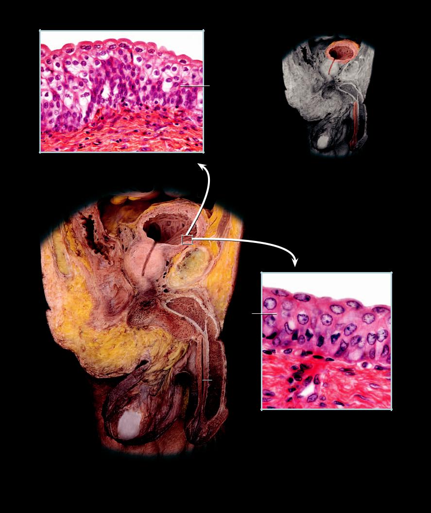
Bladder and Urethra
The bladder is the convenience organ of the urinary system that
stores the urine, which is continually being produced by the kidneys, until it is convenient to remove it from the body. Arising from the inferior surface of the bladder is the drain for the bladder called the urethra. It is a short tube in females and a much longer tube in males. The male urethra not only transports urine, but also is the passageway for sperm as it exits during ejaculation.
Female |
Male (opposite page) |
1 |
Bladder |
1 |
Bladder |
2 |
Urethra |
2 |
Prostatic urethra |
3 |
Clitoris |
3 |
Spongy urethra |
4 |
Vagina |
4 |
Prostate |
5 |
Uterus |
5 |
Penis |
6 |
Rectum |
6 |
Testis |
7 |
Pubis |
7 |
Scrotum |
8 |
Anus |
8 |
Rectum |
9 |
Labia majora |
9 |
Anus |
|
|
10 |
Pubis |
|
|
11 |
Transitional epithelium of tunica mucosa |
5
1
4
7
6
2
3
8
9
Sagittal section of female pelvis
Medial view
318
11
Section of relaxed mucosal lining of bladder
400X
1
4
 2 10
2 10
8
11
9
3
7
5
6
Section of distended mucosal lining of bladder
640X
Sagittal section of male pelvis and penis
Medial view
1 |
Kidney |
8 |
Diaphragm |
15 |
Inferior mesenteric artery |
2 |
Ureter |
9 |
Esophageal hiatus |
16 |
Common iliac artery |
3 |
Bladder |
10 |
Celiac artery |
17 |
Common iliac vein |
4 |
Renal artery |
11 |
Left gastric artery |
18 |
Posas major muscle |
5 |
Renal vein |
12 |
Splenic artery |
19 |
Iliacus muscle |
6 |
Aorta |
13 |
Common hepatic artery |
20 |
Ductus deferens |
7 |
Inferior vena cava |
14 |
Superior mesenteric artery |
21 |
Femoral nerve |
6
9
8
8
11
10
13
12
7
5
6
21
3
 20
20
Dissection of urinary system
Anterior view

20 Reproductive Systems
The organs of the male and female reproductive (genital) systems have, as their primary role, the responsibility of producing the specialized cells called gametes and making it possible for these cells to unite to form a new individual. The male gametes, the sperm, arise in the testes from meiotic divisions in the walls of the numerous seminiferous tubules. From here hundreds of millions of sperm make their way during ejaculation through a series of tubes — rete testis, efferent ductules, epididymis, ductus deferens, ejaculatory duct, prostatic urethra, intermediate urethra, spongy urethra — that move the sperm out of the male genital system and introduce them into the female genital system. During this passage secretions are added to the sperm by the prostate, seminal, and bulbourethral glands to help protect and nurture the sperm in their journey to unite with the
female gamete.
The sperm are introduced by the male intromittent organ, the penis, into the female vagina, which serves the dual function of being a penile receptacle and the birth canal. Sperm deposited in the fornices of the vagina then enter the os of the uterine cervix and propel themselves to the top of the uterine cavity. Here the sperm enter the openings into the uterine tubes where they continue their journey toward the ovulated female gamete.
After rupturing the surface of the ovary in an event called ovulation, the female gamete, the primary oocyte, is swept into the ostium of the uterine tube by the fingerlike fimbriae. Ciliary action of the uterine tube mucosa carry the the oocyte down the uterine tube where the sperm and oocyte make contact. If a sperm penetrates the oocyte’s surrounding cells and membranes, then fertilization occurs and the DNA of the two cells unite to form a new individual called a zygote. Cell divisions give rise to the embryo, and ciliary actions and muscular contractions in the wall of the tube move the embryo into the uterus, the mammalian equivalent of a nest, where the remainder of development will occur.
Find more information about the reproductive system in
R E A L A N AT O M Y