

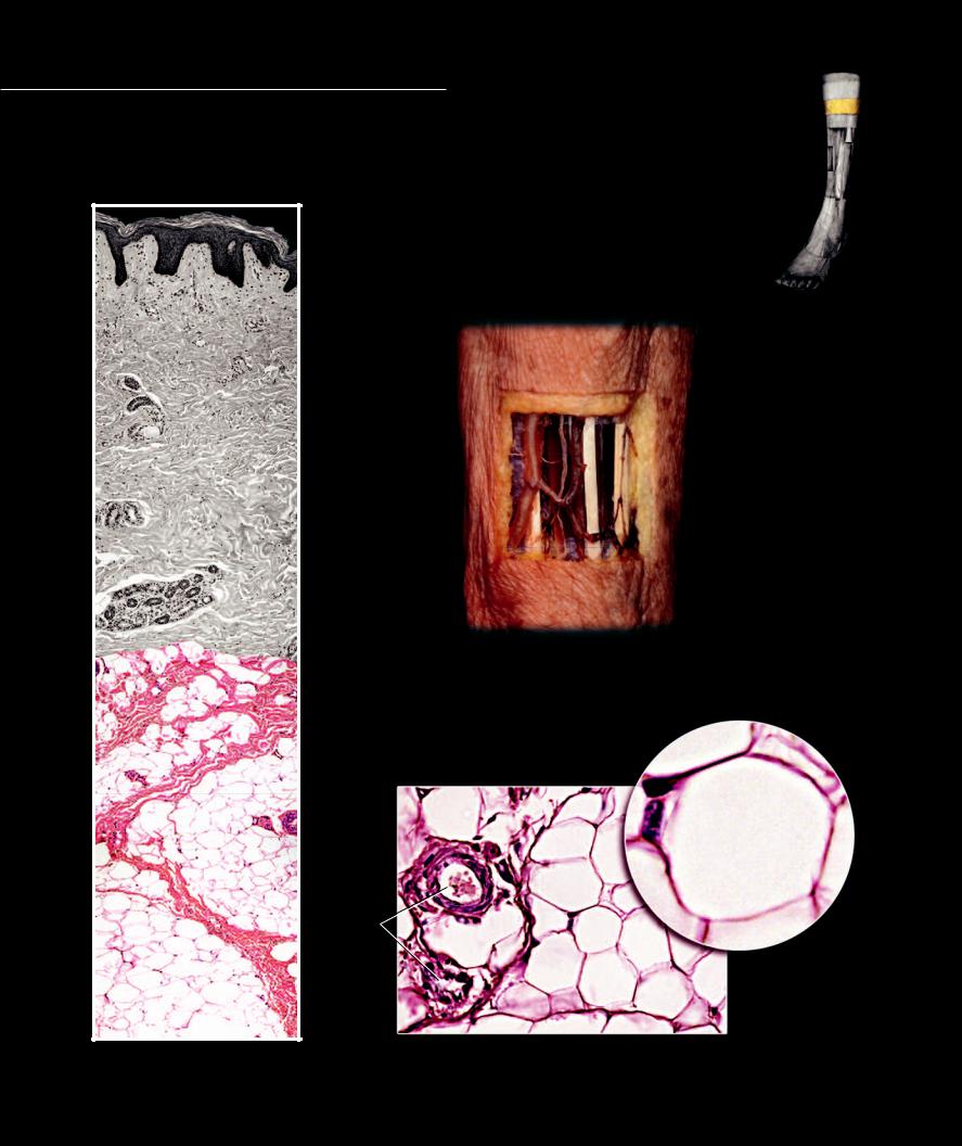
The subcutaneous layer, also called the hypodermis, is a layer of variable thickness that ranges from a thin layer of loose connective tissue to a
thick fi broadipose layer. This layer is a prominent location of fat storage in the body. In addition, it functions as an insulative layer and is the site of distribution of the main venous drainage channels of the integument and the cutaneous nerves that supply the skin.
1 |
Epidermis of skin |
7 |
Muscle |
2 |
Subcutaneous layer |
8 |
Retinaculum cutis |
3 |
Fascia |
9 |
Adipose cell membrane |
4 |
Superficial veins |
10 |
Nucleus of adipose cell |
5 |
Cutaneous nerve |
11 |
Fat storage vacuole of adipose cell |
6 |
Tendon |
12 |
Blood vessel |
1
2
3
4 5
3 7
2
6
4
Superficial veins and cutaneous nerves in the subcutaneous layer
Step dissection of antebracial integument, anterior view
8
9
8
10
11
9
12
8
Subcutaneous layer of integument |
Subcutaneous adipose tissue (left), adipose cell (callout) |
|
Section of subcutaneous layer, 200x and 640x |
||
100x |
||
|
24
4 Skeletal System
The skeletal system forms the internal framework for the soft tissues of the body. This is not a static framework, but a highly dynamic internal scaffolding. It is dynamic in many ways. On one hand, because of its jointed design, it shows extreme flexibility of movement when acted upon by muscles.
At another extreme, the cells of skeletal tissue are constantly monitoring and changing the micro-structure of this amazing tissue called bone, providing it with maximal strength, toughness, and resilience. In addition to its dynamic role of support, it also serves a protective role for many organs of
the body. This dynamic framework also exhibits a tremendous capacity for growth and repair. It is a storehouse of calcium ions, ions that play a significant role in many of the body’s functions.
The skeleton consists of 206 separate bones, ignoring various sesamoid bones and the fact that some bones represent the fusion of multiple bones. These bones range in size from the small ear ossicles measuring a few millimeters in length to the large femur measuring up to fifty centi-
meters. The skeleton is divisible into two portions, the axial skeleton and the appendicular skeleton. The axial skeleton includes the cranium, vertebral column, ribs, and sternum. The appendicular skeleton consists of the bones of the limbs and their girdles. The individual bones of the skeleton come in a variety of shapes. Some are long and tubular, while others have the spread-winged appearance of a butterfly. Bones can be grouped into four shape categories. Although not that meaningful, the four categories descriptively group the bones. The four shape categories are: long bones, short bones, flat bones, and irregular bones. Long bones are unique in hav-
ing a diaphysis or shaft with a medullary cavity. The other bone types lack this hollow tubular region. The short, flat, and irregular bones are similar in having outer plates of compact bone surrounding internal centers of spongy bone. In general, long bones and short bones are found in the appendicular skeleton, while flat bones and irregular bones occur in the axial skeleton. In the right hands, the skeleton can be a library of information. Its markings, foramina, landmarks, and canals each tell a story about the soft tissues of the body. A strong foundation of skeletal anatomy is an important starting point in understanding anatomy.
This chapter covers bone tissue and the general structure of bones and the skeleton. In the two chapters that follow you will explore the two subdivisions of the skeleton — the axial skeleton and the appendicular skeleton.
Find more information about the skeletal system in
R E A L A N AT O M Y
25
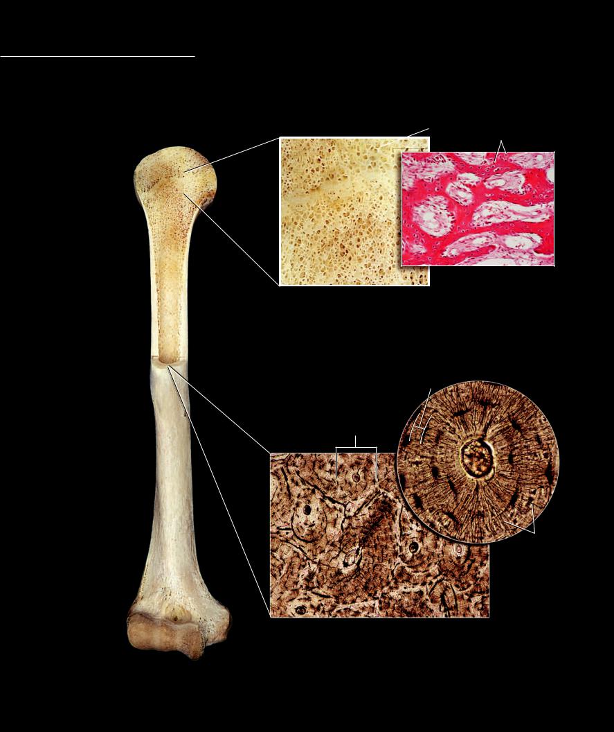
The tissue bone has two general forms — trabecular or spongy bone and compact bone. Trabecular bone is an internal bone that always resides deep to the more dense compact bone. Like its name implies, trabecular bone has many small beams of bone tissue con-
nected together in complex array around obvious spaces in the tissue. To the unaided eye this gives the bone a spongy appearance. Bone marrow fills the spaces in the trabecular bone. The second type of bone tissue, compact bone, is very dense and solid looking to the unaided eye. Compact bone forms the outer surface of all bones and can range in thickness from paper thin to many centimeters thick. Microscopic analysis of this dense bone reveals that it has many microscopic spaces containing cells
and blood vessels in circular arrangements called osteons.
 3
3
10
3 |
3 |
|
1
4
3
3
Trabecular bone
200x
Trabecular bone
Frontal section
2 |
1 |
Trabecular bone |
6 |
Central canal |
|
2 |
Compact bone |
7 |
Lacuna |
||
|
|||||
|
3 |
Trabecula |
8 |
Canaliculi |
|
|
4 |
Bone marrow |
9 |
Lamella |
|
|
5 |
Osteon |
10 |
Nucleus of osteocyte |
9
7
5
6
7
7
8
Compact bone and callout of osteon
Transverse section, 100x and 400x
Sectioned humerus
Anterior view, proximal half frontal section
26
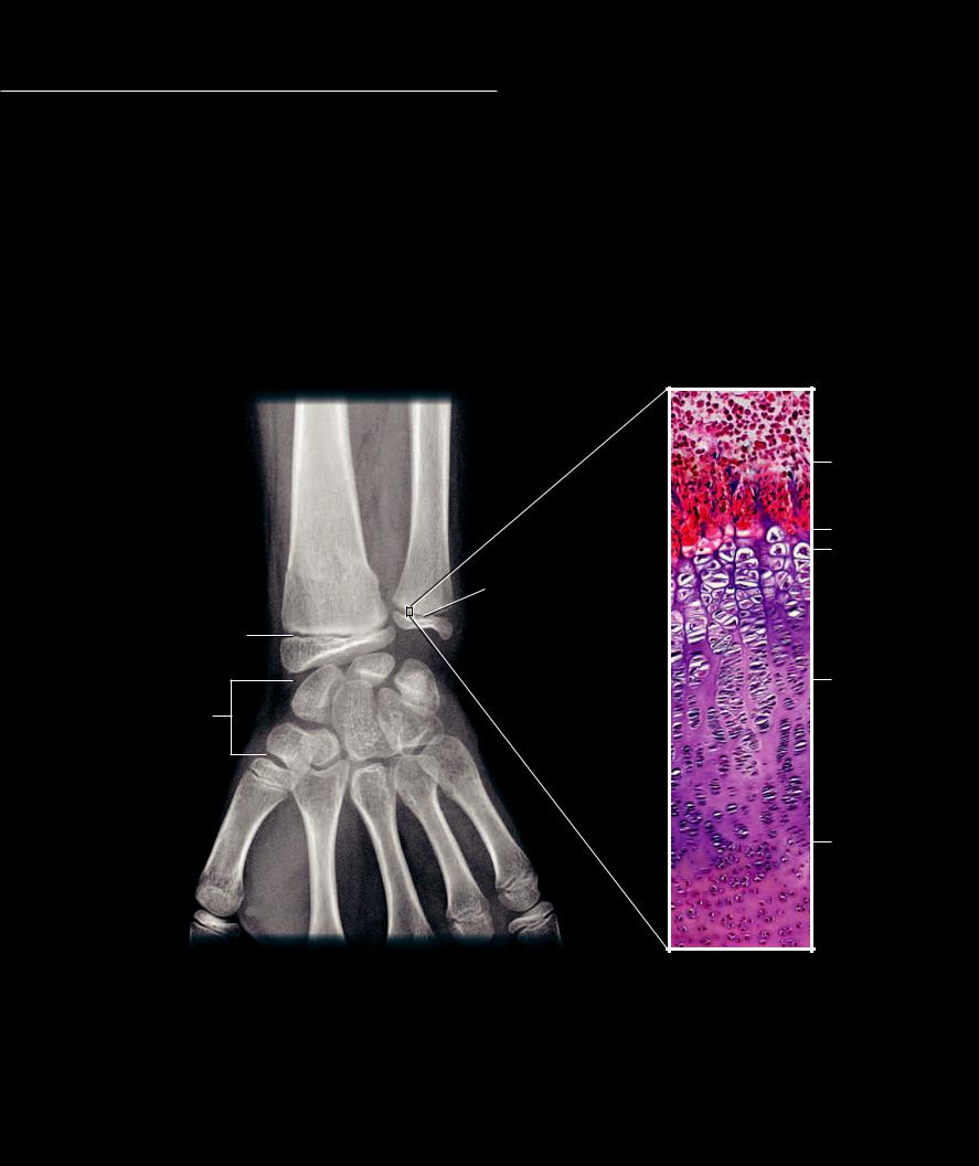
Bone tissue forms during development by either replacing cartilage tissue precursors (endochondral ossifi cation) or by developing within mesenchymal connective tissue
(intramembranous ossifi cation). In endochondral ossifi cation cartilaginous growth plates remain between developing bone centers to allow a bone to increase in length and size. During an individual’s young life, the growth plates are evident on a radiograph and are a clear indication that the individual is still growing.
1 |
Radial diaphysis |
7 |
Metacarpal bones |
2 |
Radial epiphysis |
8 |
Developing diaphysial bone |
3 |
Ulnar diaphysis |
9 |
Zone of calcified cartilage |
4 |
Ulnar epiphysis |
10 |
Zone of hypertrophied cartilage |
5 |
Growth plate |
11 |
Zone of proliferating cartilage |
6 |
Carpal bones |
12 |
Zone of resting cartilage |
1 |
3 |
8 |
|
|
|
|
|
9 |
5
10
4
5
2
6
11
7
7
77
12
Radiograph of the wrist region of a child |
Growth plate |
|
Posterior view |
||
200x |
||
|
27
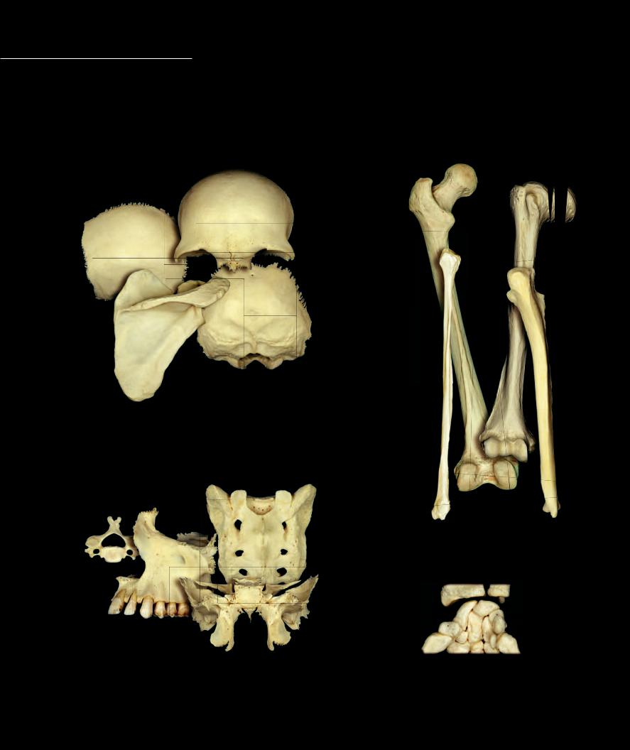
The bones of the skeleton come in a variety of sizes and shapes. The form of each bone emerges from its position and functional role in the skeletal system. In an effort to classify the different bones of the body anatomists defi ne four general categories of bones based
on their size and shape. Long bones, as their name suggests, are longer in one dimension than any other dimension. The long bones range in size from the short phalanges of the digits to the long proximal humerus and femur of the limb skeletons. Conversely, short bones are small, block-like bones. Like the long bones, short bones occur in the limb skeletons where they form the bones of the wrist and ankle. Flat bones are plate-like bones and are common in the cranium. The fi nal category, irregular bones, is a mixed group of bones that have a variety of shapes and locations within the skeleton.
Flat bones
Long bones
Irregular bones |
Short bones |
|
28

All bones share basic features in common. Compact bone tissue forms all the visible outer surface of the bone and can vary from a paper-thin covering to a thick wall of bone. Trabecular bone tissue
occupies the core of the bone beneath the compact bone. Areas of compact bone covered by articular cartilage form smooth subchondral compact bone surfaces. These subchondral bone surfaces mark the joint surfaces of bones. The photos below illustrate the basic parts and features of a long bone.
1 |
Epiphyses |
5 |
Subchondral bone |
2 |
Diaphysis |
6 |
Trabecular bone |
3 |
Metaphysis |
7 |
Medullary cavity |
4 |
Compact bone |
8 |
Epiphysial line |
1
1
3
1
2
3
11
Femur
Anterior view
5
1
8
3
6
4
2
Proximal end of femur
Frontal section
4
7
2
4
Femur |
3 |
6 |
Frontal section
8
1
5
Distal end of femur
Frontal section
29