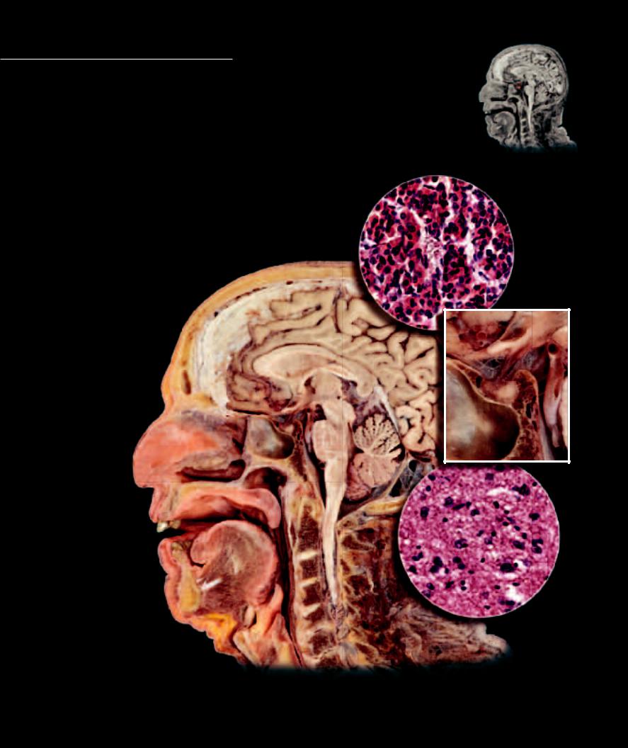
The pituitary gland, or hypophysis, “hangs” from the base of the brain via a connecting stalk, the infundibulum, which connects it to the hypothalamus. The infundibulum
contains numerous nerve fibers that relay from the hypothalamus to the posterior portion of the pituitary gland. In addition to this nervous pathway between the hypothalamus and the pituitary, numerous small blood vessels pass between the two organs. The pituitary gland has two anatomically and functionally distinct lobes, the neurohypophysis (posterior lobe) and the adenohypophysis (anterior lobe). The posterior lobe arises as an outgrowth of the embryonic brain. It is composed of nervous tissue and forms a neural link with the hypothalamus through the infundibulum. The anterior lobe arises from the epithelial lining of the embryonic pharynx. It consists of glandular epithelial tissue and forms a vascular link with the hypothalamus via the small blood vessels that pass between the two regions.
1 |
Pituitary gland |
|
|
2 |
Infundibulum |
Photomicrograph of anterior pituitary |
|
3 |
Adenohypophysis |
200x |
|
4 |
Neurohypophysis |
||
|
5Parenchyma consisting of acidophils, basophils, and chromophobes
6 Capillary with red blood cells
7 Parenchyma consisting of 5 6 axons and pituicytes
8 Hypothalamus
9 Cerebrum
10Falx cerebri
11Midbrain
12Pons
13 |
Cerebellum |
9 |
8 |
|
14 |
Medulla oblongata |
|||
|
|
|||
15 |
Spinal cord |
10 |
|
16Nasal septum
17Soft palate
18 Tongue |
2 |
19Epiglottis
20Atlas
21 |
Axis |
|
|
|
3 |
4 |
|
8 |
|
|
|||
22 |
Intervertebral disc |
|
11 |
|
|
|
23 |
Sphenoid sinus |
|
|
|
|
|
24 |
Occipital bone |
|
1 |
|
|
|
|
|
|
|
|
|
|
|
16 |
23 |
|
12 |
|
|
|
|
|
|
13 |
|
|
|
|
|
|
|
|
|
|
|
|
|
14 |
|
|
|
|
17 |
20 |
|
24 |
|
|
|
|
|
|
||
|
|
|
|
|
|
|
|
|
|
|
|
20 |
|
|
|
|
21 |
15 |
7 |
|
|
|
|
|
|
||
|
|
18 |
22 |
|
|
|
|
|
|
|
|
|
19
Photomicrograph of posterior pituitary
200x
Sagittal section of head and neck with enlarged callout of pituitary gland
Medial view
251

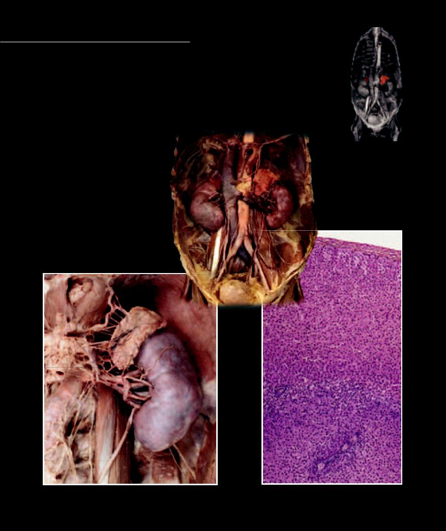
The pineal gland, a small reddish-gray body covered with pia mater, is a midline epithelial outgrowth of the embryonic midbrain positioned in a depression between the two superior
colliculi on the midbrain’s dorsal surface. The distal end of this outgrowth becomes a small mass of secretory cells that resemble the shape of a pine cone. It is from this appearance that it derives its name. The pia mater sends septa into the pineal gland that divide it into cords of secretory cells that are intermingled with numerous blood capillaries. The secretory cells of the pineal gland, called pinealocytes, have arm-like processes that contact both neighboring capillaries and the ependymal cells that line the third ventricle. Hormonal secretions produced in the body of the cell are moved through the arm-like processes where they are released by exocytosis into the capillaries and cerebrospinal fluid. Projecting into these cords of tissue are sympathetic postganglionic neurons from the superior cervical sympathetic ganglion. The gland plays a role in integrating photoperiod and affecting circadian rhythms.
1 |
Pineal gland |
|
|
2 |
Adenohypophysis |
|
|
3 |
Neurohypophysis |
10 |
|
4 |
Thalamus |
||
11 |
|||
5 |
Superior colliculi |
||
|
|||
6 |
Inferior colliculi |
|
|
7 |
Medial geniculate nucleus |
|
|
8 |
Cerebral peduncle |
|
|
9 |
Medulla oblongata |
|
|
10 |
Falx cerebri |
|
|
11 |
Corpus callosum |
1 |
12Pons
13Cerebellum
14Sphenoid sinus
15 |
Occipital bone |
2 |
3 |
|
16 |
Atlas |
|
||
|
|
|
||
17 |
Axis |
|
12 |
13 |
18 |
Soft palate |
14 |
19Nasopharynx
20Tongue
21Middle cerebellar peduncle
22 Fourth ventricle |
|
|
15 |
9 |
|
|
|
15 |
|
|
|
|
|
|
|
4 |
|
19 |
|
|
|
|
|
|
|
|
1 |
16 |
|
|
|
18 |
|
|
|
5 |
|
16 |
|
7 |
|
|
|
8 |
17 |
|
|
|
6 |
|
20 |
|
|
Sagittal section of brainstem and diencephalon in situ
Medial view
21 22
9
Dissection of brainstem and diencephalon
Posterior view
252
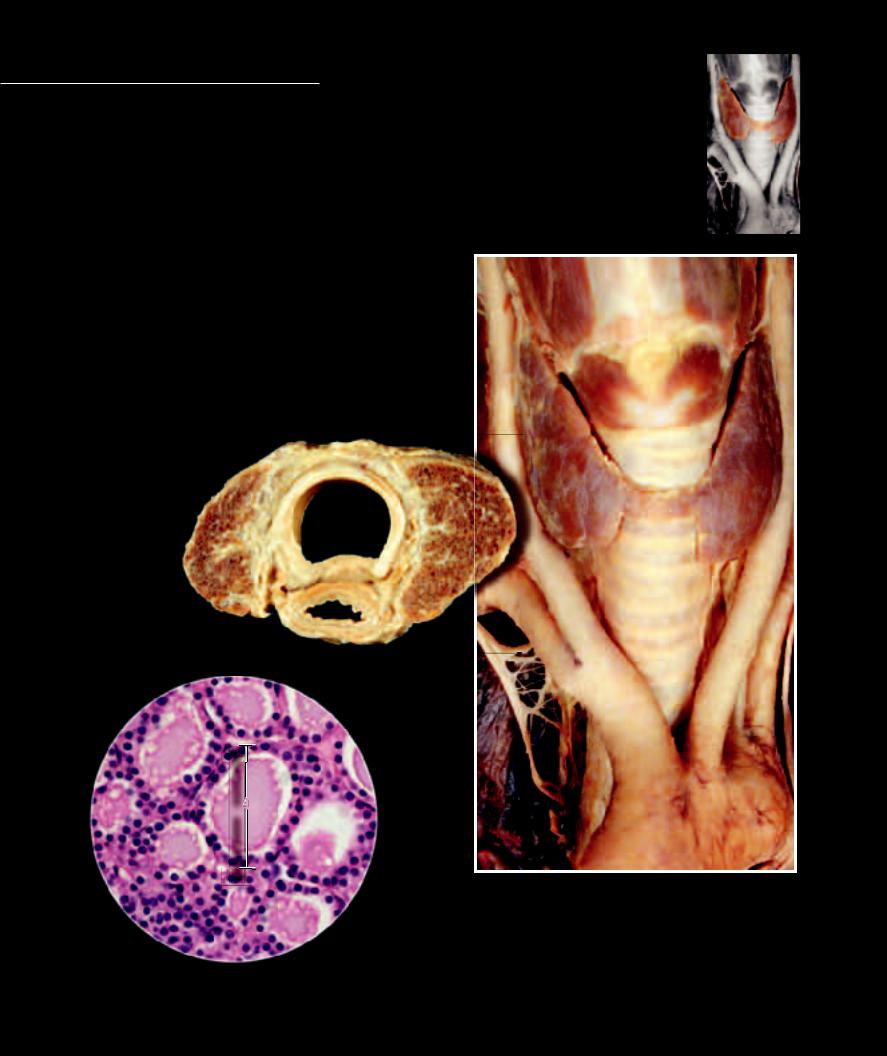
The thyroid gland is a bilobed organ positioned in the anterior neck. This highly vascular organ consists of two lateral lobes of endocrine tissue joined in the middle by a narrow portion of the
gland called the isthmus. It is red-brown in color and is enveloped by a thin layer of connective tissue. This connective tissue capsule sends extensions into the gland that divide the vascular and epithelial core into masses of irregular shape and size. The epithelial cells within the compartments of the thyroid gland form the secretory tissues of the organ. The major thyroid secretory cells are arranged into hollow spheres, each of which forms a functional unit called a follicle. In a microscopic section the follicles appear as rings of follicular cells enclosing an inner lumen fi lled with colloid, a substance that serves as an extracellular storage site for thyroid hormones. Interspersed in the interstitial spaces between the follicles are other secretory cells, the C cells, so called because they secrete the peptide hormone calcitonin.
1 |
Right lobe of thyroid gland |
|
|
|
|
|
|
||
2 |
Left lobe of thyroid gland |
|
|
|
|
|
|
||
3 |
Isthmus of thyroid gland |
|
|
|
19 |
11 |
|||
4 |
Thyroid follicle |
|
|
|
|
||||
|
|
|
|
|
|
||||
5 |
Follicular cell |
|
|
|
|
|
|
||
6 |
Thyroglobulin (TGB) |
|
|
|
|
|
|
||
7 |
Parafollicular (C) cell |
|
|
|
|
|
|
||
8 |
Trachea |
|
|
|
|
|
|
||
9 |
Fibromuscular membrane of trachea |
|
|
|
|||||
10 |
Esophagus |
|
|
|
|
|
|
||
11 |
Thyroid cartilage |
|
|
|
|
|
13 |
||
12 |
Cricoid cartilage |
|
|
|
|
|
|||
|
|
|
|
|
|
||||
13 |
Cricothyroid muscle |
|
|
|
|
|
12 |
||
14 |
Brachiocephalic artery |
|
|
|
|
|
|
||
15 |
Common carotid |
artery |
|
|
|
|
|
|
|
16 |
Subclavian artery |
|
|
|
1 |
2 |
|||
17 |
Aortic arch |
|
|
|
|||||
8 |
|
|
|
||||||
18 |
Vagus nerve |
|
|
|
|||||
|
|
|
|
|
|
||||
19 |
Thyrohyoid muscle |
|
|
|
|
|
|
||
|
|
|
|
|
|
|
|
|
3 |
|
1 |
|
|
|
2 |
|
|
||
|
|
|
|
9 |
|
|
8 |
||
|
|
|
|
|
|
|
15 |
||
|
|
|
|
|
|
|
15 |
||
|
|
|
|
10 |
|
|
|||
|
|
|
|
16 |
|
||||
|
|
|
|
|
|
|
18 |
16 |
|
|
|
|
|
|
|
|
|
|
|
|
|
|
|
|
Transverse section of thyroid gland |
|
|
|
|
|
|
|
|
|
|
Inferior view |
|
|
|
|
|
|
|
|
|
|
|
|
14 |
|
|
|
|
|
|
|
|
|
18 |
|
6 |
|
|
|
|
7 |
|
|
|
|
|
|
|
|
|
|
|
||
|
5 |
|
|
|
|
|
|
||
|
|
|
|
|
|
|
|||
|
|
|
|
|
|
|
|
|
|
|
5 |
|
|
4 |
|
|
|
|
|
|
|
|
|
|
|
|
|
17 |
|
|
|
|
|
|
|
|
|
|
|
5
Thyroid gland in situ
Anterior view
Photomicrograph of thyroid gland
240x
254
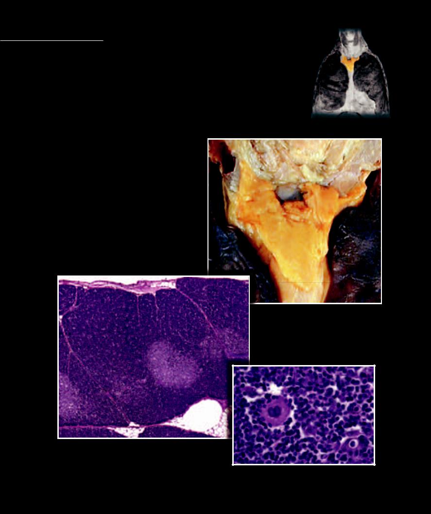
The parathyroid glands are small, oval, light brown glands situated on the posterior border of the two lateral lobes of the thyroid gland. The parathyroid glands sit just beneath the connective tissue capsule
of the thyroid gland. There are four parathyroid glands, two superior and two inferior. The endocrine cells of the parathyroid glands are called chief or principal cells. The chief cells form interconnecting columns of cells separated by fenestrated capillaries. The chief cells produce the parathyroid hormone.
1 |
Superior parathyroid gland |
7 |
Chief cell |
2 |
Inferior parathyroid gland |
8 |
Oxyphil cell |
3 |
Left lobe of thyroid gland |
9 |
Capillary |
4 |
Right lobe of thyroid gland |
10 |
Arteriole |
5 |
Isthmus of thyroid gland |
11 |
Venule |
6 |
Pyramidal lobe of thyroid gland |
|
|
8
11
10
9
7
Photomicrograph of parathyroid gland
240x
1
6
3
4
2
5
Thyroid and parathyroid glands (exposed on left)
Posterior view
255