

- •Preface
- •Content
- •Tissues
- •Nerve Tissue
- •Skin - Epidermis
- •Skin - Dermis
- •Skin - Glands
- •Subcutaneous Layer
- •Skeleton
- •Axial Skeleton
- •Cranium
- •Cranial Bones – Inferior Nasal Concha
- •Vertebral Column
- •Sacrum and Coccyx
- •Ribs
- •Sternum
- •Clavicle
- •Scapula
- •Humerus
- •Ulna
- •Radius
- •Metacarpals and Phalanges
- •Pelvis - Male
- •Femur
- •Tibia
- •Fibula
- •Tarsal Bones - Cuboid and Navicular
- •Phalanges
- •Patella
- •Skeletal Muscles
- •Transversospinales Muscles
- •Cervical Hypaxial Muscles
- •Thoracic and Abdominal Hypaxial Muscles
- •Shoulder Muscles - Rotator Cuff
- •Shoulder Muscles - Prime Movers
- •Anterior Brachial Muscles
- •Posterior Brachial Muscles
- •Posterior Thigh Muscles
- •Thigh Muscles
- •Lateral Leg Muscles
- •Posterior Leg Muscles
- •Spinal Nerves
- •Dorsal Rami
- •Intercostal Nerves
- •Cutaneous Nerves
- •Autonomic Nerves
- •Spinal Cord
- •Brain
- •Cerebrum
- •Cerebellum
- •Meninges
- •Hypothalamus
- •Pituitary Gland
- •Pineal Gland
- •Thymus
- •Pancreas
- •Ovaries
- •Testes
- •Blood
- •Heart
- •Lymphatics
- •Larynx
- •Lungs
- •Cast of Trachea and Bronchial Tree
- •Esophagus
- •Stomach
- •Pancreas
- •Large Intestine
- •Mesenteries
- •Omenta
- •Female Reproductive Organs
- •Ovary
- •Vagina
- •Ductus Deferens and Spermatic Cord
- •Penis
- •Index
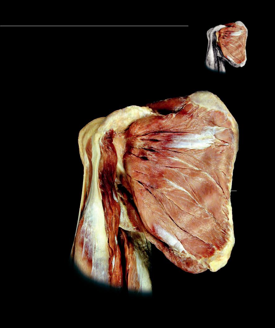
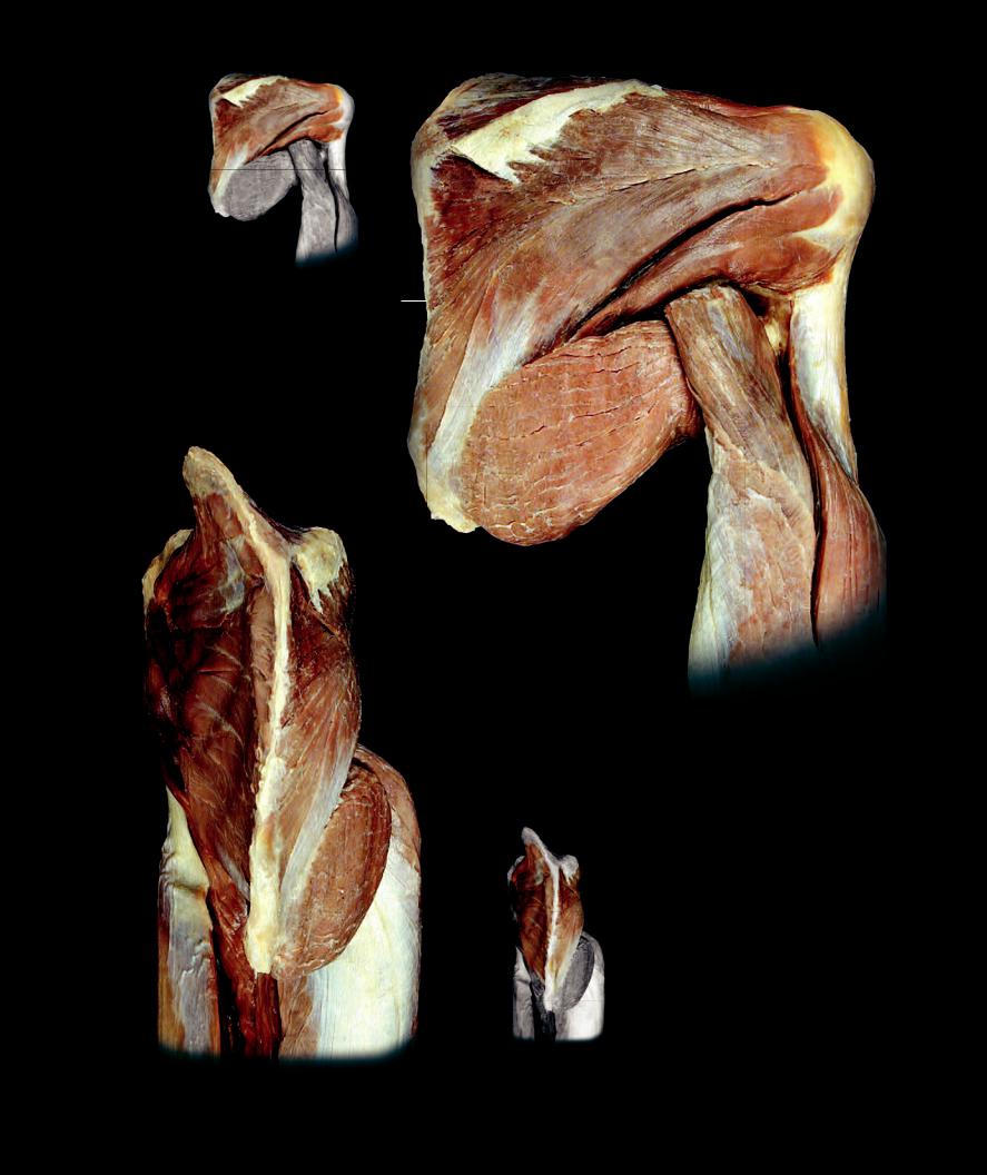
Shoulder Muscles - Rotator Cuff
The rotator cuff muscles are an important muscle group that play a critical role in stabilizing the shoulder joint. The four muscles (supraspinatus, infraspinatus, teres minor, and subscapularis) have thick, fl at tendons of insertion that form a strong musculotendinous cuff around all but the inferior aspect of the glenohumeral joint. These tendons are intimately applied to the fi brous membrane of the joint capsule. Individually each muscle contributes little to the total range of motion of the humerus at the glenohumeral joint. However, they play a prominent role in stabilizing the joint and positioning and stabilizing the head of the humerus in the glenoid cavity. When the rotator cuff muscles are compromised by injury, the shoulder joint loses stability and becomes highly susceptible to dislocation.
Rotator Cuff Muscles |
|
||
1 |
Supraspinatus |
|
|
2 |
Infraspinatus |
10 |
|
3 |
Teres minor |
||
4 |
Subscapularis |
1 |
|
|
|
||
Other Muscles and Structures |
|
||
5 |
Biceps brachii |
|
|
6 |
Coracobrachialis |
9 |
|
7 |
Triceps brachii |
||
|
|||
8 |
Teres major |
|
|
9 |
Coracoid process of scapula |
|
|
10 |
Superior angle of scapula |
|
|
11 |
Inferior angle of scapula |
14 |
|
12Spine of scapula
13Medial border of scapula
14Greater tubercle of humerus
4
6
13
|
8 |
5 |
11 |
|
|
|
7 |
Deep dissection of the right shoulder muscles
Anterior view
182
10
112
14
2
3
13
8
10
7
11
1 |
7 |
12
9
2
4
13
Deep dissection of the right shoulder muscles
Posterior view
8
611
7
5
Deep dissection of the right shoulder muscles
Medial view
183
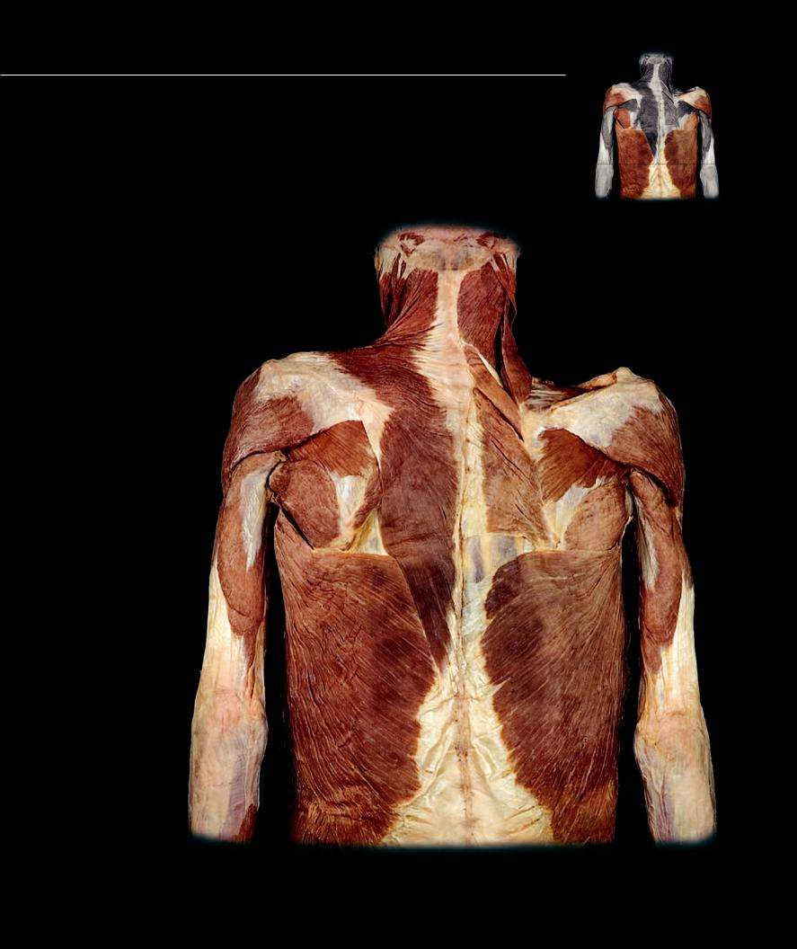
Shoulder Muscles - Prime Movers
The prime movers of the shoulder joint are the muscles that share a common attachment on the intertubercular groove (pectoralis major, teres major, and latissimus dorsi) and the deltoid muscle. These large muscles are superficial to the muscles of the rotator cuff and form extensive attachments on the pectoral girdle and axial skeleton. Inserting more distally on the humerus then the muscles of the rotator cuff, they have a better mechanical advantage and produce the major movements of the shoulder joint. The intertubercular groove muscles also form the anterior and posterior walls of the axilla. The large pectoralis major forms the anterior wall of the axilla, while the sheet-like latissimus dorsi and thick, round teres major form the posterior axillary wall.
Shoulder Prime Movers |
|
||
1 |
Deltoid |
|
|
2 |
Pectoralis major |
|
|
3 |
Teres major |
|
|
4 |
Latissimus dorsi |
|
|
Other Muscles and Structures |
|
||
5 |
Levator scapulae |
13 |
|
6 |
Rhomboideus minor |
||
|
|||
7 |
Rhomboideus major |
|
|
8 |
Supraspinatus |
5 |
|
9 |
Infraspinatus |
||
10Teres minor
11Triceps brachii
12 |
Trapezius |
|
6 |
8 |
34 |
13 |
Spleneus capitis |
|
|
|
|
|
|
|
|
||
14 |
Serratus anterior |
1 |
|
|
|
15 |
Pectoralis minor |
|
|
|
|
16 |
External intercostal |
|
|
|
|
17 |
Internal intercostal |
|
|
9 |
|
18 |
Rectus abdominis |
|
|
|
10 |
19 |
Coracobrachialis |
12 |
7 |
|
|
20 |
Biceps brachii |
|
|
|
|
3 |
|
|
|
||
|
|
|
|
|
21Brachialis
22Posterior scalene
23Middle scalene
24Anterior scalene
25Omohyoid
26 Sternohyoid |
11 |
27Sternothyroid
28Thyrohyoid
29Sternocleidomastoid
30External oblique
31 Brachioradialis |
4 |
|
32Clavicle
33Humerus
34Spine of scapula
35Thoracolumbar fascia
36Linea alba
37Common carotid artery
35
Muscles of neck, shoulder, brachium, and back
Posterior view
184
|
|
28 |
|
|
|
37 |
|
|
5 |
|
12 |
|
26 |
29 |
|
|
|
|
|
|
25 |
|
|
|
23 |
24 |
|
|
|
|
|
22 |
|
|
1 |
32 |
|
27 |
|
|
|
||
|
16 |
|
|
33
19
15
17
2
11
20
14
20 4
21
30 |
18 |
31
36
31
Muscles of neck, shoulder, brachium, and chest
Anteror view
185