- •Preface
- •Content
- •Tissues
- •Nerve Tissue
- •Skin - Epidermis
- •Skin - Dermis
- •Skin - Glands
- •Subcutaneous Layer
- •Skeleton
- •Axial Skeleton
- •Cranium
- •Cranial Bones – Inferior Nasal Concha
- •Vertebral Column
- •Sacrum and Coccyx
- •Ribs
- •Sternum
- •Clavicle
- •Scapula
- •Humerus
- •Ulna
- •Radius
- •Metacarpals and Phalanges
- •Pelvis - Male
- •Femur
- •Tibia
- •Fibula
- •Tarsal Bones - Cuboid and Navicular
- •Phalanges
- •Patella
- •Skeletal Muscles
- •Transversospinales Muscles
- •Cervical Hypaxial Muscles
- •Thoracic and Abdominal Hypaxial Muscles
- •Shoulder Muscles - Rotator Cuff
- •Shoulder Muscles - Prime Movers
- •Anterior Brachial Muscles
- •Posterior Brachial Muscles
- •Posterior Thigh Muscles
- •Thigh Muscles
- •Lateral Leg Muscles
- •Posterior Leg Muscles
- •Spinal Nerves
- •Dorsal Rami
- •Intercostal Nerves
- •Cutaneous Nerves
- •Autonomic Nerves
- •Spinal Cord
- •Brain
- •Cerebrum
- •Cerebellum
- •Meninges
- •Hypothalamus
- •Pituitary Gland
- •Pineal Gland
- •Thymus
- •Pancreas
- •Ovaries
- •Testes
- •Blood
- •Heart
- •Lymphatics
- •Larynx
- •Lungs
- •Cast of Trachea and Bronchial Tree
- •Esophagus
- •Stomach
- •Pancreas
- •Large Intestine
- •Mesenteries
- •Omenta
- •Female Reproductive Organs
- •Ovary
- •Vagina
- •Ductus Deferens and Spermatic Cord
- •Penis
- •Index
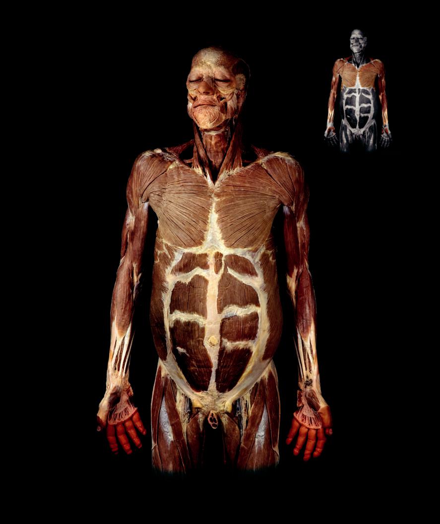
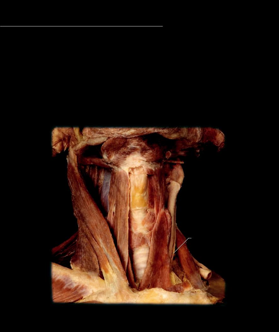
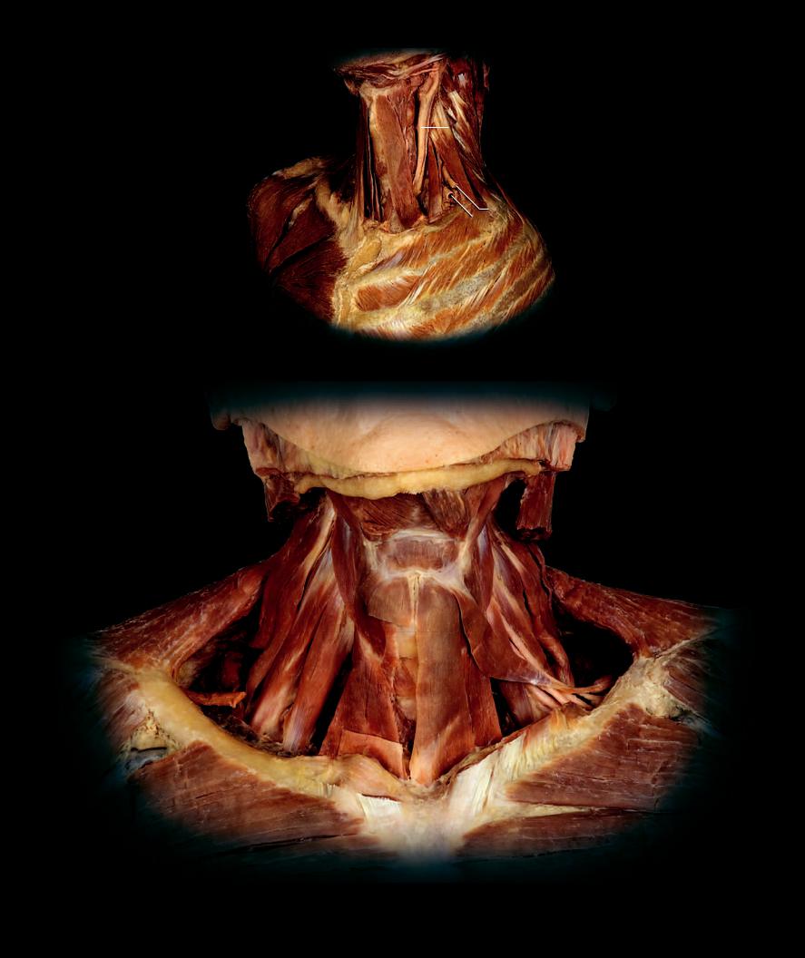
The muscular wall of the neck arises from the hypomeres of the cervical somites and develops in accordance with the anterior and lateral
body wall muscle pattern. A close scrutiny of the cervical hypaxial muscles reveals a ventral muscle, which has split into numerous subdivisions, a four-layered lateral muscle wall where the muscles have lost their sheet-like structure, and a subvertebral muscle on the anterior surface of the neck vertebrae. The cervical trunk muscles have a variety of functions. Some of the muscles function to stabilize and move the cervical vertebral column. Some of the muscles assist in raising the upper ribs. Some are annexed by the upper limb to support the pectoral girdle. The strap-like ventral muscles, which run from sternum to larynx to hyoid bone to mandible, are active during mastication, swallowing, respiration, and sound production. These seemingly varied muscles are all innervated by the anterior rami of the cervical spinal nerves.
Cervical Hypaxial Muscles |
Other Muscles and Structures |
21 |
Subclavian artery |
||
1 |
Sternohyoid muscle |
11 |
Anterior digastricus muscle |
22 |
Root of brachial plexus |
2 |
Sternothyroid muscle |
12 |
Mylohyoid muscle |
23 |
Common carotid artery |
3 |
Thyrohyiod muscle |
13 |
Sternocleidomastoid muscle |
24 |
Vagus nerve |
4 |
Omohyoid muscle |
14 |
Trapezius muscle |
25 |
Thyroid cartilage |
5 |
Geniohyoid muscle |
15 |
Deltoid muscle |
26 |
Thyroid gland |
6 |
Anterior scalene muscle |
16 |
Pectoralis major muscle |
27 |
Trachea |
7 |
Middle scalene muscle |
17 |
Serratus anterior muscle |
28 |
External intercostal muscle |
8 |
Posterior scalene muscle |
18 |
Cricothyroid muscle |
29 |
Internal intercostal muscle |
9 |
Levator scapulae muscle |
19 |
Stylohyoid muscle |
|
|
10 |
Longus colli muscle |
20 |
Posterior digastricus muscle |
|
|
11 |
|
12 |
|
|
20 |
19 |
|
|
7 |
|
|
10 |
25 |
3 |
4 |
|
|
1 |
|
23 |
13
|
|
18 |
26 |
|
24 |
|
2 |
|
|
|
|
14 |
|
|
|
27 |
6 |
|
|
22
21
16
Dissection of neck muscles
Anterior view
168
|
3 |
|
|
|
|
2 |
|
|
23 |
|
|
|
||
|
|
|
|
|
|
14 |
|
7 |
|
|
13 |
|
8 |
|
|
6 |
|||
15 |
2 |
|
22 |
|
|
|
|
21 |
|
|
16 |
|
28 |
|
|
29 |
|
|
|
Dissection of cervical hypaxial muscles
Anterolateral view
|
|
19 |
|
12 |
11 |
|
|
13 |
|
|
5 |
|
10 |
|
|
9 |
|
|
8 |
14 |
|
|
|
|
3 |
|
|
7 |
22 |
|
|
|
|
|
4 |
17 |
6 |
1 |
|
|
15 |
|
|
18 |
2
26
16
Dissection of cervical hypaxial muscles
Anterior view
169
Thoracic and Abdominal Hypaxial Muscles
The muscles of the thorax and abdomen develop from the hypomere of the thoracic and abdominal somites of the embryo. Like the neck they clearly demonstrate the muscle pattern of the vertebrate body wall. The thoracic body wall differs from the abdomen in having well-developed ribs that dominate the wall and limit the movements of the vertebral column. Because of the well-developed segmental ribs, the muscles of the thoracic wall retain their segmental origins. The uniquely mammalian diaphragm muscle is a member of this group that plays an important role in respiration. The outermost layer of the lateral muscle wall is well developed in the thorax. Some portions of this muscle layer remain associated with the ribs, while the rhomboid muscles (depicted in the upper limb chapter that follows) and large serratus anterior muscle migrate onto the scapula to become principal stabilizers of the upper limb. The ventral ramus of each of the thoracic and upper lumbar spinal nerves innervates these muscles.
Thoracic and Abdominal Musculature |
12 |
Sternothyroid muscle |
|
26 |
Pectineus muscle |
|||||||
1 |
Rectus abdominis muscle |
13 |
Omohyoid muscle |
|
27 |
Adductor longus muscle |
||||||
2 |
Serratus anterior muscle |
14 |
Sternocleidomastoid muscle |
|
28 |
External lamina of rectus sheath |
||||||
3 |
External intercostal muscle |
15 |
Trapezius muscle |
|
29 |
Linea alba |
|
|||||
4 |
External oblique muscle (superficial lamina) |
16 |
Deltoid muscle |
|
30 |
Tendinous intersections |
||||||
5 |
External oblique muscle (deep lamina) |
17 |
Pectoralis major muscle |
|
31 |
Internal lamina of rectus sheath |
||||||
6 |
Internal intercostal muscle |
18 |
Anterior scalene muscle |
|
32 |
Semilunar line |
|
|||||
7 |
Internal oblique muscle |
19 |
Middle scalene muscle |
|
33 |
Arcuate line |
|
|||||
8 |
Innermost intercostal muscle |
20 |
Posterior scalene muscle |
|
34 |
Transversalis fascia |
||||||
9 |
Transversus abdominis muscle |
21 |
Biceps brachii muscle |
|
35 |
Inguinal ligament |
||||||
|
|
|
|
|
|
22 |
Tensor fasciae latae muscle |
|
36 |
Spermatic cord |
|
|
Other Muscles and Structures |
23 |
Gluteus medius muscle |
|
37 |
Inferior epigastric vessels |
|||||||
10 |
Platysma muscle |
24 |
Gluteus minimis muscle |
|
38 |
Cutaneous nerves |
||||||
11 |
Sternohyoid muscle |
25 |
Iliopsoas muscle |
|
|
|
|
|
||||
|
10 |
|
|
|
|
|
11 |
15 |
||||
|
|
|
|
|
|
16 |
|
13 |
|
14 |
|
16 |
|
|
|
|
|
|
|
|
|
|
|||
|
|
|
|
|
|
|
|
|
|
|
||
|
17 |
|
|
|
|
|
17 |
|
||||
|
|
|
38 |
|
|
|
|
|
|
|
|
21 |
|
|
|
|
|
|
|
|
|
||||
|
|
|
|
|
|
|
|
|
|
|
|
2 |
2 |
|
|
|
|
30 |
32 |
|
|
29 |
|
|
|
|
|
|
|
28 |
4 |
|
1 |
4 |
|
|
|||
|
|
|
22 |
22 |
|
36
36
Dissections of thoracic and abdominal hypaxial muscles
Anterior view
170
|
|
18 |
18 |
|
|
19 |
|
|
|
19 |
|
|
|
13 |
|
13 |
|
|
|
|
|
11 |
|
11 |
|
|
|
|
|
|
|
|
|
|
3 |
6 |
3 |
|
6 |
|
|
||
|
|
|
|
|
|
2 |
|
|
|
|
|
30 |
|
|
|
|
|
|
|
|
|
30 |
|
|
|
|
|
|
|
|
|
29 |
|
32 |
4 |
|
|
|
|
|
|
|
|
|
|
7 |
|
|
|
|
|
|
|
1 |
|
|
|
|
|
|
|
29 |
1 |
32 |
|
|
|
|
|
||||
|
|
|
|
|
|
|
|
|
|
|
|
5 |
|
|
|||||||
|
|
|
|
|
|
|
|
|
|
|
|
|
|
|
|
|
4 |
|
|
|
|
|
18 |
|
|
|
35 |
|
|
|
|
|
|
|
|
|
|
|
|
|
|
|
|
|
19 |
|
|
|
|
|
|
|
23 |
|
|
|
|
|
|
|
|
|
|
|
|
|
|
|
|
|
|
|
|
|
|
|
|
|
35 |
|
|
|
|
|
|
||
|
|
|
|
|
|
|
|
|
|
20 |
|
|
|
|
|
|
|
|
23 |
||
|
|
|
|
|
|
|
|
|
|
|
|
|
|
|
|
||||||
|
|
|
|
|
25 |
|
|
|
24 |
|
|
|
|
|
|
|
|
||||
12 |
36 |
|
|
36 |
|
|
|
36 |
|
|
|
|
25 |
|
|
|
|
||||
|
|
|
|
|
|
|
|
|
|
|
|||||||||||
|
|
|
|
|
|
|
|
|
|
|
24 |
||||||||||
|
|
26 |
|
|
|
|
|
|
|
|
26 |
|
|
|
|||||||
|
|
|
|
|
|
|
|
|
|
|
|
|
|
|
|
||||||
|
|
|
|
|
|
|
|
|
|
|
|
|
|
|
|
|
|
|
|
||
|
|
|
|
|
|
|
|
|
|
|
|
|
|
|
|
|
|
|
|
|
27
27
6
|
|
|
|
|
|
|
|
|
8 |
|
|
|
|
|
|
30 |
|
|
|
|
|
|
|
|
|
29 |
1 |
32 |
|
31 |
|
|
|
|
||||
|
|
|
|
7 |
|
29 |
9 |
|
|
|||
|
|
|
|
|
|
|
|
|
|
|||
|
|
|
|
|
|
33 |
|
|
|
|
||
|
|
|
|
|
|
37 |
|
|
|
|
||
|
|
|
|
|
|
34 |
|
|
|
|
||
|
|
|
|
|
|
|
|
23 |
|
|
|
23 |
|
|
|
|
|
|
|
|
|
|
|
||
36 |
|
|
|
25 |
|
|
24 |
|
|
|
||
|
|
|
|
|
|
|
||||||
|
|
|
|
|
|
|
24 |
|||||
|
|
|
|
|
|
|
||||||
|
|
|
|
|
25 |
|
|
|||||
|
|
|
|
26 |
|
|
||||||
|
|
|
|
|
26 |
|
|
|
|
|||
|
|
|
|
|
|
|
|
|
|
27
27
Dissections of thoracic and abdominal hypaxial muscles
Anteriolateral view
171
Thoracic and Abdominal Hypaxial Muscles
Again we would have you notice the rarely described deep lamina of the external oblique muscle. Notice its continuity with the external intercostal muscles, while the superficial lamina of the external oblique interdigitates with the serratus anterior muscle. Also note the similar fi ber orientations of the intercostal muscles and their homologues in the abdominal wall. The photos of the diaphragm on the opposite page clearly reveal the continuity of this internal layer muscle with its internal homologue in the abdomen – the transversus abdominis muscle.
4
28
31
33
34
19 |
|
|
3 |
|
21 |
20 |
22 |
|
|
|
|
|
23 |
|
24
25
6
29
30
32
27
8
5
1
9
6 7 7
2
8
5
4
6 |
1 |
7 7
6
28
30
36
8
10
1
1
9 |
13 |
|
26
26
Dissections of lateral muscle layers of thoracic and abdominal wall
Posterior view upper left, Lateral view upper center, Posterolateral view all others
172
17
18
10
11
16
14
15
35
Thoracic and Abdominal Musculature |
|
|
1 |
Rectus abdominis muscle |
|
2 |
Serratus anterior muscle |
|
3 |
Serratus posterior superior muscle |
|
4 |
Serratus posterior inferior muscle |
|
5 |
External intercostal muscle |
|
6 |
External oblique muscle (superficial lamina) |
|
7 |
External oblique muscle (deep lamina) |
|
8 |
Internal intercostal muscle |
|
9 |
Internal oblique muscle |
41 |
10 |
Innermost intercostal muscle |
11Subcostal muscle
12Diaphragm
13Transversus abdominis muscle
14Quadratus lumborum muscle
15Psoas major muscle
16Psoas minor muscle
Other Muscles and Structures
|
17 |
Longus capitis muscle |
|
|
18 |
Longus colli muscle |
|
|
19 |
Splenius capitis muscle |
|
|
20 |
Trapezius muscle |
|
|
21 |
Deltoid muscle |
|
|
22 |
Infraspinatus muscle |
|
|
23 |
Teres major muscle |
|
|
24 |
Triceps brachii muscle |
|
|
25 |
Latissimus dorsi muscle |
|
|
26 |
Tensor fasciae latae muscle |
|
|
27 |
Gluteus maximus muscle |
|
|
28 |
Gluteus medius muscle |
|
|
29 |
Gluteus minimis muscle |
|
29 |
30 |
Piriformis muscle |
13 |
31 |
Superior gemellus muscle |
||
|
32 |
Obturator internus muscle |
|
|
33 |
Inferior gemellus muscle |
|
|
34 |
Quadratus femoris muscle |
|
|
35 |
Iliacus muscle |
|
|
36 |
Sacrotuberous ligament |
|
Dissection of hypaxial subvertebral muscles
12
43
10
|
|
Anterior view |
|
|
|
Dissection revealing diaphragm |
||
|
|
|
|
|
|
|||
37 |
|
Lateral view |
||||||
|
|
|
||||||
|
|
|
|
|
45 |
|
|
|
44 |
43 |
37 |
Sternum |
|||||
|
|
|
|
|
|
38 |
Thoracic vertebra |
|
|
|
|
|
|
|
39 |
Spinal cord |
|
|
|
|
|
|
|
40 |
Ribs |
|
|
|
|
|
|
|
41 |
Costal pleura |
|
40 |
|
|
|
|
12 |
42 |
Mediastinal pleura |
|
|
|
|
|
|||||
46 |
43 |
Diaphragmatic pleura |
||||||
|
||||||||
|
44 |
Parietal pericardium |
||||||
|
|
|
|
|
|
|||
48 |
|
45 |
Fibrous pericardium |
|||||
|
46 |
Inferior vena cava |
||||||
|
|
|
|
|
|
|||
47 |
|
47 |
Thoracic aorta |
|||||
|
48 |
Esophagus |
||||||
42 |
|
|
|
|||||
|
|
|
|
|
||||
|
|
|
|
|
||||
38 |
|
|
|
2
25
39
41
Dissection revealing diaphragm
Superior view
173
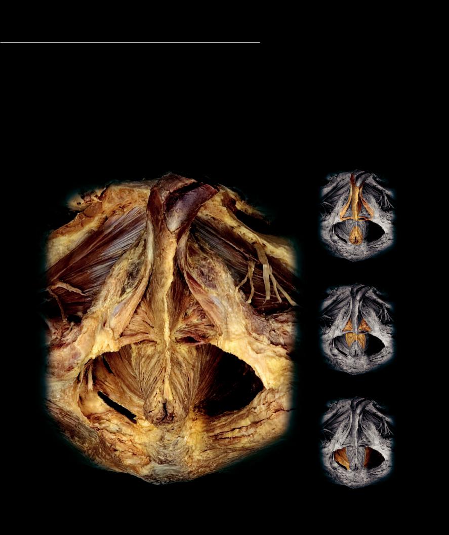
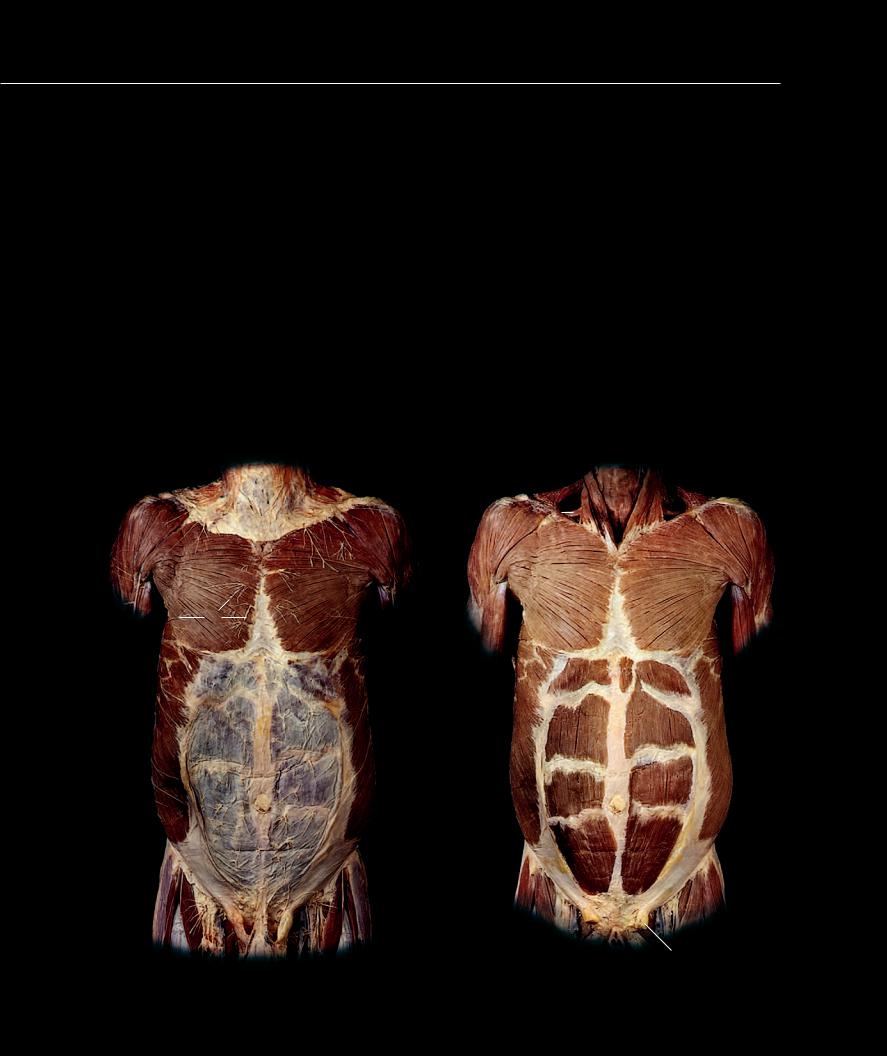
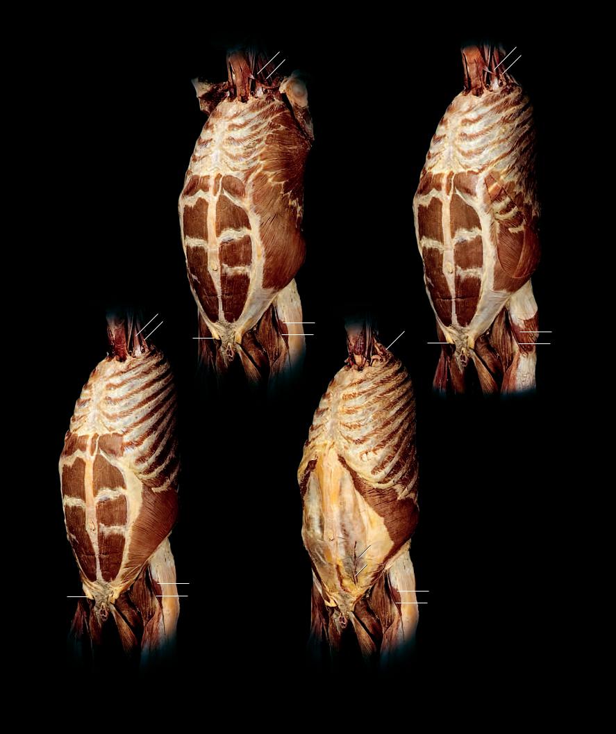
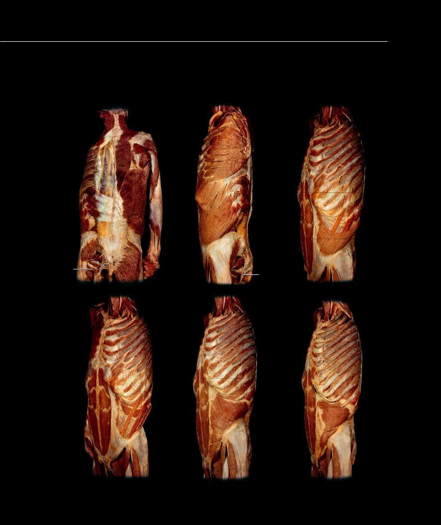
The ventral, subvertebral, and lateral supracostal muscles are either annexed by the lower limb or terminate above the pelvic region of the
trunk. Therefore, the three inner layers of the lateral wall become the major contributors to the pelvic hypaxial wall. The three muscle layers from each side pass into the bottom of the pelvis where they meet in the midline to surround the urethra, vagina, and anus. This three-layered muscle fl oor at the bottom of the pelvis is called the pelvic diaphragm (internal layer) and the perineum (middle and external layers.) The pelvic diaphragm forms a basin-shaped fl oor that supports the pelvic viscera. The perineal muscles span the diamond-shaped pelvic outlet, and are divided into an anterior urogenital triangle and a posterior anal triangle. The perineal muscles support the pelvic viscera, form important sphincter muscles that surround the urethral and anal orifi ces, assist in erectile function, and propel the sperm from the male penis during ejaculation. Additional views of these muscles in both the male and female are depicted in the reproductive system chapter.
Perineal Musculature |
6 |
Deep external anal sphincter muscle |
Other Muscles and Structures |
||
1 |
Obturator externus muscle |
7 |
Deep transverse perinei muscle |
10 |
Gluteus maximus muscle |
2 |
Ischiocavernosus muscle |
8 |
Levator ani muscle |
11 |
Penis (cut) |
3 |
Bulbospongiosus muscle |
9 |
Ischiococcygeus muscle |
12 |
Obturator nerve |
4 |
Superficial transverse perinei muscle |
|
|
13 |
Ischial tuberosity |
5 |
Superficial external anal sphincter muscle |
|
|
14 |
Coccyx |
|
|
|
|
15 |
Perineal body |
11
12
1 |
12 |
|
External perineal muscles
2
|
3 |
|
|
7 |
|
|
4 |
|
|
15 |
|
13 |
6 |
|
|
8 |
|
|
5 |
Middle perineal muscles |
9
14
10
Dissection of male perineal muscles |
Internal perineal muscles |
|
Inferior view |
||
|
174
11 Upper Limb Muscles
While the majority of the muscles of the upper limb arise as true limb muscles from the embryonic somites, some of the upper limb muscles are annexed from the body wall and head musculature to support and stabilize the scapula and suspend it from the trunk skeleton. The levator
scapulae, rhomboideus major and minor, serratus anterior, pectoralis minor, and subclavius muscles are annexed lateral body wall muscles that help suspend the scapula, while the trapezius is an annexed branchial arch muscle that is also a part of the scapular group. Unlike these annexed body wall and head muscles, the true muscles of the limb arise from mesenchymal migrations of the somites into the developing limb
bud. These migrations form two distinct muscle masses in the limb, an anterior muscle group and a posterior muscle group. As the limb develops, the two distinct muscle groups become separated by connective tissue septa and bones into anterior and posterior muscle compartments within the different sections of the limb. As the ventral rami of the associated spinal nerves grow into the developing upper limb bud, a nerve network, or plexus, develops. From this plexus posterior divisions of the network send branches into the posterior muscle compartments and anterior divisions of the network send branches into the anterior muscle compartments. At the proximal end of the limb, some of the true limb muscles from the anterior and posterior compartments increase in size and migrate back onto the trunk. As they spread onto the trunk, they cover the body wall muscles and attach to the axial skeleton. This muscular expansion of the proximal limb muscles increases their mechanical advantage at the shoulder joint. Because of this interesting arrangement of body wall muscles and true limb muscles at the shoulder end of the
superior limb, a clear compartment organization is not evident. For this reason, we will group these muscles into groups that share some common feature, such as a common attachment or function. In the limb
proper we group the muscles into their developmental anterior and posterior muscle compartments. This greatly simplifies the learning process
because most of the muscles in a compartment share common attachments, actions, and nerves. Grouping things in this way can help to simplify the learning process.
Find more information about the muscles of the upper limb in
R E A L A N AT O M Y
175
This chapter depicts the interesting array of muscles of the upper limb. Because of its weak ligamentous association with the axial skeleton, the upper limb annexed muscles from the
outer layer of the trunk wall and head to help suspend it from the axial skeleton. This scapular muscle sling, which has no homologous counterpart in the lower limb, is the major difference between the muscles of the upper and lower limbs. On the pages that follow we present the muscles of the upper limb and organize them primarily by developmental groups, with the exception of the muscles of the shoulder joint (see the outline below). The opposite page and the two pages that follow show anterior and posterior views of the upper limb muscles and their relationships to the trunk musculature.
Pectoral Girdle Muscles
(Annexed from head muscles (trapezius) and outermost layer of lateral trunk muscles to support and stabilize scapula)
Trapezius Levator scapulae
Rhomboideus major Rhomboiedus minor Serratus anterior Pectoralis minor Subclavius
Shoulder Joint Muscles Rotator cuff muscles
(Muscles with a ligamentous role that function as stabilizers of the weakly ligamentous shoulder joint)
Supraspinatus
Infraspinatus Teres minor Subscapularis
Intertubercular groove muscles
(Muscles that share an insertion on the intertubercular groove and are prime movers of the shoulder joint)
Pectoralis major Latissimus dorsi Teres major
Deltoid
Anterior Brachial Muscles
(Nerve supply - musculocutaneous nerve; function as flexors of the shoulder and elbow)
Coracobrachialis Brachialis Biceps brachii
Posterior Brachial Muscles
(Nerve supply - radial nerve, like all posterior compartment muscles; functions as extensor of shoulder and elbow)
Triceps brachii
Anterior Antebrachial Muscles
(Nerve supply - median and ulnar nerves; function as flexors of wrist and digits)
Superficial muscles Pronator teres Flexor carpi radialis Palmaris longus Flexor carpi ulnaris
Flexor digitorum superficialis Deep muscles
Flexor digitorum profundus Flexor pollicis longus Pronator quadratus
Posterior Antebrachial Muscles
(Nerve supply - radial nerve; function as extensors of the wrist and digits)
Lateral muscles Brachioradialis
Extensor carpi radialis longus Extensor carpi radialis brevis Extensor digitorum
Extensor digiti minimi Extensor carpi ulnaris Anconeus
Supinator Radial muscles
Abductor pollicis longus
Extensor pollicis longus Extensor pollicis brevis Extensor indicis
Hand Muscles
(All intrinsic hand muscles arise from anterior muscles of embryonic limb and are innervated by the median and ulnar nerve from the anterior divisions of the plexus)
Thenar Muscles
(All supplied by the median nerve except adductor pollicis) Abductor pollicis brevis
Flexor pollicis brevis Opponens pollicis Adductor pollicis
Hypothenar Muscles
(All supplied by the ulnar nerve) Palmaris brevis
Abductor digiti minimi Flexor digiti minimi Opponens digiti minimi
Intermetacarpal Muscles
(All supplied by the ulnar nerve except first two lumbricals) Lumbricales
Palmar interossei Dorsal interossei
176
1 |
Trapezius |
|
|
2 |
Deltoid |
|
|
3 |
Pectoralis major |
|
|
4 |
Biceps brachii |
|
|
5 |
Triceps brachii |
|
|
6 |
Serratus anterior |
11 |
|
7 |
Anterior antebrachial muscles |
|
|
8 |
Posterior antebrachial muscles |
|
|
9 |
Thenar muscles |
|
|
10 |
Hypothenar muscles |
13 |
|
11 |
Temporalis |
||
12 |
|||
12 |
Masseter |
13Muscles of facial expression
14Sternocleidomastoid
15Cervical body wall muscles
16Rectus abdominis
17External oblique
18 |
Anterior thigh muscles |
1 |
|
19 |
Medial thigh muscles |
15 |
14 |
|
20Linea alba
21Tendinous intersection
22 |
Spermatic cord (cut) |
2 |
23 |
Penis (cut) |
|
24 |
Inguinal ligament |
|
25 |
Umbilicus |
3 |
26 |
Palmar aponeurosis |
5
46
8 |
16 |
20 |
|
21 |
17 |
7 |
|
|
25 |
10 |
24 |
9
22
26
23
19
18
Muscles of the upper limb
Anterior view
177
Upper Limb Muscles
Upper Limb Muscles |
|
|
|
1 |
Trapezius |
|
15 |
2 |
Deltoid |
|
|
|
|
||
3 |
Pectoralis major |
|
|
4 |
Biceps brachii |
|
|
5 |
Triceps brachii |
|
|
6 |
Serratus anterior |
|
15 |
7 |
Teres major |
|
|
8 |
Infraspinatus |
|
16 |
9 |
Teres minor |
|
17 |
10 |
Latissimus dorsi |
|
|
|
|
||
11 |
Posterior antebrachial muscles |
|
|
12 |
Anterior antebrachial muscles |
|
1 |
13 |
Hypothenar muscles |
|
|
|
|
||
14 |
Intermetacarpal muscle |
|
|
Other Muscles and Structures |
|
|
|
15 |
Muscles of mastication |
|
2 |
16 |
Muscles of facial expression |
|
|
17 |
Sternocleidomastoid |
8 |
|
18 |
Rectus abdominis |
|
|
|
|
||
19 |
External oblique |
7 |
|
20 |
Gluteal muscles |
3 |
|
|
|
|
21Posterior thigh muscles
22Thoracolumbar fascia
23Antebrachial fascia
24Iliotibial tract
5 4
6
23
19
18
11
20
14
Muscles of the upper limb
Right lateral view
178
16
17
1
2
9 8
7
5
10
23
11 |
22 |
|
|
|
12 |
13
20
24 21
Muscles of the upper limb
Posterior view
179
The muscles that insert on the scapula and anchor it to the trunk form an extensive muscular sling. During development the upper limb annexes these
muscles from the head and trunk wall. They share the common functional goal of moving the scapula, stabilizing it, and anchoring it to the axial skeleton. These muscles are some of the larger muscles of the upper limb, yet produce visibly minor movements of the skeleton. Realize, however, that their major role is to stabilize and anchor the scapula to the axial skeleton. With the exception of the pectoralis minor, the nerves that supply these muscles arise from the roots of the brachial plexus.
Scapular Musles
1 Trapezius
2 Levator scapulae
3 Rhomboideus minor
4 Rhomboideus major
5 Serratus anterior
6 Pectoralis minor
7 Subclavius
Other Muscles and Structures
8 Sternocleidomastoid
9 Omohyoid
10Clavicle
11Deltoid
12Coracobrachialis
13Pectoralis major (cut)
14External intercostal
15Internal intercostal
16Biceps brachii
17Brachialis
18Triceps brachii
19Latissimus dorsi
20Supraspinatus
21Infraspinatus
22Teres major
23External oblique
24Rectus abdominis
25Brachioradialis
26Extensor carpi radialis longus
27Serratus posteror inferior
28Teres minor
29External oblique aponeurosis
30Trachea
31Spine of scapula
32Greater tubercle of humerus
33Rib
9 |
8 |
|
|
|
10 |
7
11
14
12
6
5
18 |
16 |
19
17
23
25
26
Muscles of right brachium, shoulder, and chest
30
13
15
29
24
Anterior view
180
8
1
11
21
4
22
18
19
2
320
31
32
21 228
4
22
18
5
33
33
33
27
Muscles of neck, shoulder, and back
Posterior view
181