Добавил:
kiopkiopkiop18@yandex.ru Вовсе не секретарь, но почту проверяю Опубликованный материал нарушает ваши авторские права? Сообщите нам.
Вуз: Предмет: Файл:
Скачиваний:
8
Добавлен:
03.02.2024
Размер:
76.04 Mб
Скачать
Anterior Brachial Muscles

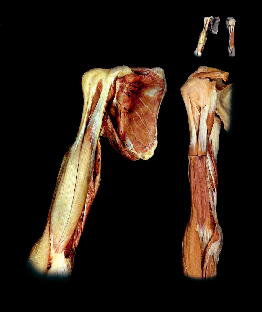
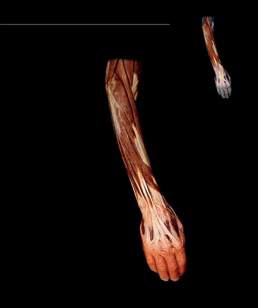
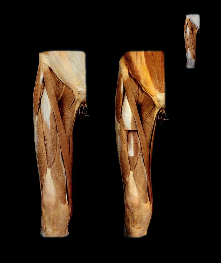
The anterior muscle compartment of the brachium consists of

three muscles — the coracobrachialis, brachialis, and biceps brachii. The coracobrachialis and brachialis each cross a single joint, the shoulder joint and elbow joint respectively. The biceps brachii crosses three joints, the shoulder, and the humero-ulnar and radio-ulnar joints of the elbow. The muscles share in common the actions of fl exion of the shoulder and elbow. All three muscles are innervated by the musculocutaneous nerve.

Anterior Brachial Muscles


1

Coracobrachialis

11

6

12

2

Brachialis


3

Biceps brachii - long head

14


4

Biceps brachii - short head


5

Triceps brachii


Other Muscles and Structures


14


15

6

Supraspinatus


7

Subscapularis

1


8

Teres major


9

Brachioradialis


10

Pronator teres


7

11

Coracoid process


12

Superior angle


1

13Inferior angle

14Greater tubercle

15Lesser tubercle

8

3 4

13

34

2

5

10

9

9

Muscles of the right brachium and scapula

Deep muscles of the right brachium

Anterior view

Anterior view


186

Posterior Brachial Muscles

The three headed triceps brachii muscle is the sole muscle of the posterior

compartment of the brachium. This large muscle extends the shoulder and elbow joints and is innervated by the radial nerve.

Posterior Brachial Muscles


1

Triceps brachii - medial head

7


2

Triceps brachii - lateral head

13

8

3

Triceps brachii - long head


4

Biceps brachii - long head


5

Beceps brachii - short head


12

6

Brachialis


9


8

Other Muscles and Structures


7

Supraspinatus


8

Infraspinatus


9

Teres minor


10

Teres major


11

11

Humerus


12Greater tubercle

13Spine of scapula

14 Brachail artery

10


2

3

4

5

6

11

14

1

1

2

3

Transverse section of right midbrachim

Muscles of the right brachium and scapula

Inferior view

Posterior view

187

Anterior Antebrachial Muscles

The muscles of the anterior antebrachium form three

distinct muscle layers. The superficial group has four superfi cial muscles (pronator teres, fl exor carpi radialis, palmaris longus, and fl exor carpi ulnaris) covering the intermediate fl exor digitorum superfi cialis. All fi ve of these muscles share a common attachment on the medial epicondyle of the humerus. The three deep muscles (fl exor digitorum profundus, fl exor pollicis longus, and pronator quadratus) do not cross the elbow joint. Other than the two pronators, all the muscles are fl exors of either the wrist or digits. The median nerve innervates all but the fl exor carpi ulnaris and the ulnar half of the fl exor digitorum profundus, both of which are supplied by the ulnar nerve.

Anterior Antebrachial Muscles 1 Pronator teres

2 Flexor carpi radialis

3 Palmaris longus

4 Flexor carpi ulnaris

5 Flexor digitorum superficialis

6 Flexor digitorum profundus

7 Flexor pollicis longus

8 Pronator quadratus

Other Muscles and Structures

9 Brachialis

10Palmar aponeurosis

11Brachial artery

12Radial artery

13Ulnar artery

14Anterior interosseous artery

15Interosseous membrane

16Abductor pollicis brevis

17Flexor pollicis brevis

18Lumbricals

19Adductor pollicis

20Flexor digiti minimi brevis

21Abductor digiti minimi

22Palmaris brevis

23Supinator

24Superficial transverse metacarpal ligament

1

3

25

4

7

8

24

16

22

17 20

10

Superficial muscles of the right antebrachium

Anterior view, hand pronated

188

9

11

23

12

13

6

7

4

2

5


21

16


20

17


1918

18

18

18


Deep muscles of the right antebrachium

Anterior view, superficial muscles removed and hand pronated

9

11

23

12

13

14

15

8

2 4

6

5


21

16


20

17


19


18

18

18

18


Deep muscles of the right antebrachium

Anterior view, muscles removed to expose pronator quadratus

189

Posterior Antebrachial Muscles

There are two muscle groups in the posterior

antebrachium — the eight muscles of the lateral group that share a common attachment on or near the lateral epicondyle of the humerus and the four muscles of the radial group that course along the distal aspect of the radius to insert on the thumb and fi rst fi nger. Like the triceps of the posterior brachial compartment, all the muscles of the posterior antebrachium receive innervation via the radial nerve. With a few exceptions, the muscles are extensors of either the elbow, wrist, or digits.

Posterior Antebrachial Muscles 1 Brachioradialis

2 Anconeus

3 Supinator

4 Extensor carpi radialis longus

5 Extensor carpi radialis brevis

6 Extensor digitorum

7 Extensor digiti minimi

8 Extensor carpi ulnaris

9 Abductor pollicis longus

10Extensor pollicis longus

11Extensor pollicis brevis

12Extensor indicis

Other Muscles and Structures

13Biceps brachii

14Brachialis

15Triceps brachii

16Flexor carpi radialis

17Pronator teres

18Flexor pollicis longus

19Abductor digiti minimi

20Dorsal interossei

15 14

13

1

2

4

5

6

9

7

11

8

10

20 20

19

20

Superficial muscles of the right antebrachium

Posterior view, hand pronated

190

15

13

14

3

17

9

10 11

12

19

20 20

20 20

13

3

17

16

18

9

11

10

20

Deep muscles of the right antebrachium

Deep muscles of the right antebrachium

Posterior view, lateral group muscles removed and hand pronated

Anterolateral view, lateral group muscles removed and hand pronated

191

Hand Muscles

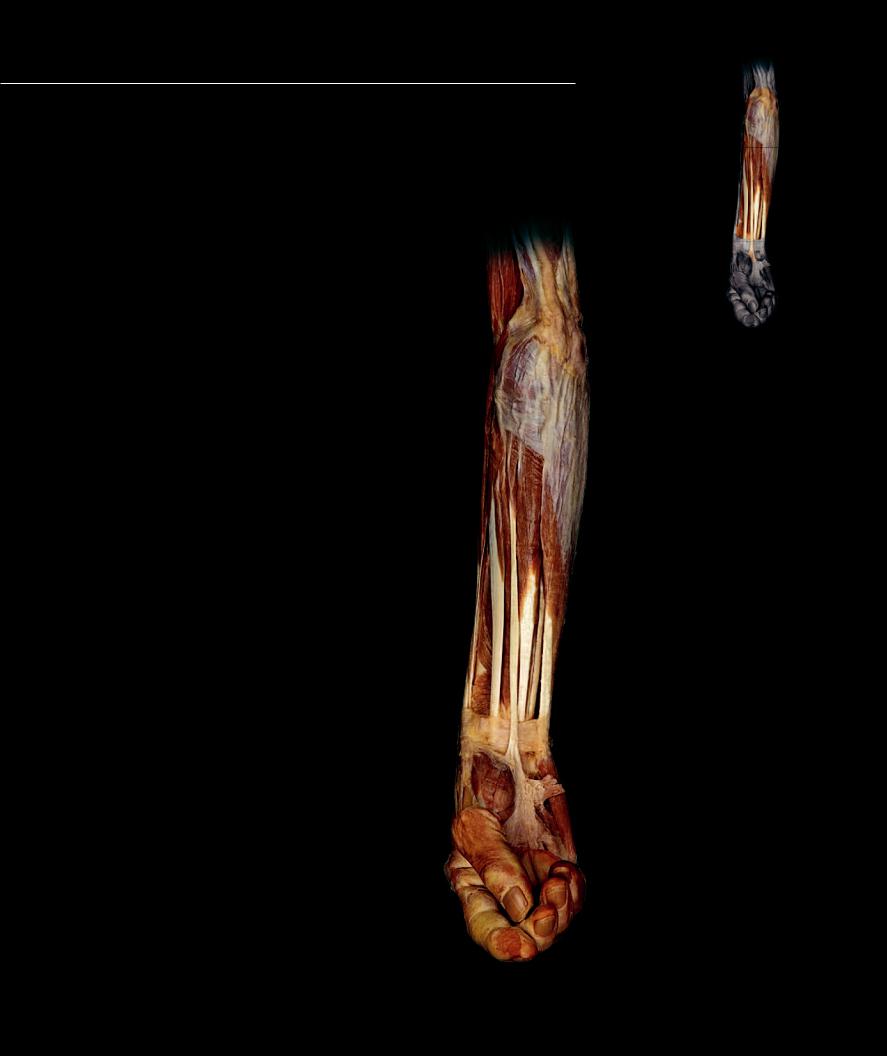
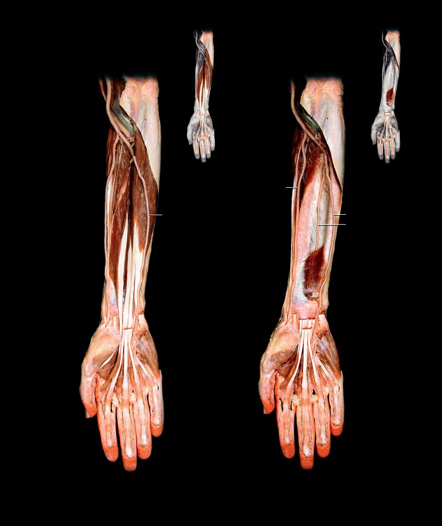
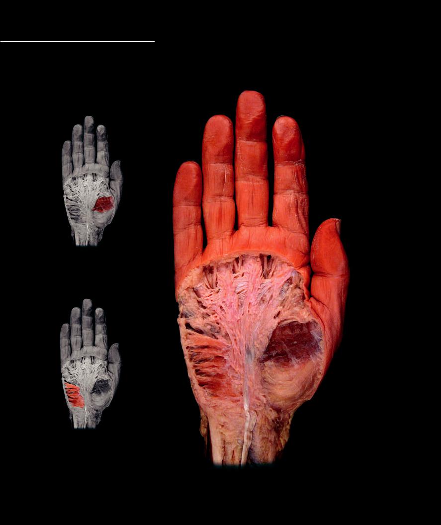
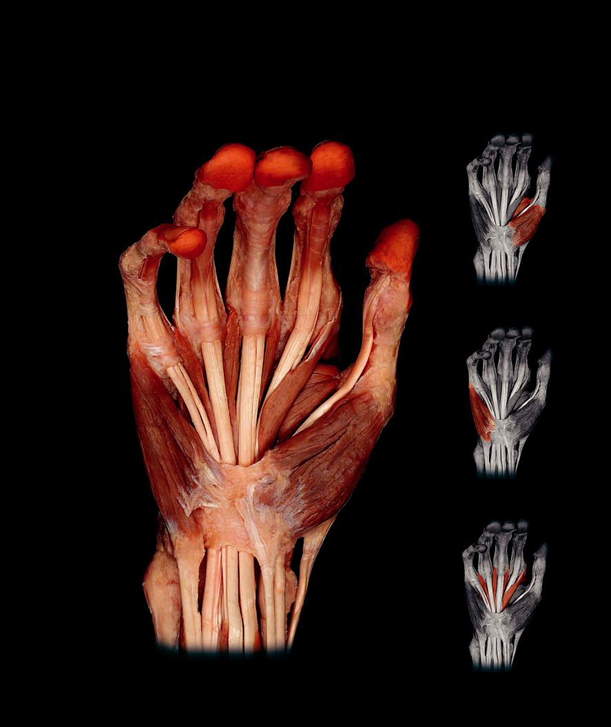
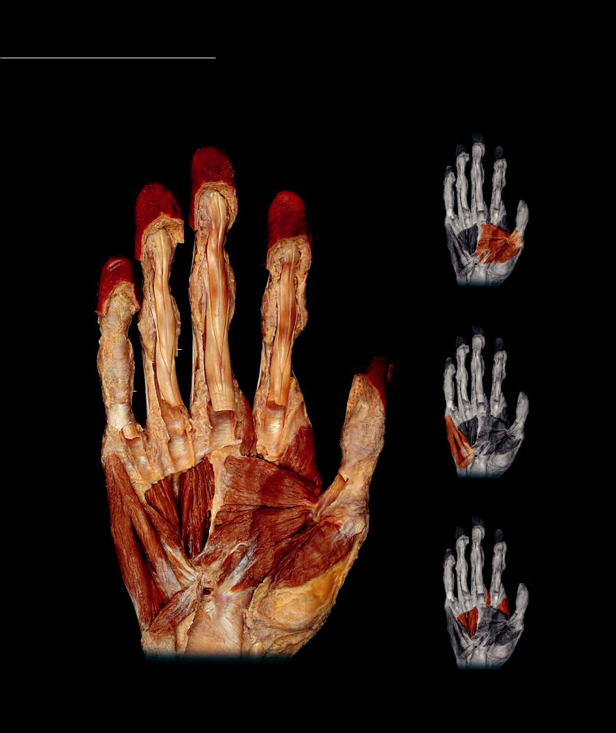
There are three muscle groups in the hand — the muscles of the thenar eminence at the base of the thumb, the muscles of the hypothenar eminence at the base of the little fi nger, and the three layers of intermetacarpal muscles that occupy the

spaces between the metacarpal bones. All of these muscles arise from the anterior muscles of the embryonic limb bud and receive anterior division nerve supply from the median and ulnar nerves as they pass from the anterior antebrachium into the hand. While the median nerve supplies the majority of the muscles of the anterior antebrachium, the ulnar nerve supplies all but three of the muscles in the hand.

Muscles of the thenar eminence

16

2

1

6

Muscles of the


hypothenar eminence

13

12

Superficial muscles of the right hand

Anterior view

192

Hand Muscles

7

Lumbricals

12

Palmaris longus

1

Abductor pollicis brevis

8

Palmar interossei

13

Flexor carpi ulnaris

2

Flexor pollicis brevis

9

Dorsal interossei

14

Flexor pollucis longus

3

Adductor pollicis


15

Flexor carpi radialis

4

Abductor digiti minimi

Other Muscles and Structures

16

Palmar aponeurosis

5

Flexor digiti minimi brevis

10

Flexor digitorum superficialis

17

Flexor retinaculum

6

Palmaris brevis

11

Flexor digitorum profundus

18

Ulna

11

11

Muscles of the thenar eminence


7

10


9


7


7

10


10


10

7

3

16


8


5

4

2


1

Muscles of the hypothenar eminence

17

10 10

12

18

13

15

11

Intermediate muscles of the right hand

Intermetacarpal muscles

Anterior view

193

Hand Muscles

1

Abductor pollicis brevis (cut)

10

Palmar interossei


Hand Muscles

9

Lumbricals (cut)


2

Flexor pollicis brevis (cut)

11

Dorsal interossei


3

Opponens pollicis


4

Adductor pollicis

Other Muscles and Structures


5

Abductor digiti minimi

12

Flexor digitorum superficialis


6

Flexor digiti minimi brevis

13

Flexor digitorum profundus


7

Opponens digiti minimi

14

Carpal tunnel


8

Palmaris brevis


13


13

13


9


9


12


12

12


12


11


11


11


11

10


10


4

67

5

3

2

8

1


14

Muscles of the thenar eminence

Muscles of the hypothenar eminence

Deep muscles of the right hand

Intermetacarpal muscles

Anterior view


194

12 Lower Limb Muscles

The design of the inferior limb musculature is similar to that of the true limb muscles of the superior limb. The major difference between the two limbs is that the proximal end of the lower limb forms a direct skeletal attachment to the vertebral column via the strong sacro-iliac joint, unlike the unattached scapula of the superior limb. Because of this difference, the inferior limb does not require body wall muscles to support, stabilize, and suspend it from the axial skeleton. There are two additional features that are important to keep in mind when studying this powerful locomotor limb. First, during development of the lower limb the embryonic posterior muscles rotate and reposition themselves to the anterior aspect of the limb. For this reason the knee and ankle move directly opposite the elbow and wrist. The second notable feature is that there are three muscle compartments in the thigh and leg, as compared

to just two in the brachium and antebrachium. One of the two original compartments in each lower limb segment (thigh and leg) splits to give rise to an additional compartment. The thigh has an anterior compartment and a posterior compartment, but the posterior compartment is subdivided into posterior and medial compartments. The leg has a large posterior compartment and a smaller anterior compartment and the anterior compartment is subdivided into anterior and lateral compartments. As with the upper limb, we present the muscles of the lower limb proper in their muscle compartments. Again, this greatly simplifies the learning process because most of the muscles in a compartment share simi-

lar attachments, perform common actions, and have a common nerve supply. Unlike the compartmental muscles of the lower limb proper, the proximal muscles of the lower limb that surround the hip joint are a more diverse group of muscles. Some are true limb muscles, while others are annexed muscles from the trunk wall. We organize these hip muscles into three groups — the deep hip rotator muscles, the gluteal muscles, and the hip flexors.

Find more information about the muscles of the lower limb in

R E A L A N AT O M Y

195

Lower Limb Muscles

The muscles of the lower limb share similarities with their upper limb counterparts, yet have important differences. As you will notice in the groups below there are no homologues in

the lower limb to the scapular muscles of the upper limb. Like the shoulder muscles, the muscles surrounding the hip joint are a varied group of muscles, with some annexed from the body wall of the abdominopelvic region. In the limb proper the muscles develop in muscular compartments as they do in the upper limb; however, the embryonic posterior aspect of the limb rotates to an anterior position. As a result, the nerves that arise from the posterior divisions of the lumbosacral plexus innervate the anterior muscle compartments, and the nerves from the anterior divisions of the plexus innervate the posterior muscle compartments. The developmental groups of muscles and their nerve supply are outlined below.

Hip Muscles Gluteal muscles

(Nerve supply - gluteal nerves, superior to maximus and inferior to the other three; arise from lateral aspect of ilium and are prime movers and stabilizers of hip joint)

Gluteus maximus Gluteus medius Gluteus minimus Tensor fasciae latae

Deep hip rotator muscles

(All are lateral rotators of the hip joint and insert on the medial aspect of greater trochancter)

Piriformis Obturator internus Obturator externus Superior gemellus Inferior gemellus Quadratus femoris

Hip flexor muscles Psoas major Iliacus

Anterior Thigh Muscles

(Nerve supply - femoral nerve; major extensor group of the knee) Sartorius

Quadriceps femoris Rectus femoris Vastus lateralis Vastus intermedius Vastus medialis

Articularis genu

Medial Thigh Muscles

(Nerve supply - obturator nerve with exception of pectineus, which is supplied by femoral nerve and condylar head of adductor magnus, which is supplied by tibial nerve)

Pectineus Adductor brevis Adductor longus

Adductor magnus Adductor minimis Gracilis

Posterior Thigh Muscles

(Nerve supply - Tibial nerve with exception of short head of biceps femoris, which is supplied by common fibular nerve)

Biceps femoris Semitendinosus Semimembranosus

Anterior Leg Muscles

(Nerve supply - deep fibular nerve) Tibialis anterior

Extensor digitorum longus Extensor hallucis longus Peroneus tertius

Lateral Leg Muscles

(Nerve supply - superficial fibular nerve) Peroneus longus

Peroneus brevis

Posterior Leg Muscles (Nerve supply - tibial nerve)

Triceps surae Gastrocnemius Soleus

Plantaris

Popliteus Tibialis posterior

Flexor digitorum longus Flexor hallucis longus

Dorsal Foot Muscles

(Nerve supply - deep fibular nerve) Extensor hallucis brevis Extensor digitorum brevis

Plantar Foot Muscles

(Nerve supply - tibial nerve via its terminal branches, medial plantar nerve supplies first lumbrical, abductor hallucis, flexor hallucis brevis, and flexor digitorum brevis; lateral plantar nerve supplies all the others)

First layer Abductor hallucis

Flexor digitorum brevis Abductor digiti minimi

Second layer Quadratus plantae Lumbricales

Third layer

Flexor halluci brevis Adductor hallucis

Flexor digiti minimi brevis Fourth layer

Plantar interossei Dorsal interossei

196

1

Tensor fasciae latae

9

Vastus lateralis

17

Soleus

2

Iliacus

10

Vastus medialis

18

Tibialis anterior

3

Psoas major

11

Gluteus maximus

19

Fibularis longus

4

Pectineus

12

Adductor magnus

20

Fibularis brevis

5

Adductor longus

13

Biceps femoris

21

Iliotibial tract

6

Gracilis

14

Semitendinosus

22

Calcaneal tendon

7

Sartorius

15

Semimembranosus

23

Quadriceps tendon

8

Rectus femoris

16

Gastrocnemius

24

Flexor digitorum longus

2

13

4

5

67

8

9

10

23

16

17

18

24

11

16

22

12

21

13

14

15

19

17

20

Muscles of the lower limb

Muscles of the lower limb

Posterior view

Anterior view


197

Hip Muscles

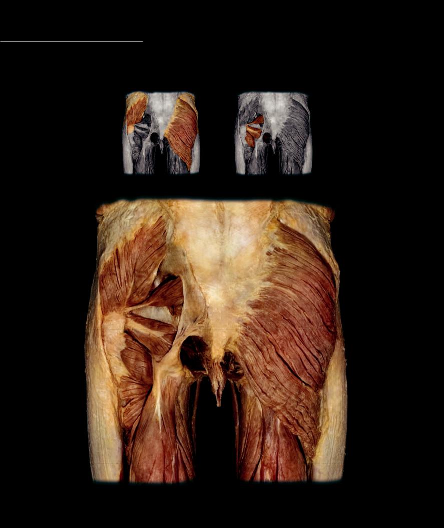
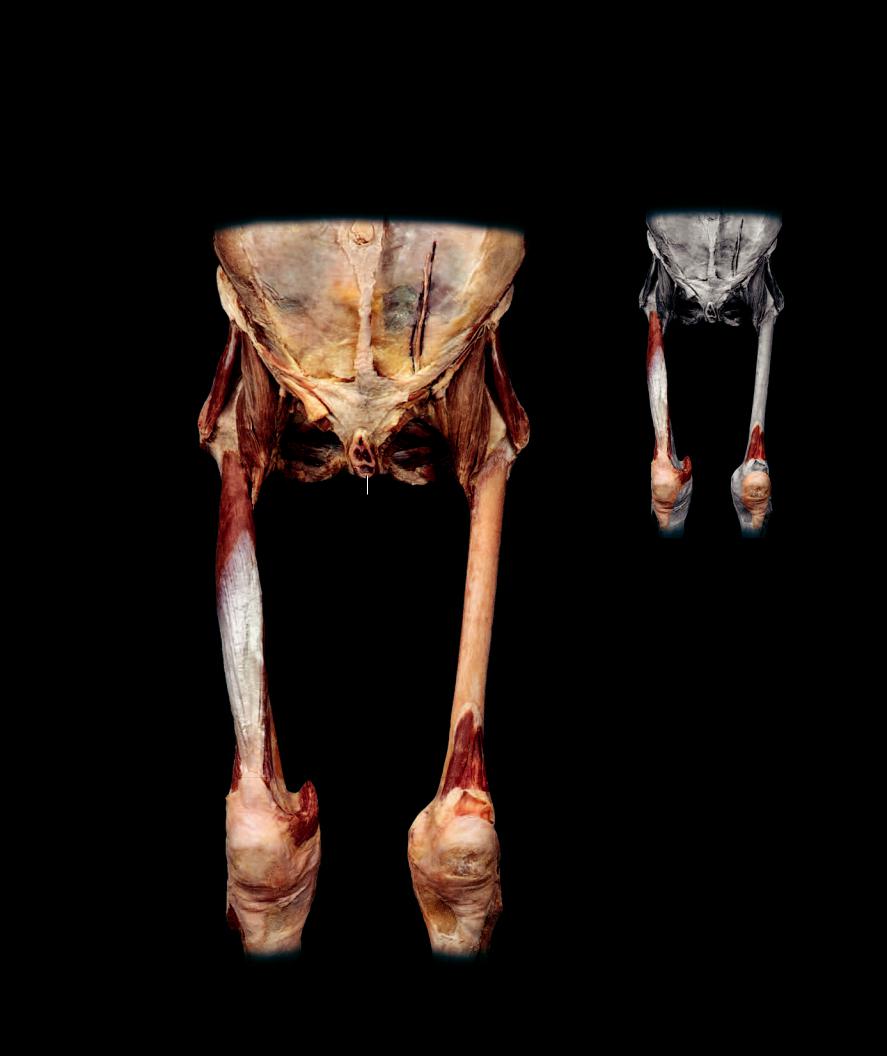
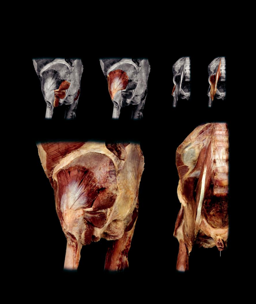
The muscles that surround the hip joint form three groups. The gluteal muscles arise from the posterior musculature of the embryonic limb bud and are prime movers of the hip joint. They create the characteristic profi le of the human buttocks. The deep hip

rotator muscles are closely associated with the body wall of the pelvic region. Five of the six muscles sit deep to the gluteal musculature on the posterior aspect of the hip joint. The hip fl exors are deep body wall muscles of the abdominal wall that have been annexed by the lower limb during development. These muscles, the psoas major and iliacus, form a pulley over the superior ramus of the pubis on their descent onto the lesser trochanter of the femur.

Gluteal muscles

Deep hip rotator muscles

228

4

5


1

6

726

8

20

7

9

17

18

16

25

13

14

15

Muscles of the gluteal region, gluteus maximus removed on left

Posterior view

198

Gluteal Muscles

7

Obturator internus

Other Muscles and Structures

21

Transversus abdominis

1

Gluteus maximus

8

Inferior gemellus

13

Biceps femoris

22

Quadratus lumborum

2

Gluteus medius

9

Quadratus femoris

14

Semitendinosus

23

Psoas minor

3

Gluteus minimis

10

Obturator externus

15

Semimembranosus

24

Pectineus (cut)

4

Tensor fasciae latae


16

Adductor magnus

25

Iliotibal tract


Hip Flexor Muscles

17

Adductor minimus

26

Sacrotuberous ligament

Deep Hip Rotator Muscles

11

Psoas major

18

Gracilis

27

Penis (cut)

5

Piriformis

12

Iliacus

19

Vastus intermedius

28

Ilium

6

Superior gemellus


20

Pelvic diaphragm

29

Femur

Deep hip rotator

Gluteal muscles

muscles


21

28

3

5

76

26

8

9

29

Muscles of gluteal region, gluteus maximus and medius removed

Posterolateral view

Gluteal muscles

Hip flexor muscles

22 23

21

11

11

12

3

24

10

9

27

19

Deep dissection of iliopsoas muscles

Anterior view

199

Anterior Thigh Muscles

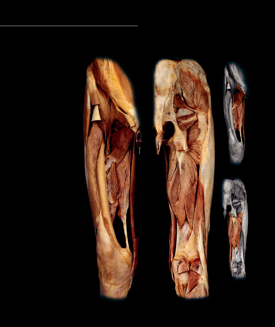
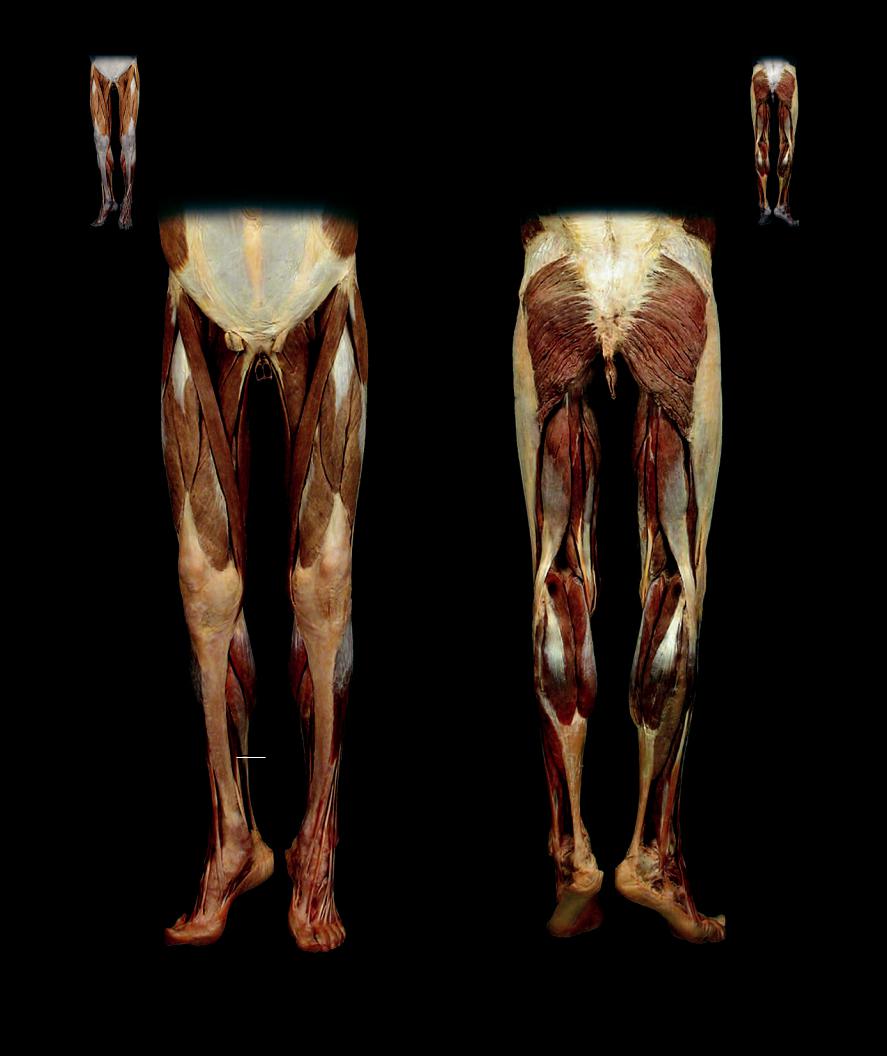
The four major muscles of the anterior compartment form the quadriceps femoris muscle group.The four muscles of this

group converge to form the strong quadriceps tendon that surrounds all but the posterior surface of the patella. As the sole extensors of the knee, the quadriceps are essential for running, jumping, and kicking. The sartorius, which is the longest muscle in the body, is a knee fl exor. The small articularis genus raises the suprapatellar bursa during extension of the knee. All of the muscles in this compartment receive their innervation via the femoral nerve from the posterior divisions of the lumbar plexus.

26


8


9


19


11


7

12


11


10


20

7


12


13


13

20


5


14


25

1


14


25


22


1


5


15


3


15


2


2

1

4

4

Muscles of the thigh

Muscles of the thigh, rectus femoris cut

Anterior view, left thigh

Anterior view. left thigh

200

Anterior Thigh Muscles

Other Muscles and Structures

13

Pectineus

20

Spermatic cord

1

Rectus femoris

7

Tensor fasciae latae

14

Adductor longus

21

Linea alba

2

Vastus lateralis

8

Transversus abdominis

15

Gracilis

22

Iliotibial tract

3

Vastus intermedius

9

Rectus abdominis

16

Gluteus minimis

23

Femur

4

Vastus medialis

10

Pyramidalis

17

Obturator externus

24

Inferior epigastric vessels

5

Sartorious

11

Iliacus

18

Quadratus femoris

25

Penis (cut)

6

Articularis genus

12

Psoas major

19

Inguinal ligament

26

Rectus sheath

21 24

16

11

20

12


17

18

25

3

23

6

4

Deep muscles of the thigh

Anterior view

201

Medial Thigh Muscles

The six muscles of the medial compartment are all capable of adducting the hip joint. The pectineus and four adductor muscles all originate from a medial position on the pubis

and ischium and project laterally to insert on the posterior surface of the femur. The gracilis muscle differs from the others in the group by crossing the knee joint in addition to the hip. It courses with the sartorius muscle as a fl exor of the knee. With the exception of the pectineus and condylar part of the adductor magnus, all the muscles are innervated by the obturator nerve, which arises from the anterior divisions of the lumbar plexus.

Medial Thigh Muscles 1 Pectineus

2 Adductor longus

3 Adductor brevis

4 Adductor magnus

5 Adductor minimis

6 Gracilis

Other Muscles and Structures 7 Sartorius

8 Iliacus

9 Psoas major

10Tensor fasciae latae

11Rectus femoris

12Obturator externus

13Vastus lateralis

14Articularis genus

15Gluteus medius

16Piriformis

17Superior gemellus

18Obturator internus

19Inferior gemellus

20Quadratus femoris

21Biceps femoris (short head)

22Gastrocnemius

23Plantaris

24Soleus

25Pelvic diaphragm

26Transversus abdominis

27Rectus abdominis

28Spermatic cord

29Sacrotuberous ligament

30Femur

31Penis (cut)

202


26


27


7


10

8

9

11


1


28


12


2


1

3


3121

13

4

2

3020

6

14

7

Dissection of medial thigh muscles

Anterior view, right thigh

15

16

17 18

29

25 19

18

20

5

6

4

21

7

22

23

24

Dissection of medial thigh muscles

Posterior view, right thigh