

Extending from the brainstem is a long slender rod of nerve tissue, the spinal cord. The cord exits the foramen magnum of the skull and descends within the vertebral canal of the boney vertebral column. It is about 45 cm long (18 inches) and ends between the fi rst
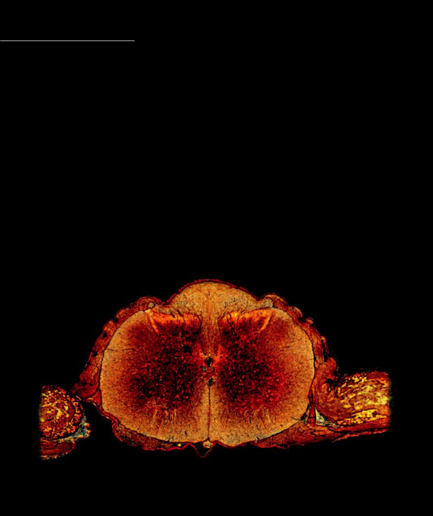
and second lumbar vertebrae. Although there are some slight regional variations, the cross-sectional anatomy of the spinal cord is generally the same throughout its length. The gray matter of the spinal cord forms a butterfl y-shaped region in the center of the cord that is surrounded by the white matter. As is the theme throughout the central nervous system, gray matter consists primarily of neuronal cell bodies and their dendrites, short interneurons, and glial cells. The white matter is organized into tracts, which are bundles of myelinated nerve fi bers (axons of long interneurons and sensory neurons) that communicate between the gray circuit centers at all levels of the spinal cord and brain.
Each side of the H-shaped gray matter of the spinal cord has a dorsal horn and a ventral horn sandwiching an intermediate gray region. Entering the dorsal horns from the dorsal rootlets are the axons of the afferent neurons, which synapse with small interneuron pools to form segmental integration centers for that level of the body. The dorsal horn and intermediate gray matter contain numerous small interneurons. The intermediate gray also contains, at certain levels, the preganglionic efferent neurons of the autonomic output. The ventral horns are primarily populated by the efferent neurons to the skeletal muscles of their respective spinal levels. The white matter tracts are grouped into columns of myelinated axons that extend the length of the cord. Each of these tracts begins or ends within a particular area of the cord and brain, and each is specific in the type of information that it transmits. Some are ascending tracts that carry signals derived from sensory input. For example, one tract carries information derived from pain and temperature receptors, whereas another carries information regarding touch. Other tracts are descending tracts that relay messages from the brain to motor neurons in the ventral horn.
Both the white and gray matter exhibit regional differences throughout the length of the spinal cord. There is relatively more white matter at the cranial end of the spinal cord than at the caudal end. Notice that the gray matter, especially the ventral horn, is the largest at lower cervical levels and at lower lumbar-upper sacral levels. These levels correspond to upper and lower limb anatomy respectively, where large amounts of muscle tissue require motor innervtion from the ventral horn motor neuron pools.
1 |
Dorsal horn of gray matter |
7 |
Central canal |
13 |
Conus medullaris |
2 |
Lateral horn of gray matter |
8 |
Dorsolateral fasciculus |
14 |
Cauda equina |
3 |
Ventral horn of gray matter |
9 |
Dorsal root of spinal nerve |
15 |
Dorsal rami of spinal nerve |
4 |
Posterior funiculus of white matter |
10 |
Dorsal root ganglion |
16 |
Cerebrum |
5 |
Lateral funiculus of white matter |
11 |
Ventral root of spinal nerve |
17 |
Cerebellum |
6 |
Anterior funiculus of white matter |
12 |
Spinal cord |
18 |
First lumbar vertebra |
|
4 |
|
8 |
|
1 |
|
2 |
5 |
7 |
9 |
|
|
3 |
10
6
11
Photomicrograph of spinal cord
50x
232

 15
15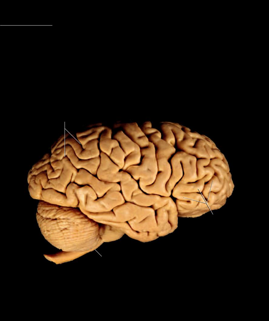
 4 13
4 13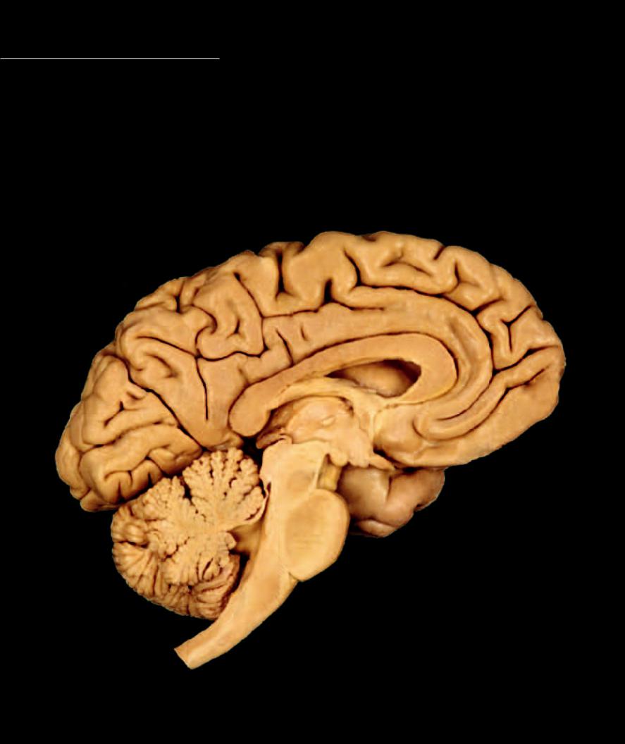
As the spinal cord ascends through the foramen magnum to enter the skull, the cranial central nervous system gradually expands in size to form the large central processing circuitry we call the brain. The increasing size of the brain results from
the addition of more and more gray processing centers to the basic cord-like brain stem. The caudal part of the brain, called the brain stem, consists of the medulla oblongata, pons, and midbrain. Though all of these structural regions exhibit their own specializations, they have certain fi ber tracts in common and all have nuclei for the cranial nerves. Added to the brain stem are the more rostral portions of the brain — the cerebellum, diencephalon, and cerebral hemispheres. These large processing centers greatly increase the size of the brain. The images on the facing page show the principal parts of the brain.
1 |
Spinal cord |
8 |
Superior colliculus |
15 |
Frontal lobe of cerebrum |
2 |
Medulla oblongata |
9 |
Thalamus of diencephalon |
16 |
Parietal lobe of cerebrum |
3 |
Pons |
10 |
Hypothalamus of diencephalon |
17 |
Occipital lobe of cerebrum |
4 |
Cerebellum |
11 |
Interthalamic adhesion |
18 |
Temporal lobe of cerebrum |
5 |
Fourth ventricle |
12 |
Pineal gland |
19 |
Corpus callosum |
6 |
Midbrain |
13 |
Mammillary body |
20 |
Lateral ventricle |
7 |
Inferior colliculus |
14 |
Optic tract |
21 |
Fornix |
16
15
|
|
19 |
|
|
|
20 |
|
|
|
21 |
|
|
|
9 |
|
|
|
11 |
|
|
12 |
10 |
|
|
8 |
||
17 |
13 |
||
6 |
|||
|
14 |
||
|
7 |
|
|
|
|
18 |
4
3
5
2
1
Sagittal section of the brain
Medial view
236
