Добавил:
kiopkiopkiop18@yandex.ru Вовсе не секретарь, но почту проверяю Опубликованный материал нарушает ваши авторские права? Сообщите нам.
Вуз: Предмет: Файл:
Скачиваний:
8
Добавлен:
03.02.2024
Размер:
76.04 Mб
Скачать

Blood In the histology chapter we learned that the fl uid material we call blood has been historically classifi ed as a connective tissue. This classifi cation was a result of the fact that, like other connective tissues, blood

has more extracellular matrix than cells. More recently, however, blood has been placed in a tissue category of its own — the hematolymphoid complex. The extracellular portion of the blood is a water solution that gives rise to its liquid nature. Blood is closely related to other aqueous fl uids within the body, in fact most of the other body fl uids, such as interstitial fl uid, lymph, cerebrospinal fl uid, and aqueous humor, arise from the blood. These extracellular fl uids are the water environment that nourish, protect, and exchange with every cell of the body. This water environment is derived from the blood, renewed by the blood, and returned to the blood. Dispersed in the blood plasma are the three groups of blood cells — erythrocytes (red blood cells), leukocytes (white blood cells), and thrombocytes (platelets). The blood smear below depicts the three cell categories.

1 Erythrocyte (red blood cell)

2 Leukocyte - neutrophil (white blood cell)

3 Leukocyte - monocyte (white blood cell)

4 Thrombocyte (platelet)

5 Blood plasma

1

2

4

2

1

5

4

1

4

1

1

1

5

4

2

3

Blood smear

700x

262

Heart From its origin in the embryo as a simple pumping tube, the heart develops into a strong fi bromuscular organ. During its development the original tubular pump is folded and subdivided into a four chambered organ that has a pyramidal or conical form. It is approximately the size of a closed fi st and weighs

approximately 300 grams in males and a little less than this in females. For its small size, comprising only one half of one percent of the total body mass, it is an important and functionally amazing organ. The wall of the heart consists of three structural layers that each play significant roles in its function as an effi cient pump. While the tissue makeup of this wall is similar at any location in the heart, the thickness can vary considerably. Internally a septum and series of valves divide the heart into four chambers through which the blood moves in a unidirectional flow. The chambers differ in structure and function, which is primarily reflected in the anatomy of their walls. Embedded within the walls of heart is a special electrical conduction system that helps regulate its coordinated pumping action.

1

Right atrium

18

Right coronary artery


2

Left atrium

19

Conus arteriosus branch


3

Right ventricle

20

Marginal branch


4

Left ventricle

21

Anterior interventricular artery


5

Right auricle

22

Lateral branches


10

6

Left auricle

23

Circumflex branch

8

9


7

Aorta

24

Posterior interventricular artery


8

Brachiocephalic artery

25

Anterior cardiac vein


9

Left common carotid artery

26

Great cardiac vein


10

Left subclavian artery

27

Posterior vein of left ventricle


11

Pulmonary trunk

28

Middle cardiac vein


12

Right pulmonary artery

29

Small cardiac vein

15


13

Left pulmonary artery

30

Right superior pulmonary vein


14


14

Ligamentum arteriosum

31

Right inferior pulmonary vein


15

Superior vena cava

32

Left superior pulmonary vein


13

16

Inferior vena cava

33

Left inferior pulmonary vein


17 Coronary sinus


7


11

32


10


33


8


9


31


15

5


19


6


14


7


20

21


1

18


26

22


12


3


13


25


30


32


20


31


4


33


2


25


1


235


17

16


27


Heart


Anterior view

4


29

24


28


3


Heart

Posterior view

263

Heart

1

Parietal pericardium


26


2

Fibrous pericardium


3

Visceral pericardium


4

Epicardium


5

Myocardium


30

3


6

Endocardium


2


7

Right atrium


1

8

Right auricle


9

Interatrial septum

41

8


10

Fossa ovalis


11

Crista terminalis


13


12

Valve of inferior vena cava


11


13

Pectinate muscle


9


14

Tricuspid valve


15

Chordae tendineae


10


16


14

16

Trabeculae carnae


17

Papillary muscle


23

18

Right ventricle


12

15


19

Pulmonary valve


17


20Left atrium

21Left auricle

22Bicuspid valve

23Left ventricle

24 Aortic valve

40

25Apex

26Aorta

27Brachiocephalic artery

28

Left common carotid artery

Dissection of heart and pericardial sac

29

Left subclavian artery

Anterolateral view

30

Pulmonary trunk


31

Left pulmonary artery


32

Ligamentum arteriosum

35

33

Anterior interventricular artery

34Lateral branches of interventricular artery

35Superior vena cava

36Right coronary artery

37Left coronary artery

38

Right pulmonary veins

26

39

Left pulmonary veins


40

Diaphragm

38

41

Lung


13 8


16


7


6


4


36


5

15


14


18


23


17


15

29

27 28


32


31


30


39

19

21


33


34


23

16

25

Transverse section of heart comparing ventricle thickness

Dissected heart showing interior of chambers

Inferior view, left ventricle at right

Anterior view

264

24

19

36

Aortic valve


Superior view

13 37

24

13

20

14

7

9

22

Heart dissection with atria and arteries removed

Superior view, anterior at top

14

15

18

16

17

Dissection of heart revealing tricuspid valve

Anterior view

265

Blood Vessels

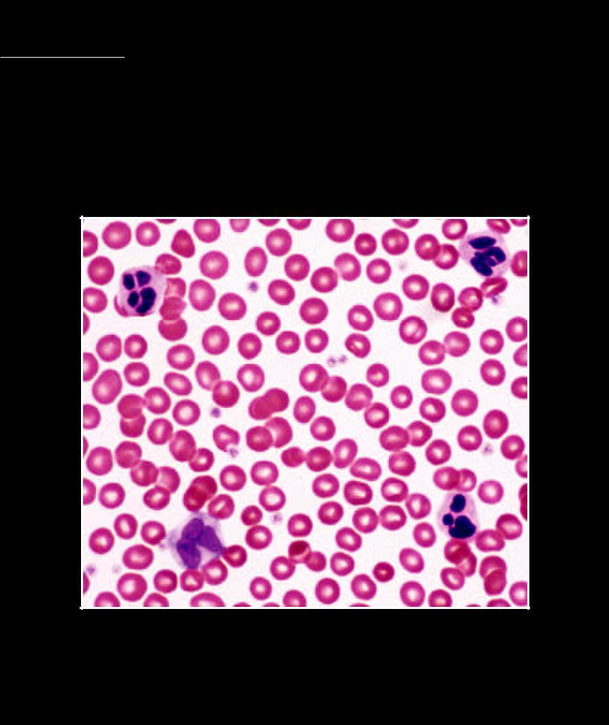
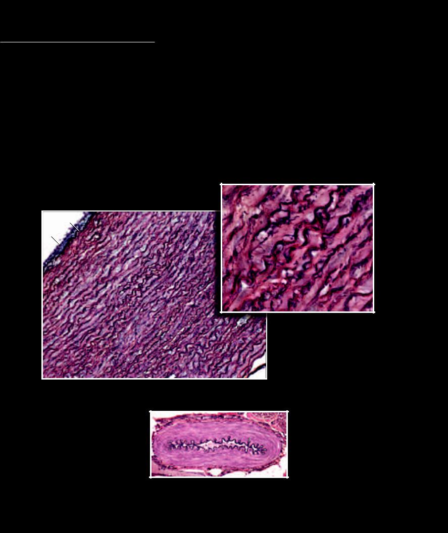
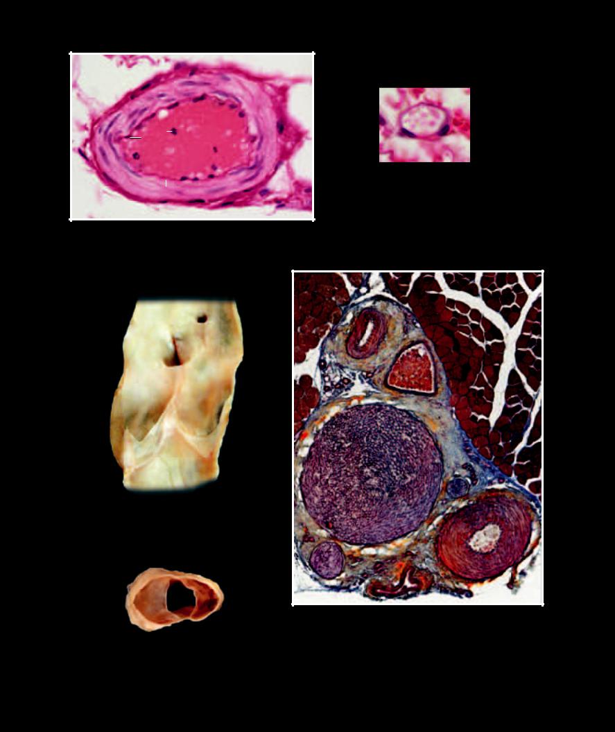
Like all tubes in the body, blood vessels have a basic pattern of design that involves three structural tunics, or layers. The inner layer of the vessel is the tunica intima. This consists of the luminal endothelium and a thin network of underlying elastic

connective tissue. The middle layer of the vessel is the tunica media, which consists of varying amounts of smooth muscle and elastic connective tissue. Variations in the tunica media defi ne the different types of blood vessels. The outer layer, the tunica externa, is a dense connective tissue outer coat. The designations — elastic arteries, muscular arteries, arterioles, venules, and veins — are based on size differences and the differences in the vessels’ tunica media. Elastic arteries have a thick elastic tunica media. Muscular arteries have a tunica media dominated by smooth muscle. Arterioles are tiny arteries with a muscular tunica media. All the venous vessels have a thin, almost non-existent tunica media. The smallest blood vessels, the capillaries, loose all the layers of their wall except the inner endothelium. These microscopic, thin walled tubes become the exchange vessels of the system.

1

Endothelium of tunica intima

6

Red blood cells

2

Internal elastic membrane of tunica intima

7

White blood cells

3

Elastic lamellae of tunica media

8

Venous valves

4

Smooth muscle cells of tunica media

9

Nerve

5

Connective tissue of tunica externa

10

Striated skeletal muscle

3

2

1

3


3

3

3

Elastic lamellae of aorta

640x

Section of aorta — large elastic artery

100x

5

42

Muscular artery

100x

266

5

4

1

6

7

1

1

6

Capillary

1000x

2

Arteriole

500x

88

Longitudinal section of vein showing valves

Anterior view

8

Transverse section of vein showing valves

Superior view

10

54

10

6

9

9

4

9

5

Neurovascular bundle — note thin-walled vein filled with red blood cells (6) compared to thick-walled muscular arteries (4)

100x

267

Pulmonary Circuit

The vascular system consists of two long circular loops of continuous branched tubing that each begin and end with the heart. Leaving the right ventricle and returning to the left atrium is the smaller pulmonary

circulation. This circular loop courses through the lung tissues where its smallest vessels form an extensive interface with the small air sacs of the lungs. This important interface is the site of exchange of O2 and CO2 between the blood and air.

1

Heart

16

Superior vena cava

23


2

Pulmonary trunk

17

Inferior vena cava


3

Right pulmonary artery

18

Trachea


4

Left pulmonary artery

19

Right principal bronchus


5

Right superior pulmonary vein

20

Left principal bronchus

13 12

6

Right inferior pulmonary vein

21

Esophagus

7

Left superior pulmonary vein

22

Thyroid gland


8

Left inferior pulmonary vein

23

Vagus nerve


9

Aorta

24

Pulmonary plexus


10

Right coronary artery

25

Posterior vagal trunk


11

Left coronary artery

26

Esophageal plexus


12

Right common carotid artery

27

Anterior vagal trunk


13

Right subclavian artery

28

Anterior scalene muscle


14

Left common carotid artery

29

Cricothyroid muscle

3

15

Left subclavian artery


6

25

17

18

19 20

7

4

3

5

11

2

9

16

10

1


Dissections of pulmonary trunk, arteries, and veins

Anterosuperior view below, anterior view above

268

29

23

22

28

14

18

15

9

4

20

24

21 8

26

27

Systemic Circuit

The left ventricle pumps blood into the much larger systemic circulation, which is distributed throughout all the body’s tissues. Unlike the smaller pulmonary

circuit, the extensive systemic circuit serves a multitude of functions before returning to the right atrium:

(1) it distributes the necessary nutrients and other supplies to all the body cells while removing their metabolic wastes; (2) it acquires metabolic fuel through the lining of the digestive system to distribute throughout the body; (3) it expels wastes and excess water and adjusts the body’s electrolyte composition through its association with the tubes of the kidney; (4) it distributes generated heat throughout the body and plays an important role in adjusting heat loss to the external environment as it courses through the skin; and (5) it distributes hormones, regulatory chemical-messenger molecules secreted by endocrine glands, to various sites of action throughout the body.

1

Aorta


21

Common iliac arteries


2

Brachiocephalic artery


22

Internal iliac arteries

3


6


3

Right common carotid artery

23

External iliac artery


4

Right subclavian artery


24

Femoral artery

4


7


5

Right internal thoracic artery

25

Deep femoral artery


6

Left common carotid artery

26

Popliteal artery


2


8


7

Left subclavian artery


27

Azygos vein


1


8

Left axillary artery


28

Thyroid gland


9

Left brachial artery


29

Trachea


10

Left ulnar artery


30

Ligamentum arteriosum


11

Left radial artery


31

Vagus nerve


36


12

Left radial recurrent artery

32

Phrenic nerve

27


13

Coeliac trunk


33

Anterior scalene muscle


9


14

Common hepatic artery

34

Brachialis muscle

34


15

Left gastric artery


35

Brachioradialis muscle


1


16

Splenic artery


36

Innermost intercostal muscles


17

Superior mesenteric artery

37

Quadratus lumborum muscle


15


18

Right renal artery


38

Psoas major muscle

13


16


12


14


19

Left renal artery


39

Clavicle

35


20

Inferior mesenteric artery

40

First rib


18


19


17


37


20


11


10


21


38


33


22


23


28


7


39


4


3


24


4


40


8


29


7


25


2


31


6


5


1


32


30


26


Dissection of aortic arch and its branches

Dissection of major arterial pathways

Anterior view

Anterior view

269

Heart Vessels

The coronary arteries are the fi rst branches of the aorta. These important vessels provide the constantly needed blood supply to the heart. The left coronary artery is, on average, larger than the right coronary artery and supplies a greater percentage

of the heart tissue. Accompanying the branches of the coronary arteries, a series of cardiac veins emerge from the capillaries of the heart to return blood to the right atrial chamber, either by entering directly or by joining the large coronary sinus, which enters the right atrium from the posterior side.

1

Coronary sinus

15

Pulmonary trunk


2

Right coronary artery

16

Superior vena cava

27

3

Conus arteriosus branch

17

Left atrium

25


4

Marginal branch

18

Right atrium

26


5

Anterior interventricular artery

19

Right ventricle


6

Lateral branches

20

Left ventricle


7

Circumflex branch of left coronary

21

Pulmonary veins


8

Posterior interventricular artery

22

Pulmonary artery


9

Anterior cardiac vein

23

Inferior vena cava


10

Great cardiac vein

24

Ligamentum arteriosum

16


11

Posterior vein of left ventricle

25

Brachiocephalic artery


24


12

Middle cardiac vein

26

Left common carotid artery


13

Oblique vein

27

Left subclavian artery

22

14

Aorta


14


15


3


7


16


6


14


4

10


2


24


18


5


22


9


22


19


21


4

20


21


9


17

21


4


21


18


13


7


7


Dissection of coronary arteries and cardiac veins


1


23


Anterior view


11

11


11


20

8


12


Dissection of coronary arteries, coronary sinus, and cardiac veins

Posterior view

270

Head Vessels

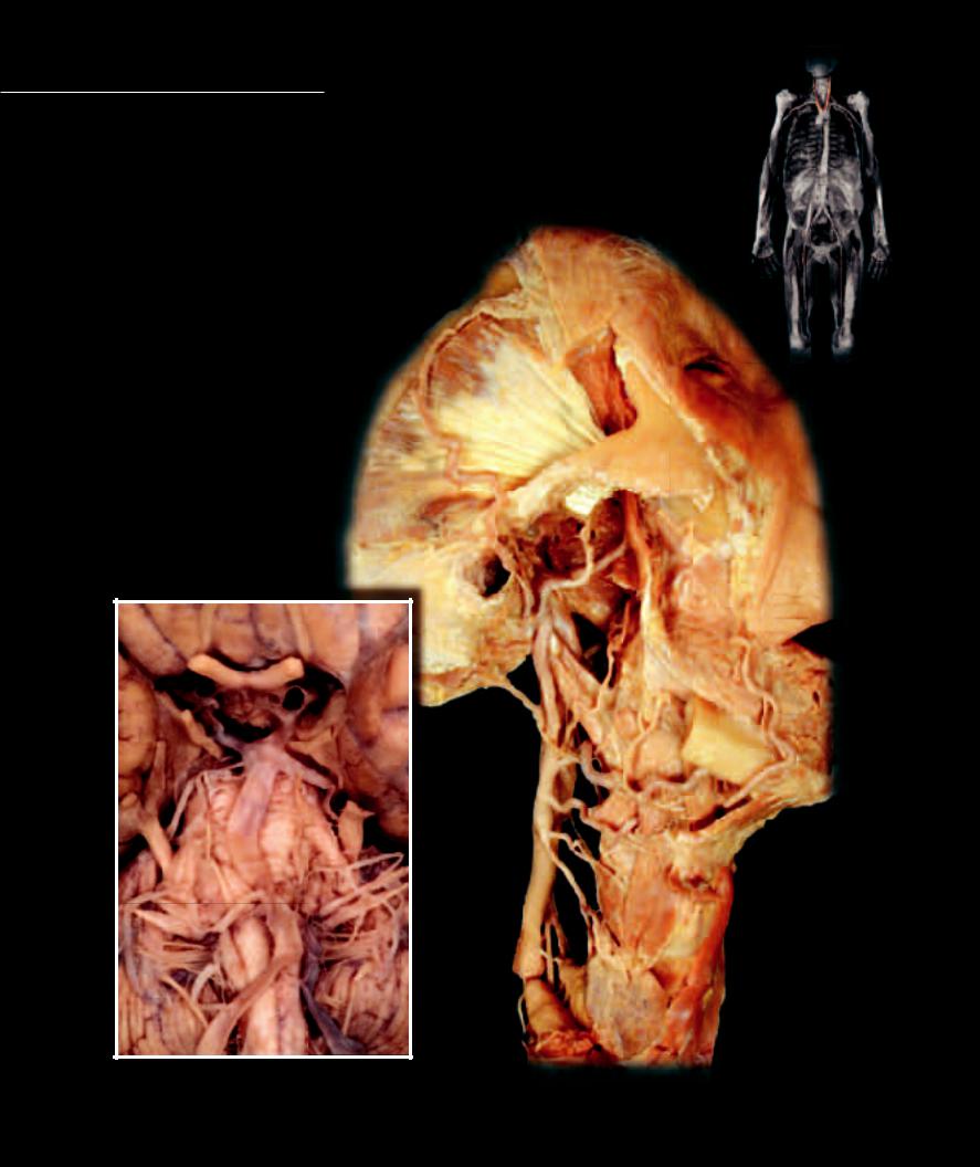
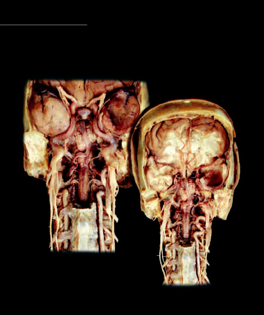
Like the heart, which needs a constant, uninterrupted blood supply, the brain tissue also must be guaranteed of a continuous perfusion in order to maintain its crucial functions. The

common carotid arteries, arising from the aortic arch, bifurcate into external and internal carotids. The external carotid supplies all tissues of the head except the brain, while the function of the internal carotid is to supply the brain. Because of the brain’s critical vascular needs the internal carotid artery has a partner, the vertebral artery, which courses cranially from the subclavian artery to assist with the essential blood supply to the brain.

1

Internal carotid artery


2

Basilar artery


3

Vertebral artery


4

Posterior cerebral artery


5

Posterior communicating artery


6

Middle cerebral artery


7

Posterior inferior cerebellar artery


8

Posterior superior cerebellar artery


9

Common carotid artery


10

External carotid artery


11

Superior thyroid artery


12

Ascending pharyngeal artery


13

Lingual artery


14

Facial artery


15

Occipital artery


16

Posterior auricular artery


17

Superficial temporal artery

25

18

Transverse facial artery

17


19Maxillary artery

20Optic chiasm

21Thyroid gland

22Trigeminal nerve

23

Lateral pterygoid muscle


18 19


24 Temporal lobe of cerebrum


25

Zygomatic arch


23


1


16


6

20


1

15


12


24


5

24


14


4

10

14

14


22

2

22

11

13


8

9

7

21

3

3

Dissection of basilar artery

Dissection of branches of external carotid artery

Inferior view

Lateral view

271

Head Vessels

1

Internal carotid artery

8

Cerebral veins

15

Confluence of the sinuses

22

Pituitary gland

2

Vertebral artery

9

Cerebellar veins

16

Dura mater

23

External acoustic meatus

3

Basilar artery

10

Superior sagittal sinus

17

Pia-arachnoid mater

24

Pons

4

Middle cerebral artery

11

Transverse sinus

18

Spinal cord

25

Frontal lobe of cerebrum

5

Anterior cerebral artery

12

Inferior sagittal sinus

19

Vertebral body

26

Vagus nerve

6

Anterior communicating artery

13

Sigmoid sinus

20

Cervical transverse process

27

Cervical sympathetic trunk

7

Posterior communicating artery

14

Opening of straight sinus

21

Temporal lobe of cerebrum

28

Superior cervical ganglion

25

1

22

21


1

25


3


24


5


1

2


6


4


1


21

1


18


7


3


24


28

23

2


20

19


2


2


2

26


18

27


20


28


20

19


2

26


27


20

Deep dissection series of head with facial and anterior neck anatomy


removed exposing internal carotid and vertebral arteries


Anterior views


272

Unlike the internal and external carotid arteries, the internal and external jugular veins form a wide array of collateral circuitry. The major structural difference of the venous pathways in the head is the existence of dural venous sinuses within the skull. The dural venous sinuses are non-collapsible, endothelial lined spaces within the tough meningeal dura mater. All the smaller veins draining capillaries within the brain tissue enter into the dural venous sinuses. These dural sinuses converge with one another throughout the skull to exit the cranial vault via the internal jugular vein.

8

16

17

10

8


14


15

11

11


9

12

8

8

8

17

11

913

Dissections of dural venous sinuses and cerebral veins

Posterior view (top), lateral view (bottom)

273

Superior Limb Vessels

The arterial pathway into the upper limb consists of a single, major arterial roadway that gradually tapers as

it gives rise to the various branches that supply the tissues of the limb. This large arterial roadway begins as the subclavian artery, takes on regional names — the axillary artery and brachial artery — as it tapers distally, then branches into the radial and ulnar arteries, which course through the antebrachium, paralleling the bones of the same names. The radial and ulnar arteries terminate as the collateral arches in the hand. This central pathway through the limb is the sole blood supply to this region, supplying the integument, muscles, bones, joints, and connective tissues of the upper limb. The deep venous pathways follow the arteries and have similar names. However, superfi cial veins that have no arterial counterparts aid the deep veins in returning blood to the heart.


28


13


9


30


38


37


31


8


29

16


14


15


4


27


12


10


11

39


21


10


13


13


10


22


18


17


19


32


24


33


20


23


34


35


Dissection of subclavian and axillary arteries

Anterior view

7

36

65

3

2

1

26 25

274

1

Brachiocephalic artery

11

Superior thoracic artery

21

Posterior circumflex humeral artery

31

Pectoralis major muscle

2

Common carotid artery

12

Thoracoacromial trunk

22

Anterior circumflex humeral artery

32

Subscapularis muscle

3

Vertebral artery

13

Pectoral artery

23

Brachial artery

33

Teres major muscle

4

Subclavian artery

14

Acromial artery

24

Deep artery of arm

34

Latissimus dorsi muscle

5

Thyrocervical trunk

15

Clavicular artery

25

Internal thoracic artery

35

Serratus anterior muscle

6

Inferior thyroid artery

16

Deltoid artery

26

Internal thoracic vein

36

Phrenic nerve

7

Ascending cervical artery

17

Lateral thoracic artery

27

Anterior scalene muscle

37

Brachial plexus

8

Suprascapular artery

18

Subscapular artery

28

Middle scalene muscle

38

Clavicle

9

Dorsal scapular artery

19

Circumflex scapular artery

29

Deltoid muscle

39

First rib

10

Axillary artery

20

Thoracodorsal artery

30

Pectoralis minor muscle

40

Suprascapular nerve


38


8


28

7


29


40


37

3


9


27


6


36


4


15


5


14

10


8


31

30


4


12


11

39


2


13


32


1


17


34

20

35

Dissection of subclavian and axillary arteries

Anterosuperior view

275

Superior Limb Vessels

1

Brachial artery

8

Deep palmar arch

15

Interosseous membrane

22

Triceps brachii muscle

2

Ulnar artery

9

Cephalic vein

16

Transverse carpal ligament

23

Pectoralis major muscle

3

Radial artery

10

Median cubital vein

17

Supinator muscle

24

Deltoid muscle

4

Anterior interosseous artery

11

Basilic vein

18

Pronator quadratus muscle

25

Deltopectoral groove

5

Superficial palmar arch

12

Median antebrachial vein

19

Flexor digitorum superficialis tendons

26

Serratus anterior muscle

6

Common digital artery

13

Accessory cephalic vein

20 Flexor digitorum profundus tendons

27

Brachioradialis muscle

7

Proper digital artery

14

Brachial vein

21

Biceps brachii muscle

28

Coracobrachialis muscle

22

2

3

21

1

16

17

2

3

4

5

15

18

18 18

18

6


2


3


7 7

19

8 18

Dissection of palmar arterial arch and branches to digits

Anterior view

Dissection of antebrachial arteries

Anterior view

276

Within the upper limb there are two sets of veins: deep veins that accompany the arteries, and superficial veins that course through the hypodermis without arterial counterparts. The deep veins, running with the arteries of the upper limb, have the same names as their arterial counterparts. These veins are significantly smaller than the arteries they accompany and form vena comitans with anastomotic channels around the arteries. The superficial veins of the upper limb are large and numerous. There are three major superficial veins into which all the other superfi cial veins fl ow; they are the basilic vein, cephalic vein, and median cubital vein. The median cubital vein is a connecting vein between the cephalic vein and the basilic vein. The cephalic and basilic veins eventually pass deep to join the axillary vein at the proximal end of the limb. Most of the venous return from the upper limb passes through the superficial veins.

24

25

24

24

22

28

9


23

22


26

21

21

22

11


10


Dissection of cephalic vein


27

11

Anterior view

12


13


9


Dissection of superficial vein of upper limb

Medial view of left upper limb

277

Thoracic Vessels

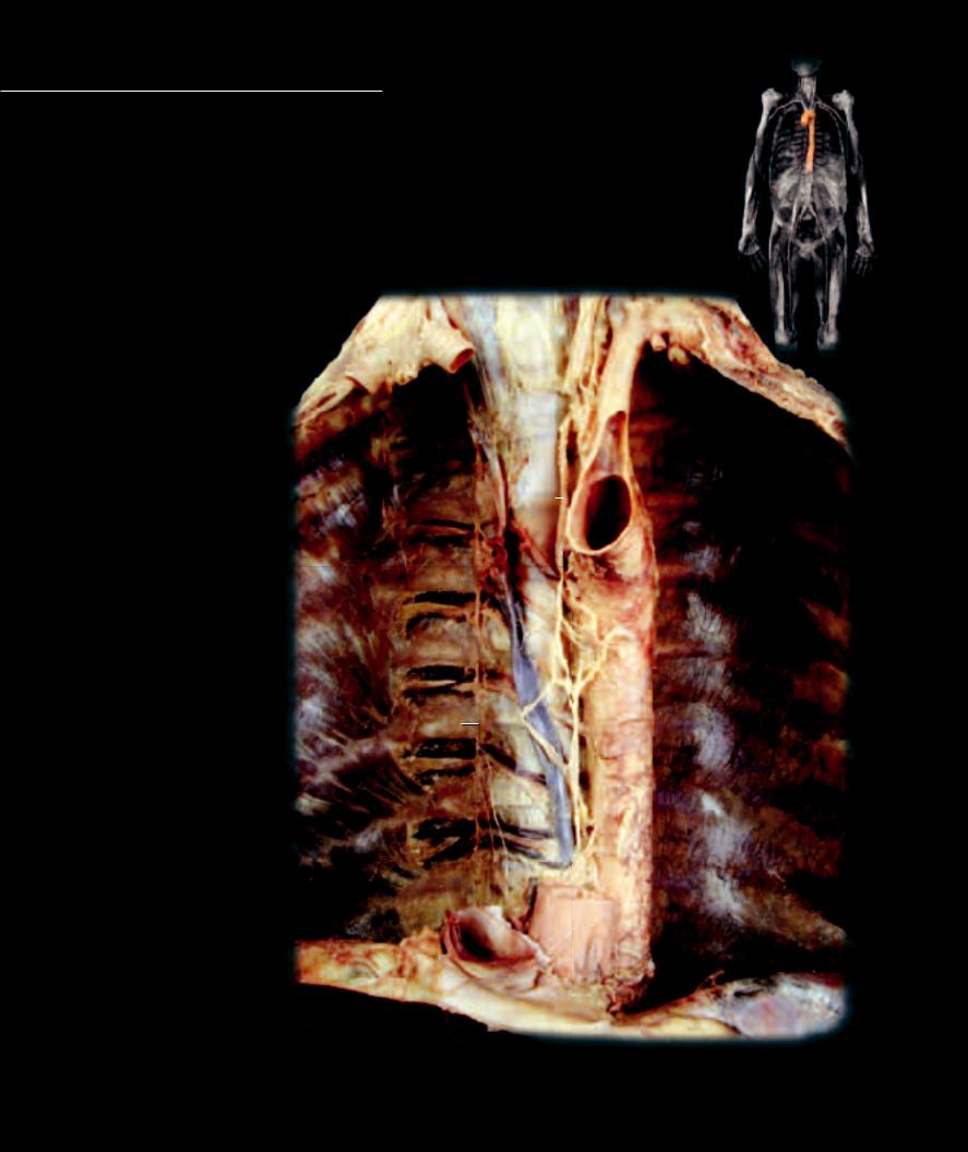
The branches of the aorta that supply the thoracic region can be divided into two principal groups — those that supply the thoracic body wall and those

that supply thoracic viscera. Two arterial supply routes carry blood into the thoracic body wall. Posteriorly the aorta courses vertically down the vertebral column, while anteriorly the internal thoracic arteries arise from the subclavian arteries and course vertically down the inside of the sternum. Between these anterior and posterior supply arteries are interconnecting collateral arteries. These collateral vessels are the anterior intercostal arteries and the posterior intercostal arteries, which supply the tissues of the intercostal spaces and form collateral circuits between the anterior and posterior arterial pathways. All thoracic viscera receive their blood supply from branches of the aorta. The thoracic viscera include the heart, lungs with their associated bronchial tubes, and the esophagus.

1 Aorta

2 Posterior intercostal artery

3 Posterior intercostal vein

4 Azygos vein

5 Hemi-azygos vein

6 Accessory hemi-azygos vein

7 Superior vena cava

8 Brachiocephalic vein

9 Subclavian vein

10Internal jugular vein

11Inferior vena cava

12 Right atrium (cut)

13

13Left subclavian artery

14Left common carotid artery

15Right common carotid artery

16

Hepatic vein

24


17

Trachea

1

18

Diaphragm

20


19

Esophageal hiatus

2


20

Subcostal muscle


21

Innermost intercostal muscle


22

Esophagus


23

Sympathetic trunk nerve


24

Thoracic lymphatic duct


4


23

3

21

2

22

18

11


Dissection of vessels of posterior thoracic wall

Anterior view

278

Like the arterial supply to the thoracic wall, the venous drainage returns via both anterior-wall and posterior-wall drainage veins. The veins of the anterior wall have the same names as their arterial counterparts, while the veins of the posterior wall differ in name and structure. Unlike the aorta, which is the posterior-wall supply artery, the superior vena cava and inferior vena cava diverge from the posterior thoracic wall to enter the thoracic cavity and return their contents to the heart. In the absence of vena cavae in the posterior thoracic wall, an azygos system of veins is formed to drain the body wall and the thoracic viscera. These azygos veins communicate with the superior vena cava to return their contents to the heart. With the exception of the azygos veins, the veins are similar to the arteries in name and distribution.

15

10

10


17


14

9

9


8


8

71


20


6

16


23


4

3


19


18


3


11


Dissection of vena cavae and tributaries

21


3

3


Anterior view


5


Dissection of azygos veins

Anterior view

279

Abdominal Vessels

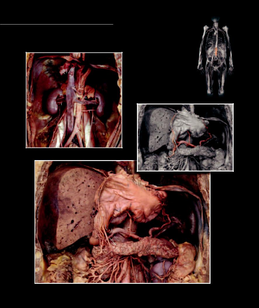
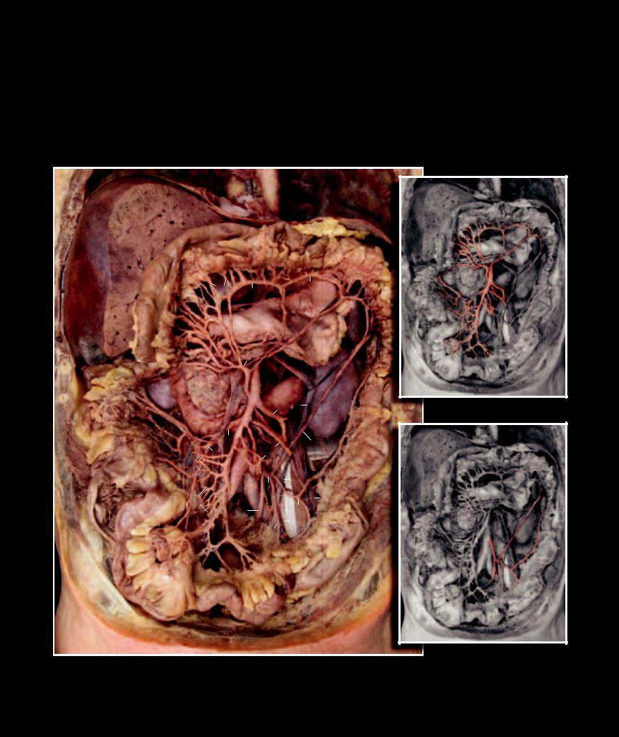
Like the thorax, the abdomen has somatic arteries that supply the abdominal muscle wall and visceral arteries that supply the viscera of the abdominal cavity. These vessels follow the same

pattern observed in the thoracic region; that is, the abdominal body wall has both anterior (epigastric arteries) and posterior (aorta) supply pathways that form interconnecting collateral arteries, while the viscera receive branches from the aorta — celiac artery to the foregut, superior mesenteric artery to the midgut, inferior mesenteric artery to the hindgut, and renal arteries to the kidneys.

46


27


3


4


5


39


39


23


2


12


33

23


35


26


28

35


23

1

23

47

19

25

34

48

Deep dissection of abdomen showing renal vessels

Anterior view


Branches of celiac artery


37


46


36


7


8


41


6


9

5


3


4


2

32


29


11

10


40


23


12


44


31

35


43

30


1


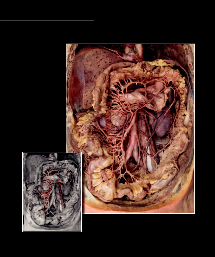
Dissection of abdomen showing celiac branches and supply of foregut viscera

Anterior view, stomach reflected upward

280

1

Aorta

13

Middle colic artery

25

Common iliac artery

37

Stomach

2

Celiac artery

14

Marginal artery

26

Inferior vena cava

38

Transverse colon

3

Splenic artery

15

Right colic artery

27

Hepatic vein

39

Suprarenal gland

4

Common hepatic artery

16

Ileocolic artery

28

Renal vein

40

Pancreas

5

Left gastric artery

17

Jejunal arteries

29

Hepatic portal vein

41

Spleen

6

Right gastric artery

18

Ileal arteries

30

Superior mesenteric vein

42

Duodenum

7

Left gastro-omental artery

19

Inferior mesenteric artery

31

Inferior mesenteric vein

43

Ascending colon

8

Right gastro-omental artery

20

Left colic artery

32

Splenic vein

44

Descending colon

9

Proper hepatic artery

21

Sigmoid artery

33

Suprarenal vein

45

Ileum

10

Gastroduodenal artery

22

Superior rectal artery

34

Testicular vein

46

Diaphragm

11

Superior pancreaticoduodenal artery

23

Renal artery

35

Kidney

47

Ureter

12

Superior mesenteric artery

24

Segmental arteries

36

Liver

48

Psoas major muscle

46

38

36


24


14


14


41


37


13


35

40


12


35


43

30


17


31


42


15


20


17


15


44


16


25


31


25


19


34


18


22


20


21


47


Superior mesenteric artery

45

Inferior mesenteric artery

Dissection of abdomen showing arterial supply of midgut and hindgut viscera

Anterior view

281

Abdominal Vessels

The major difference between the arteries and veins of the abdomen is the fact that all the visceral venous return from the capillaries of the digestive system and spleen pass via the hepatic portal system to the

capillaries of the liver before returning to the heart. Within the liver, both the hepatic artery and hepatic portal vein branch to form a complex network of specialized capillaries called the hepatic sinusoids. The hepatic sinusoids then drain into the hepatic veins to return the blood to the inferior vena cava.

1

Inferior vena cava


2

Hepatic portal vein


3

Superior mesenteric vein


4

Right colic vein


5

Inferior mesenteric vein


6

Renal vein


7

Superior mesenteric artery


8

Inferior mesenteric artery


9

Middle colic artery


10

Marginal artery


11

Left colic artery


12

Common iliac artery


13

External iliac artery


14

Internal iliac artery


10


15

Superior gluteal artery


10


16Inferior gluteal artery

17Obturator artery

18Internal pudendal artery

19Lateral sacral artery

20Superior vesical artery

21Vaginal artery

22Obliterated umbilical artery

23Uterus

24

Bladder

2 9

6

25

Prostate

7

5

26

Rectum


28

27

Stomach

3


28

Kidney


29

Upper bands of sacral plexus

4


30Sympathetic trunk

31Inferior vesical artery

32

Middle rectal artery

8


33

Obturator nerve

1


11

34

Uterine artery


12


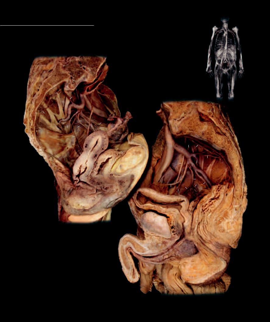
Dissection of abdomen showing arteries and veins of the intestines

Anterior view

Abdominal veins

282

Pelvic Vessels

The common iliac arteries, the terminal branches of the aorta, carry all of the blood supply to the lower limbs and pelvis. All pelvic viscera, along with the body wall anatomy

of the pelvis and perineal regions, receive their blood supply from the internal iliac artery. Numerous branches arise from the internal iliac artery to supply the pelvic wall, the perineum, and the gluteal region. Other branches course into the pelvic cavity to supply the viscera. The veins are similar in name and course with the corresponding arteries.


12


30


19


29


14


29

16


13

19


29


34

22


20


17


18


21


23


24

26

Dissection of pelvic arteries of female

Medial view, anterior at left

12


19


14


30


33


29 19


15


13


29


16


31


17

32


18


22

20

26

24

25

Dissection of pelvic arteries of male

Medial view, anterior at right

283

Inferior Limb Vessels

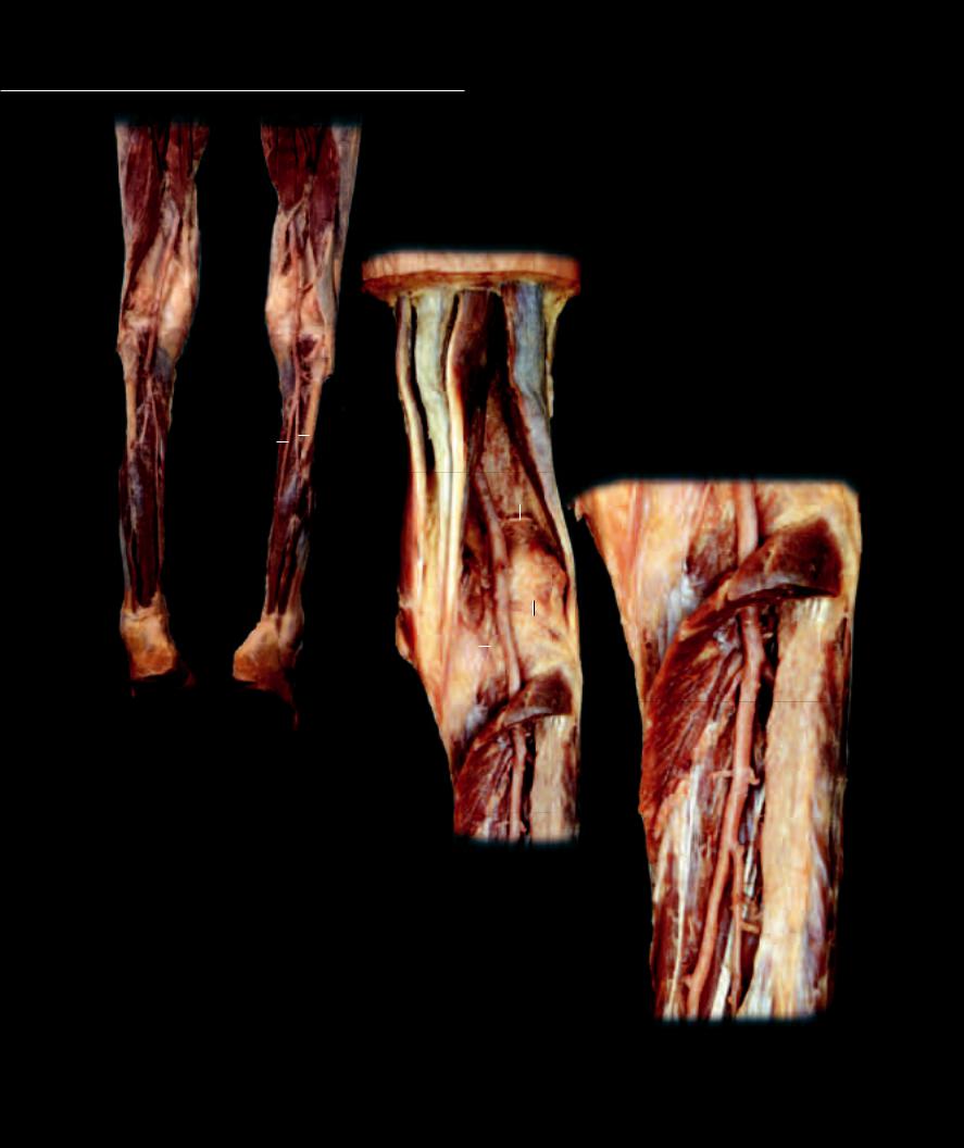
As in the upper limb, the main arterial pathway into the lower limb consists of a single, major arterial roadway that

gradually tapers as it gives rise to numerous branches on its pathway through the limb. This large arterial roadway begins as the external iliac artery in the pelvis, passes beneath the inguinal ligament to enter the thigh as the femoral artery, passes to the back of the knee to become the popliteal artery, and in the proximal aspect of the leg bifurcates into the anterior tibial and posterior tibial arteries, which course through the leg and into the foot.

1

Superior gluteal artery

10

Internal iliac artery

19

Adductor longus muscle

2

Inferior gluteal artery

11

External iliac vein

20

Rectus femoris muscle

3

Internal pudendal artery

12

Common iliac artery

21

Vastus intermedius muscle

4

Femoral artery

13

Aorta

22

Gracilis muscle

5

Deep artery of thigh

14

Gluteus maximus muscle

23

Vastus lateralis muscle

6

Muscular branches of femoral

15

Sacrotuberous ligament

24

Vastus medialis muscle

7

Femoral vein

16

Piriformis muscle

25

Fascia lata

8

Great saphenous vein

17

Spermatic cord (cut)

26

Sartorius muscle

9

External iliac artery

18

Penis (cut)

27

Iliacus muscle

14

1

16

14

2

15

3

22

Dissection of gluteal region showing gluteal arteries and nerves

Posterior view

284

27


26


17


4


8


6

7

18


6


5

19


20

23

22


13


21


12


24

27


10


9


Dissection of femoral vessels in femoral triangle


11


Anterior view

27


26


26


7


8

18

4


5


4


19


20

25

22

23

24

Dissection of vessels of inferior limb

Anterior view

285

Inferior Limb Vessels


1

Popliteal artery


2

Anterior tibial artery


3

Posterior tibial artery


4

Fibular artery


5

Superior lateral genicular artery


6

Inferior lateral genicular artery


7

Inferior medial genicular artery


8

Femoral vein

23


9

Great saphenous vein


10

Femoral artery


11

Superficial epigastric artery


12

Superficial epigastric vein


1


13

External pudendal vein


14

Superficial circumflex iliac vein


15

Superficial circumflex iliac artery


16

Femoral nerve


17

Soleus muscle


18


18

Popliteus muscle


19

Subcutaneous layer


24


20

Fascia lata


21

Sartorius muscle


22

Adductor longus muscle


23

23

Biceps femoris muscle


24

Semitendinosus muscle

3


4

5

25

Scrotum


1


18


1

6


1

7


17


2

18 17

Dissection of popliteal and crural arteries

Posterior view

3

Dissection of popliteal region revealing arteries

Posterior view

3

4

Dissection of proximal crus revealing arteries

Posterior view

286

Similar to the veins of the upper limb, the venous pathways in the lower limb consist of both deep veins that accompany the arteries, and superficial veins that course through the hypodermis. In the foot and leg, the deep veins form vena comitans with their arterial counterparts; however, the more proximal popliteal and femoral veins are large single vessels accompanying their associated arteries. Two major superfi cial venous channels receive numerous tributaries from smaller superfi cial veins throughout the lower limb. These major superficial veins are the small saphenous vein and the great saphenous vein. Unlike the upper limb, the majority of venous blood fl ow through the lower limb passes via the deep veins. Anastomosing veins between the saphenous veins and the deep veins have one-way valves. The valves direct blood fl ow to the deep veins where contractions of surrounding skeletal muscles facilitate movement of the blood toward the heart.

15

14 10 8

16

13

22

21 9


15

11


21


20

14

12

10


16

8


20

13

9

25

19

22

Dissection of femoral vein and tributaries in femoral triangle

Anterior view

Dissection of great saphenous vein

Anteromedial view

287