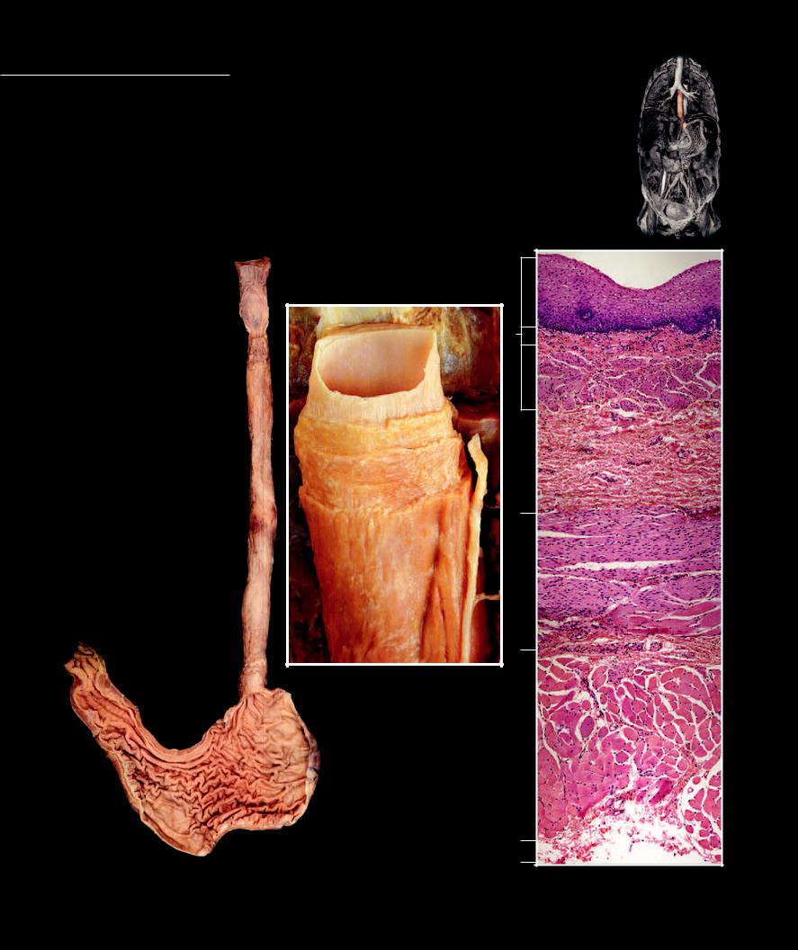
Esophagus Below the laryngopharynx the gut tube branches into an anterior respiratory tube, the larynx and a posterior digestive tube, the esophagus.
The esophagus is a narrow, collapsed muscular tube coursing from the laryngopharynx to the stomach. It is approximately 25 cm in length and begins near the level of the sixth cervical vertebra, where it runs inferiorly against the anterior surface of the thoracic vertebral column. At the level of the tenth thoracic vertebra it deviates slightly to the left passing through the esophageal hiatus of the diaphragm to enter the stomach. It functions as a muscular tube of transmission.
1 |
Esophagus |
7 |
Lamina propria |
2 |
Tunica mucosa |
8 |
Muscularis mucosae |
3 |
Tela submucosa |
9 |
Tunica adventitia |
4 |
Tunica muscularis circular layer |
10 |
Stomach |
5 |
Tunica muscularis longitudinal layer |
11 |
Pharynx - dorsal wall |
6 |
Stratified squamous epithelium |
12 |
Vagus nerve |
11 |
|
6 |
|
|
1 |
2 |
7 |
|
|
|
8 |
|
2 |
|
|
3 |
|
|
|
3 |
1 |
4 |
|
|
|
|
12 |
|
|
5 |
|
|
|
4 |
Step dissection of esophagus
Anterior view
5
10
|
9 |
Pharynx, esophagus, and stomach |
Photomicrograph of esophageal wall |
Anterior view |
|
40x |
Stomach The stomach is a J-shaped organ of variable size and shape and has the greatest diameter of any part of the gut tube. It occupies the upper left quadrant of
the abdominal cavity, where it is anchored to the posterior abdominal wall by a mesentery. The stomach performs several functions, the most important of which is to store ingested food until it can be emptied into the small intestine at a rate that allows for optimal digestion and absorption.
1 |
Stomach |
7 |
Pylorus |
13 |
Surface mucous cell |
2 |
Cardia of stomach |
8 |
Pyloric sphincter |
14 |
Lamina propria |
3 |
Fundus of stomach |
9 |
Gastric rugae |
15 |
Mucous neck cell |
4 |
Body of stomach |
10 |
Greater curvature |
16 |
Gastric glands |
5 |
Pyloric antrum |
11 |
Lesser curvature |
17 |
Liver |
6 |
Pyloric canal |
12 |
Gastric pit |
18 |
Gallbladder |
|
|
|
|
19 |
Spleen |
|
|
|
|
20 |
Greater omentum |
19
12 14
13
17
1
11
10
18
20
15
Abdominal dissection revealing stomach
Anterior view
78
8
3
11
9
16
16
|
10 |
Frontal section of stomach |
Photomicrograph of stomach mucosa |
Anterior view |
with callout above |
|
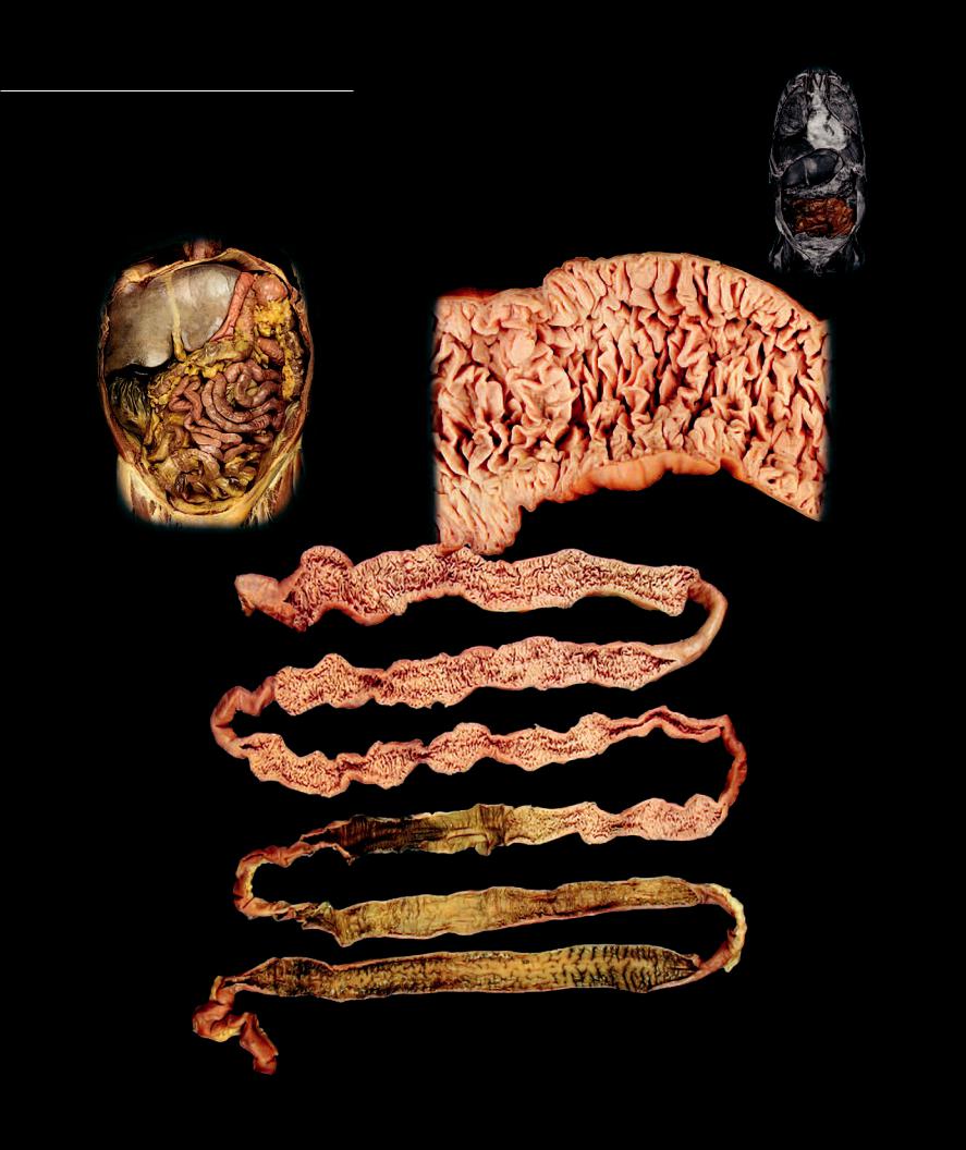
Small Intestine
The small intestine is a highly coiled tube with a fairly consistent diameter from beginning to end. It is approximately 6 to 7 meters long in the cadaver but, because of its muscle
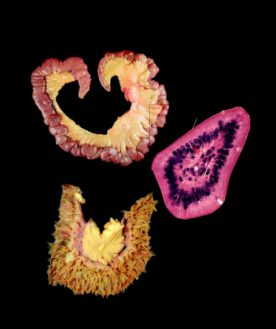
tone only around 4 to 5 meters in the living. The small intestine occupies the greater part of the midto lower abdominal cavity and consists of three regions. The retroperitoneal fi rst part is called the duodenum and is about 30 cm in length. This C-shaped region receives the secretions from the pancreas and liver. The remaining parts of the small intestine are the jejunum and ileum, which make up the bulk of the organ and are attached to the posterior wall of the abdomen by the mesentery. The small intestine is the principal site of digestion and absorption.
11
4
|
Sectioned small intestine |
|
revealing circular folds |
Small intestine in situ |
Internal view |
Anterior view |
|
1 |
|
2
Entire small intestine sectioned to show changes in internal surface from the duodenal end to the ileal end
Internal view
306
1 |
Duodenal end |
6 |
Simple columnar epithelium |
11 |
Cecum |
2 |
Ileal end |
7 |
Goblet cell |
12 |
Transverse colon |
3 |
Jejunum |
8 |
Lamina propria |
13 |
Descending colon |
4 |
Ileum |
9 |
Liver |
14 |
Mesentery |
5 |
Circular folds |
10 |
Stomach |
15 |
Microvillus brush border |
15
14
Loop of small intestine
Anterior view
7
Photomicrograph of cross-section of intestinal villus
400x
14
5
5
Loop of small intestine from unembalmed cadaver, opened to show circular folds
Anterior view