

Tissues |
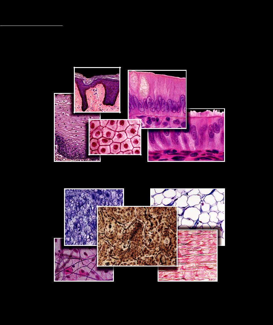
The facing pages show photomicrograph collages of the four principal tissue categories — epithelial |
tissue, connective and supporting tissue, muscle tissue, and nervous tissue. The photomicrographs |
|
|
illustrate the key structural features shared by the tissues in each category. Note the numerous |
closely packed cells of the epithelial tissues and contrast them with the scattered cells and the fi brous surrounding matrix of the connective and supporting tissues. In the muscle tissue observe the long, slender specialized cells that are designed to shorten, and in the nerve tissue the branched, wire-like cells. We will explore each of the principal tissue categories in more detail on the pages that follow.
Epithelial Tissues
Connective and Supporting Tissues
4
Muscle Tissues
Nerve Tissues
5
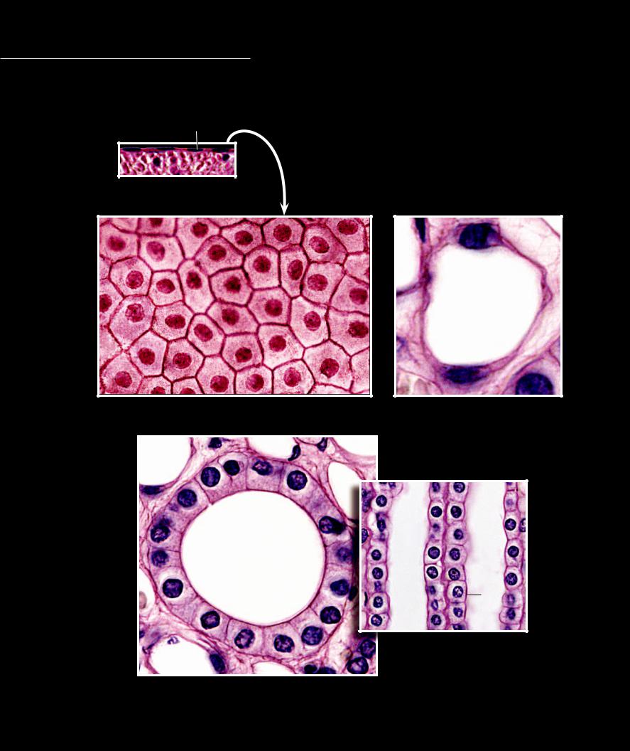
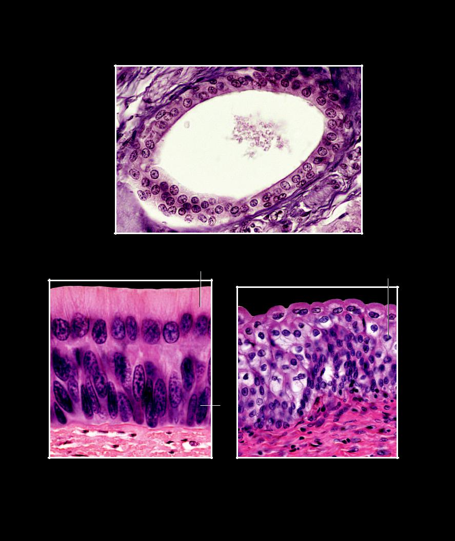
Epithelial tissues are surface tissues that consist of numerous cells, with each cell forming membrane to membrane contact with its neighbors. As a general rule, descriptions of epithelial tissues are based on the shape of
their cells and on the number of cell layers present. By combining the shape names — squamous (fl at cells), cuboidal, and columnar — with the term simple if there is a single layer of cells or the term stratifi ed if there is more than one layer of cells, almost all of the epithelial tissues can be described and named. The photomicrographs on this page and the facing page represent the simple (single cell layer) epithelial tissues.
1 |
|
|
|
|
|
1 |
Nucleus |
7 |
Mucous in goblet cell |
|
2 |
Cytoplasm |
8 |
Microvilli |
|
3 |
Cell membrane |
9 |
Basement membrane |
|
4 |
Capillary lumen |
10 |
Blood vessel with red blood cells |
|
5 |
Glandular lumen |
11 |
Cilia |
Simple squamous epithelium, mesothelium |
6 |
Connective tissue |
12 |
Basal cell |
Section of mesentery, 400x |
|
|
|
|
|
|
|
|
1 |
1 |
|
|
|
|
2 |
|
|
|
4 |
|
|
|
|
|
3 |
|
|
|
3 |
|
|
|
|
|
|
|
|
|
2 |
|
|
|
|
1 |
Simple squamous epithelium, mesothelium |
|
Simple squamous epithelium, endothelium |
||
Surface view of mesentery, 400x |
|
|
Section of capillary, 630x |
|
2
1
5
5
5
3
Simple cuboidal epithelium
Urinary tubes in kidney - transverse section, 630x (left); longitudinal section, 400x (right)
6
8
7
7
7
1
9
6
Simple columnar epithelium
Section of mucosa of small intestine, 630x
11 7
7 |
1 |
|
12
12
1
6
10
Pseudostratified columnar epithelium
Section of mucosa of larynx, 400x
7
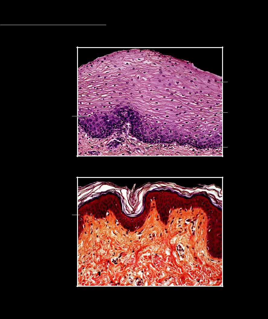
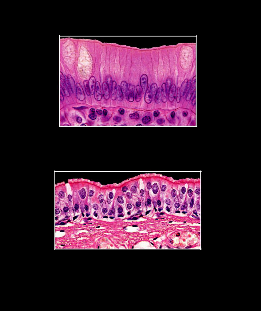
Epithelial Tissue The photomicrographs on this and the facing page illustrate the stratifi ed (more than one layer of cells) epithelial tissues. Note that the tissues range
from two layers to numerous layers and the cell shape used for the tissue name is the shape of the cells found in the surface layer.
5
4
1
1 |
Nucleus |
|
|
|
2 |
Cytoplasm |
|
|
3 |
3 |
Basal cell layer |
11 |
|
|
|
||||
|
|
|||
4 |
Intermediate cell layer |
|
|
|
5 |
Superficial cell layer |
|
|
|
6 |
Stratum basale |
Nonkeratinized stratified squamous epithelium |
|
|
7 |
Stratum spinosum |
|
|
|
Section of esophageal mucosa, 200x |
|
|
||
8 |
Stratum granulosum |
|
|
|
|
|
|
||
9 |
Stratum lucidum |
|
|
|
10Stratum corneum
11Connective tissue
12 |
Basement membrane |
|
|
10 |
|
|
10 |
||
13 |
Glandular lumen |
|
|
|
|
|
|
||
|
|
10 |
9 |
|
|
|
8 |
|
|
|
|
7 |
|
|
|
1 |
|
|
|
|
6 |
|
||
|
|
|
||
11
Keratinized stratified squamous epithelium
Section of skin, 200x
8
3
1
5
13
1
Stratified cuboidal epithelium
Section of duct of esophageal gland, 400x
5
1
2
1
1 |
|
|
3 |
|
12 |
|
|
|
11 |
11 |
|
|
|
|
|
Stratified columnar epithelium |
Transitional epithelium |
Section of pharyngeal mucosa, 400x |
Section of urinary bladder mucosa, 400x |
9
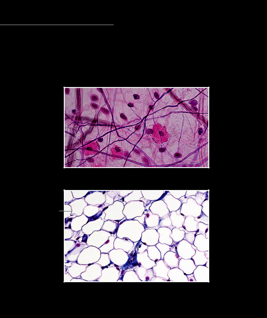
Connective tissues have relatively few cells and the cells are surrounded by an extracellular matrix of fi bers, which the cells secrete. The classifi cation and names of connective tissues arise from the type
and arrangement of the fi bers produced by the cells and secreted into the surrounding matrix. There are three named
fibers in the matrix — collagen fi bers, reticular fi bers (actually a thin form of collagen), and elastic fi bers. The fi bers are deposited in varying degrees of density and are arranged in different patterns. The tissue names are based on the different
fiber types and patterns in the matrix.
1 |
Mast cell |
7 |
Cytoplasm |
2 |
Fibroblast |
8 |
Plasma membrane |
3 |
Collagen fiber |
9 |
Lipid storage area |
4 |
Elastic fiber |
10 |
Nucleus of reticular cell |
5 |
Reticular fiber |
11 |
Nucleus of fibroblast |
6 |
Nucleus of adipose cell |
12 |
Elastic lamella |
3 |
2 |
5 |
3 |
2
3
4
1
44
1
5
2
Loose (areolar) connective tissue
Section of subcutaneous layer of integument, 400x
9
8
6
9
6
7
Adipose tissue
Section of epicardial fat, 200x
10

10
|
|
Reticular tissue |
|
|
|
Section of lymph node, 400x |
|
11 |
11 |
||
|
|
3 |
|
|
|
|
|
|
|
||
|
|
3 |
|
|
|
3 |
|
|
|
3 |
|
|
|
|
|
Dense irregular connective tissue |
Dense regular (collagenous) connective tissue |
Section of dermis, 200x |
Section of tendon, 200x |
12
12
12
12
Dense regular (elastic) connective tissue
Section of tunica media of aorta, 400x
11

The supporting tissue category consists of the skeletal tissues— cartilage and bone. Like the connective tissues, the supporting tissues have relatively few cells surrounded by a signifi cant amount of extracellular
matrix, which for the most part the cells produce. However, unlike the soft matrix of the connective tissues, the extracellular matrix of the supporting tissues is firm and rubber-like in cartilage and hard in bone tissue.
6
|
|
1 |
|
1 |
Hyaline ground substance |
|
|
2 |
Collagen fibers in ground substance |
|
|
3 |
Elastic fibers in ground substance |
|
|
4 |
Chondrocyte nucleus |
|
|
5 |
Chondrocyte in lacuna |
|
|
6 |
Perichondrium |
|
|
7 |
Bone trabecula |
4 |
|
8 |
Osteocyte |
||
5 |
|||
9 |
Red bone marrow |
||
|
10Canaliculi
11Lacuna
12Lamella
13Central canal
1
Hyaline cartilage
Section of cartilage in developing fetal bone, 200x
3
4
2
2 |
3 |
5
2
Fibrocartilage |
Elastic cartilage |
Section of intervertebral disc, 200x |
Section of cartilage from auricle of ear, 400x |
12

7
9
9
7
7
9
8
7
7
9
Spongy bone
Section of epiphysis of metacarpal bone, 200x
10
11
12
13
11 |
11 |
12
12
13
13
13
Compact bone
Section of diaphysis of fibula, 100x; callout of osteon, 400x
13
The tissues blood and lymph traditionally were classified as connective tissues because, like all connective tissues, the extracellular matrix
is a greater percentage of the tissue then are the cells. However, the extracellular matrix of blood and lymph is a liquid matrix called plasma, rather than the soft, fi rm matrix of connective tissues. The most recent Terminologia Histologica places blood and lymph in their own subcategory called the hematolymphoid complex.
|
1 |
Erythrocyte or red blood cell (rbc) |
|
|
2 |
Leukocyte or white blood cell (wbc) - neutrophil |
|
|
3 |
Leukocyte or white blood cell (wbc) - monocyte |
|
|
4 |
Thrombocyte (platelet) |
|
2 |
5 |
Plasma |
|
6 |
Crenated red blood cell |
||
|
|||
|
3 |
|
|
3 |
|
|
2
5
1
1
1
1
2
5
5
1
2
6
4 |
1 |
|
Blood smear
Wright’s stain, 200x; enlargement, 630x; individual cells, 1500x
14
Muscle cells are long, slender cells that have special arrangements of the proteins actin and myosin within the cytoplasm. The architectural design of these proteins forms the muscle cell “machinery” that allows the cell to specialize at contracting
(shortening). The names of the different types of muscle tissues arise from the arrangement of the contractile proteins within their cells. In some tissues the protein arrangement gives the cell a striated, or striped, appearance (striated muscle), while in other tissues the striped appearance is not evident (non-striated or smooth muscle).
1 Nucleus
2 Sarcoplasm
3 Smooth muscle cell
4 Cardiac muscle cell
5 Skeletal muscle cell
6 Intercalated disc
1
12
3 
1
1
Smooth (nonstriated) muscle tissue
Longitudinal section of muscular wall of intestine, 500x
2
1
5
1
1
6
6
1
2
4
Cardiac striated muscle tissue |
Skeletal striated muscle tissue |
Section of ventricle of heart, 500x |
Section of vastus lateralis muscle, 400x |
15