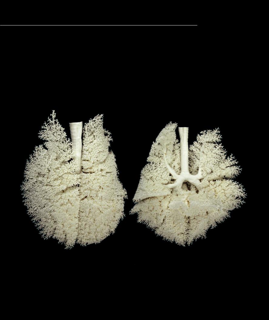
Cast of Trachea and Bronchial Tree
The cast below is from a large dog’s lungs and is approximately the same size as human lungs. The casts were created by forcing liquid latex into the respiratory passageways of the lungs and then letting the latex harden. The lungs were then placed in a weak acid until the organic tissue of the lungs was digested away. These views of the cast allow you to visualize the extensive nature of the bronchial tree as it branches out to the larger alveolar passageways within the lungs. The smaller alveolar spaces did not get incorported into the casts.
1 Trachea
2 Right main (primary) bronchus
3 Left main (primary) bronchus
4 Lobar (secondary) bronchus
5 Segmental (tertiary) bronchus
6 Branching bronchiole network
6
6
6
6
Latex cast of respiratory passageways of trachea and lungs of a dog
Anterior view at left, posterior view at right
18 Digestive System
The digestive system is the extensive environmental interface that makes it possible to transfer nutrients, water, and electrolytes from the food we eat into the body’s internal environment. This is made possible by a complex lining, which through a series of folds and a variety of small
to microscopic projections greatly increases the surface interface between the digested contents within the gastrointestinal organs and the numerous small capillaries beneath this lining. To better appreciate the degree of this surface increase, realize that the average total surface area of the skin of an adult human is about 20 square feet, while the surface area of the digestive system is approximately 2,500 square feet, or about the size of a tennis court. To make the transfer across this extensive surface area possible, the food we eat must be broken down into small molecules that can be absorbed from the digestive tract into the circulatory system, which then distributes the molecular metabolites to the cells. Therefore, the digestive organs also function in the mechanical and chemical breakdown of the food.
Developmentally the digestive system begins as a simple tube called the gut tube or gut. As this simple tube develops into the highly convoluted organs of the adult anatomy, it undergoes structural changes that account for its various functions. Though these structural changes lead to differences in the tube from one region to the next, there is a basic pattern of design throughout the
length of the tube. This structural pattern is responsible for the general function of the digestive system. Modifications of this pattern allow for the variation in structure and function along its length. This chapter will highlight the structural variation and underlying design of the digestive system.
Find more information about the digestive system in
R E A L A N AT O M Y
Digestive System Organs
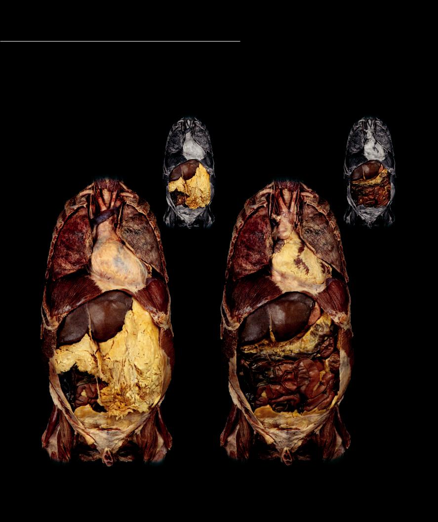
The digestive system begins at the mouth, where food and drink enter this tubular organ system to be processed by the teeth and tongue. From the mouth
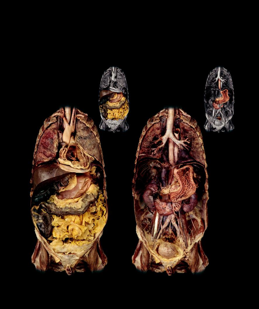
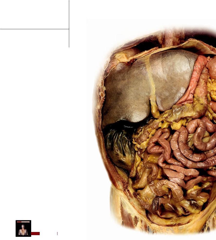
the broken-down food moves through the transport tube called the esophagus to the storage and mixing organ called the stomach. The stomach thoroughly mixes digestive juices and mucous with the food as it tosses it around to produce a softened substance called chyme. The chyme is slowly moved into the small intestine where powerful digestive chemicals are added from the pancreas. As the chyme slowly moves through the long small intestine, the digestive enzymes break it into small metabolic fuel molecules that the intestine absorbs. The material that cannot be digested and absorbed is passed into the large intestine where the nondigested remains are held until they can be removed through the anus as feces. The photos on this and the facing page depict the digestive organs and their related mesenteries.
|
23 |
|
23 |
|
|
21 |
|
21 |
|
|
|
|
|
18 |
18 |
18 |
|
18 |
|
|
|
|
19 |
20 |
20 |
20 |
|
20 |
|
|
|
|
|
|
3 |
|
3 |
2 |
|
|
|
|
17
Superficial dissection of abdominal viscera |
Intermediate dissection of abdominal viscera |
Anterior view |
Anterior view |
300
Design of the Gut Wall
The wall of the digestive tract has a basic pattern of design that is found throughout its length. This pattern consists of three tunics or layers of anatomy. The tunica
mucosa and its subdivisions, including the tela submucosa, form the inner layer of the wall and consist of an extensive epithelial lining with an underlying vascullar connective tissue. The middle layer, or tunica muscularis, consists of smooth muscle that provides for the varied types of movements that occur within the digestive organs. The majority of the organs have an outer layer, the tunica serosa, comprised of a lubricated meosthelial membrane that reduces friction as the organs move against one another. The image below, from the small intestine, illustrates the basic layers of the digestive tract wall.
1 Simple columnar epithelium
2 Lamina propria
3 Muscularis mucosae
4 Submucosal (Brunner’s) glands
5 Villi
302
5
1
1
2
2
Tunica mucosa consisting of: epithelium, lamina propria,
1 and muscularis mucosae
Tunica muscularis circular layer
Tunica muscularis longitudinal layer
Tunica serosa
Photomicrograph of small intestine wall
40x
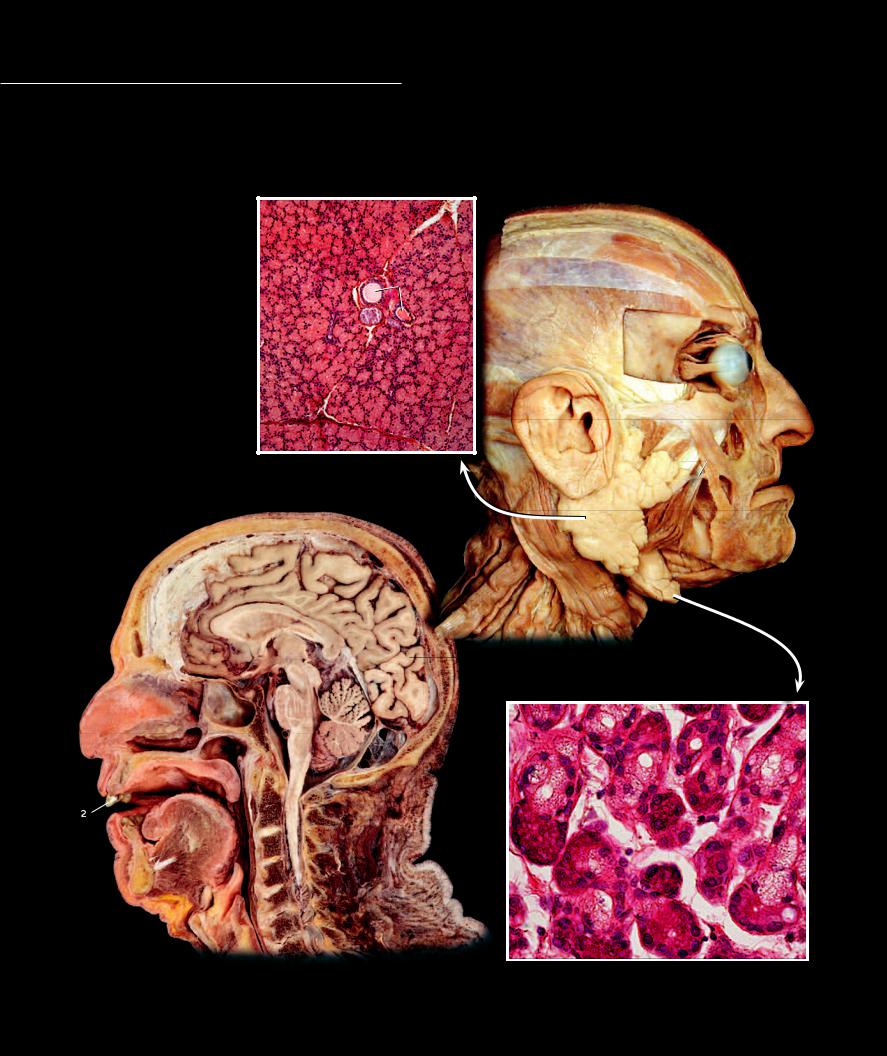
Mouth and Pharynx
The mouth, or oral cavity, is the entryway into the digestive system. In addition to serving as the portal to the tubular gut, the mouth contains structures, such as the tongue, teeth, and salivary glands,
that help initiate the digestive process. The boundaries of this region are defi ned by the lips and cheeks, which form the anterior and lateral walls, the palate, which forms the roof, and numerous muscles, the most conspicuous being the muscles of the tongue, which form the fl oor of the mouth. The pharynx is the fi rst portion of the gut tube and is divided into three regions. Each region communicates with a different cavity — the nasopharynx with the nasal cavity, the oropharynx with the oral cavity, and the laryngopharynx with the cavity of the larynx.
|
1 |
Lips |
|
|
2 |
Teeth |
|
|
3 |
Tongue |
12 |
|
4 |
Hard palate |
|
5 |
Soft palate |
|
|
6 |
Nasopharynx |
11 |
|
7 |
Oropharynx |
|
8 |
Laryngopharynx |
|
|
9 |
Parotid gland |
14 |
|
10 |
Submandibular gland |
|
12 |
|
11 |
Parotid duct |
|
|
|
12 |
Serous acini |
|
|
13 |
Mucous acini |
|
|
14 |
Vein |
12 |
|
15 |
Trabecula |
|
|
16Masseter
17Sternocleidomastoid
18 |
Sphenoid sinus |
15 |
|
19 |
Epiglottis |
|
|
20 |
Vertebral column |
|
11 |
21 |
Cerebrum |
Photomicrograph of parotid gland |
|
22 |
Spinal cord |
100x |
1 |
|
|
|
|
|
|
|
9 |
16 |
|
|
17 |
|
10
21
Dissection of head showing salivary glands
Lateral view
18
46
1 |
|
5 |
13 |
13 |
|
|
2 |
2 |
7 |
22 |
|
|
3 |
|
1 |
12 |
|
|
|
|
20 |
|
|
|
19 |
|
12 |
|
|
8 |
|
|
|
|
|
|
12 |
Sagittal section of head and neck |
Photomicrograph of submandibular gland |
Medial view |
240x |
303