

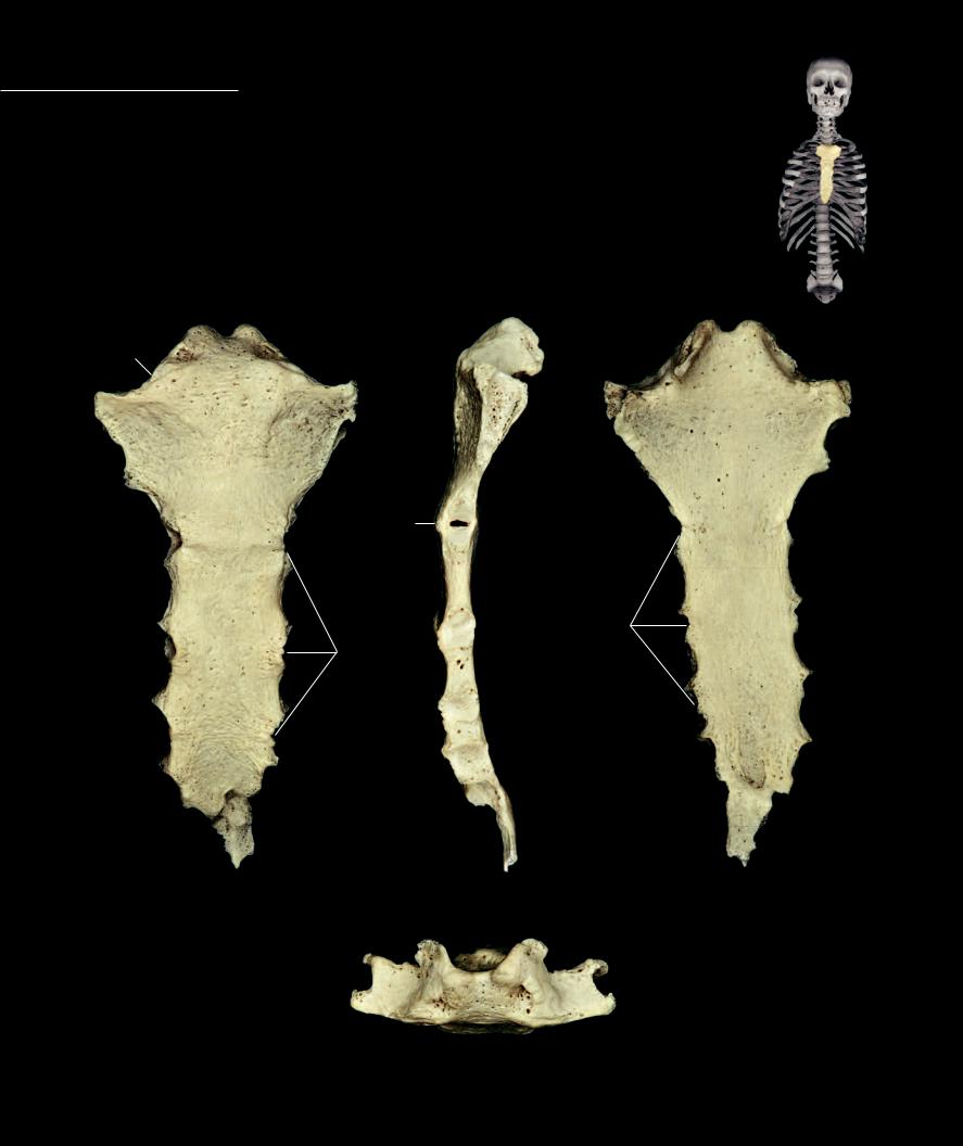
Sternum The sternum is the anterior bone of the thoracic wall. It forms from six segmental elements, or sternebrae, that fuse during development. The bone has the appear-
ance of a sword with a wide handle called the manubrium, a tapering blade or body, and a sharp point-like apex named the xiphoid process. A distinct angle forms at the junction of the manubrium and the body. This angle is called the sternal angle. A horizontal plane extended posteriorly intersects the disc between the fourth and fifth thoracic vertebrae and marks the top of the heart in the thoracic cavity. The lateral margins of the bone are notched for reception of the costal cartilages and clavicles. Its anterior surface is slightly convex, while the posterior surface is weakly concave. The sternum articulates with sixteen bones, more articulations than any other bone in the body.
1 |
Manubrium |
5 |
Body |
2 |
Clavicular notch |
6 |
Xiphoid process |
3 |
Jugular or suprasternal notch |
7 |
Costal notches |
4 |
Sternal angle |
|
|
3 |
|
|
3 |
|
|
|
|
2 |
|
2 |
|
|
|
2 |
|
|
1 |
|
|
1
1
4
7
4
7 |
7 |
5 |
|
7
5 |
|
|
|
7 |
|
|
7 |
|
6 |
6 |
6 |
|
||
|
|
Sternum |
Sternum |
Sternum |
Anterior view, superior at top |
Lateral view, anterior at left |
Posterior view, superior at top |
3
2
1
Sternum
Superior view, posterior at top
82
6 Appendicular Skeleton
The appendicular portion of the skeleton forms the framework of the limbs. It includes the limb
girdles, or fixed portion of the appendicular skeleton, and the series of bones that extend distally from the girdles into the limb proper, or free portion of the limb. The limb girdles, pectoral and pelvic, help anchor the limb to the axial skeleton. The free portion of each limb consists of a
large proximal element, the humerus and femur, forming the skeleton of the arm and thigh, respectively. Next in sequence are the ulna and radius of the forearm, and the fibula and tibia of the leg. The distalmost regions of the limbs are the hand and foot consisting of the short carpal and tarsal bones, respectively, along with the metacarpals, metatarsals, and phalanges of the digits.
As the tetrapod (land) vertebrates evolved, a major difference emerged between the two limbs. The anterior, or upper limb, evolved as
a steering device, while the posterior, or lower limb, became the locomotor limb. Accompanying these evolutionary modifications in limb function were important morphological differences. The powerful locomotor hind limb developed strong attachments to the axial skeleton. The strong iliosacral joint, with its accompanying
ligaments, transfers the powerful forces generated
by the posterior limb to the axial skeleton to propel the
body forward. On the other hand, the anterior limb developed minimal, weak skeletal attachments between the girdle and axial skeleton while becoming a more mobile limb.
As you study the skeleton of the limbs in the photos that follow, note the similarities and differences that exist between the bones of the superior and inferior limb skeletons and think about the functional differences mentioned above.
Find more information about the appendicular skeleton in
R E A L A N AT O M Y
83

Each superior limb consists of 32 bones. The proximal end of the superior limb, the clavicle and scapula, form the pectoral or shoulder girdle. This girdle of bones provides a broad base of support that is primarily
anchored to the axial skeleton by muscles rather than ligaments. The free part of the upper limb consists of the humerus, radius, ulna, and hand. The humerus forms the skeletal framework for the brachium. Distal to the brachium is the antebrachium containing the radius and ulna. The distal-most region of the superior limb is the hand consisting of a wrist region of eight carpal bones, the palm region consisting of fi ve metacarpal bones, and the fourteen phalanges of the fingers and thumb.
2
2
1 |
1 |
3 |
3 |
1 |
Scapula |
2 |
Clavicle |
3 |
Humerus |
4 |
Ulna |
5 |
Radius |
6 |
Carpals |
7 |
Metacarpals |
8 |
Phalanges |
4 |
5 |
5 |
4 |
6 |
6 |
7 |
7 |
8 |
8 |
Left upper limb |
Left upper limb |
|
Anterior view |
||
Posterior view |
||
|
84
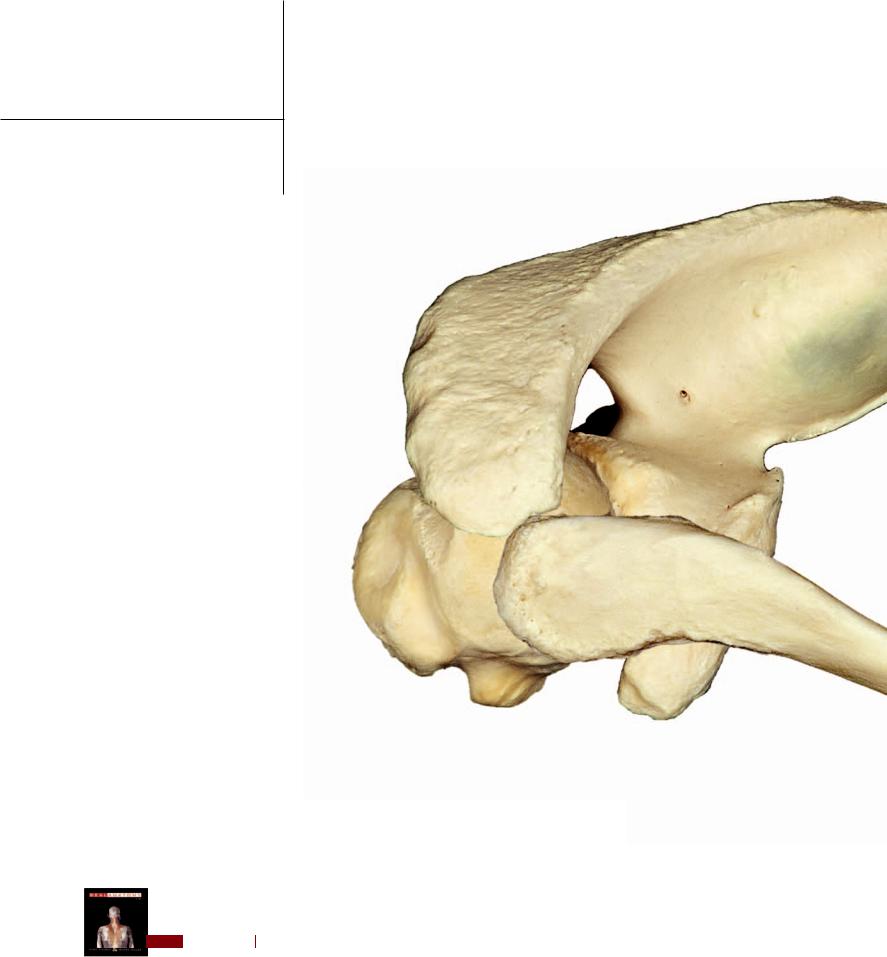
The pectoral, or shoulder, girdle consisting of the scapula and the clavicle forms the base of the upper limb skeleton. The rod-like clavicle forms a horizontal strut that links the scapula to the sternum
of the axial skeleton. The large triangular scapula presents an extensive surface area for muscle attachment and a large lateral fossa that articulates with the humerus of the free part of the upper limb. Except for the weak joint formed between the clavicle and the sternum, the pectoral girdle is essentially unattached by ligaments or joints to the axial skeleton. This was paramount in the evolutionary role of this limb as a steering device and shock absorber during locomotion.
1
1 Scapula
2 Clavicle
2
Left pectoral girdle |
2 |
Lateral view |
|
1
Left pectoral girdle
Superior view
85