

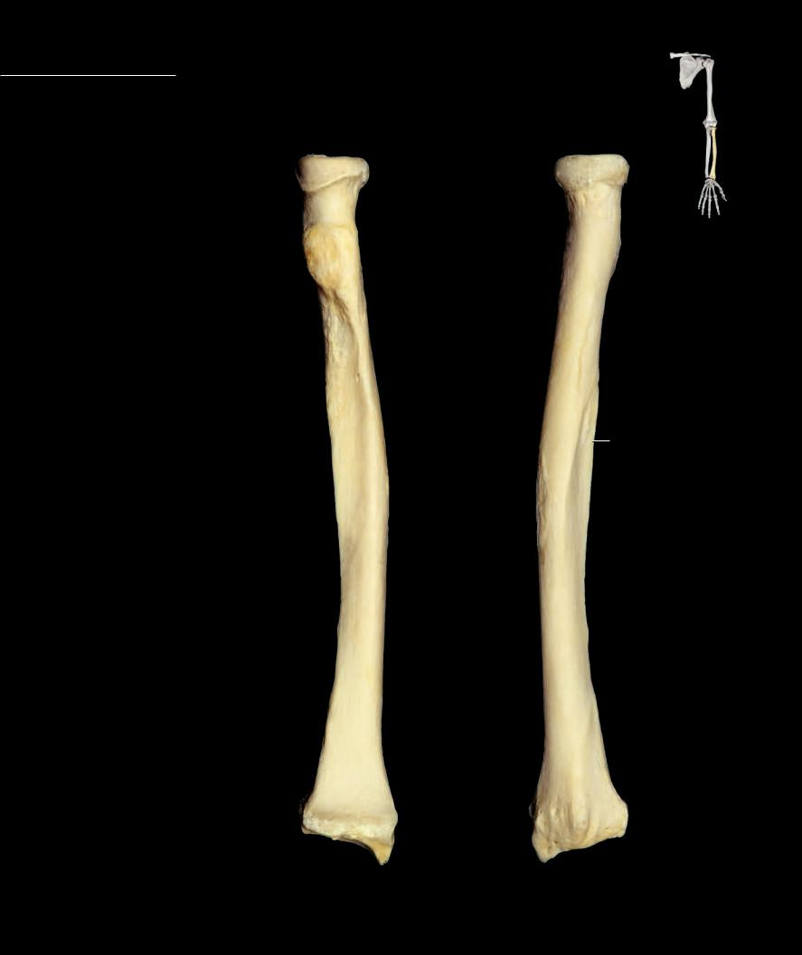
Radius The radius is the lateral, slender, rod-like bone of the antebrachium. The rod-like shaft expands at both ends. The proximal end forms a wheel-like head with a proximal con-
cavity, while the distal end expands from medial to lateral to form the widest part of the bone. The distal end is concave anteriorly and convex and grooved posteriorly. The ridge-like borders of the shaft give it a triangular shape in cross section. The radius articulates with four bones — the humerus, ulna, scaphoid, and lunate.
|
|
3 |
1 |
1 |
Head |
|
|
|
|
||
2 |
Articular facet |
|
4 |
3 |
Articular circumference |
|
|
|
|
||
4 |
Neck |
|
|
5 |
Shaft or body |
|
|
6 |
Radial tuberosity |
|
|
7 |
Pronator tuberosity |
6 |
|
8 |
Interosseous border |
|
|
9 |
Anterior border |
|
|
10Posterior border
11Radial styloid process
12Suprastyloid crest
13Dorsal tubercle
14Groove for extensor muscle tendons
15Ulnar notch
16Carpal articular surface
9
8
10
7
5
12 |
|
|
13 14 |
11 |
11 |
Left radius |
Left radius |
Anterior view, lateral to right |
Posterior view, lateral to left |
94
|
|
|
1 |
3 |
|
|
|
|
|
4 |
|
4 |
||
6 |
|
|
|
2 |
|
|
|
||
|
|
|
|
6 |
Left radius
Superior view, lateral to left
9
10
8
|
|
|
|
|
11 |
16 |
15 |
|
|
|
|
|
|
|
|
|
|
|
|
|
|
|
|
|
13 |
|
12 |
|
|
|
|
|
|
Left radius |
|
|
|
|
13 |
|
Inferior view, lateral to right |
|||
|
|
|
|
|||||
|
|
|
|
|
|
|
|
|
|
|
|
|
13 |
|
15 |
|
|
|
|
|
|
|
|
|
||
11 |
|
|
16 |
|
|
|||
|
|
|
|
|
|
|||
|
Left radius |
|
|
Left radius |
|
|
||
Lateral view, anterior to left |
Medial view, anterior to right |
|
|
|||||
|
|
|
|
|
|
|||
95
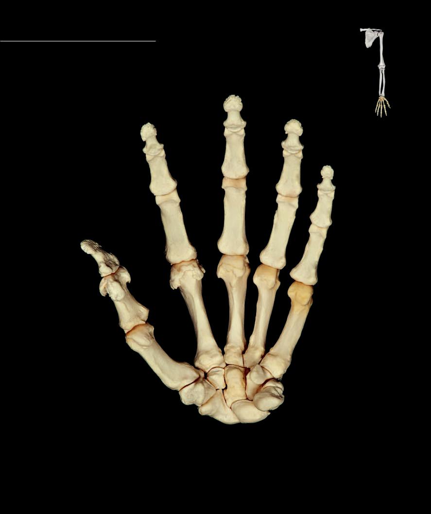
The hand is a composite structure consisting of 27 bones. The proximal end of the hand is the carpus or wrist. The carpal bones are eight in number and are arranged in two rows of four, a distal row
and a proximal row. Distal to the carpus are the fi ve digital rays. Each digit, called a fi nger of which there are four, consists of a metatarsal bone and three phalanges. The remaining digit, the thumb or pollex, has a metatarsal bone and only two phalanges. The photos of the hands below and on the opposing page are positioned as if you were looking at your own hand.
|
16 |
|
|
16 |
|
16 |
|
|
|
15 |
|
15 |
15 |
16 |
|
|
15
14
14 |
14 |
14
16
14
10 |
11 |
12 |
13
9
6 7
3
8
5
4
1 2
Left hand
Anterior view, lateral to left
96
1 |
Scaphoid |
9 |
Metacarpal I |
2 |
Lunate |
10 |
Metacarpal II |
3 |
Triquetrum |
11 |
Metacarpal III |
4 |
Pisiform |
12 |
Metacarpal IV |
5 |
Trapezium |
13 |
Metacarpal V |
6 |
Trapezoid |
14 |
Proximal phalanx |
7 |
Capitate |
15 |
Middle phalanx |
8 |
Hamate |
16 |
Distal phalanx |
16
16
16
|
|
15 |
16 |
15 |
15 |
|
|
15
14
14 |
14 |
14
16
14
11
12
10
13
9
8
7 |
6 |
5 |
3
21
Left hand
Posterior view, lateral to right
97
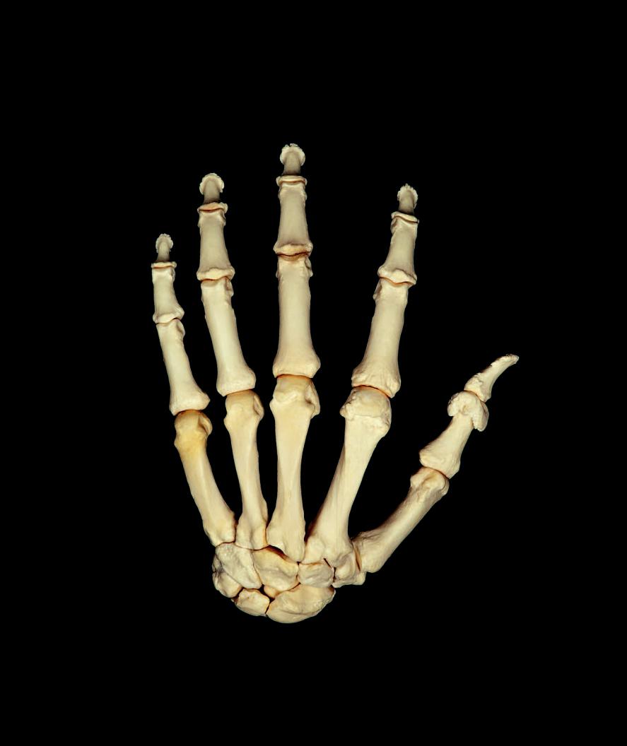
The eight carpal bones form the proximal end of the hand skeleton. The main features of this complex little series of bones are the numer-
ous articular surfaces they form with one another and with the metacarpal and antebrachial bones. The carpal bones form two rows of four bones each. The two largest bones of the proximal row, the scaphoid and the lunate, articulate with the distal end of the radius. The row of distal bones form the skeletal foundation for the fi ngers and articulate with the metacarpal bones of the fi ngers and thumb. The anterior surface of the carpal bones forms the fl oor of the carpal tunnel that supports the major digital fl exor tendons that enter the hand.
14
5
|
9 |
|
|
4 |
|
8 |
|
1 |
7 |
|
|
3 |
|
||
|
6 |
|
|
2 |
|
|
|
Left trapezium |
Left trapezoid |
10 |
|
Anterior view, lateral to left |
Anterior view, lateral to left |
||
|
25
24 |
|
30 |
31 |
22
|
26 |
32 |
|
|
|
|
27 |
29 |
|
|
|
|
|
28 |
23 |
|
|
|
|
Left lunate |
Left scaphoid |
|
Anterior view, lateral to left |
|
|
|
Anterior view, lateral to left |
|
|
15 |
16 |
|
20 |
||
|
21
12
17
19
13
18
11
Left hamate
Anterior view, lateral to left
Left capitate
Anterior view, lateral to left
35
34
33
Left pisiform
Left triquetrum
Anterior view, lateral to left
Anterior view, lateral to left
98

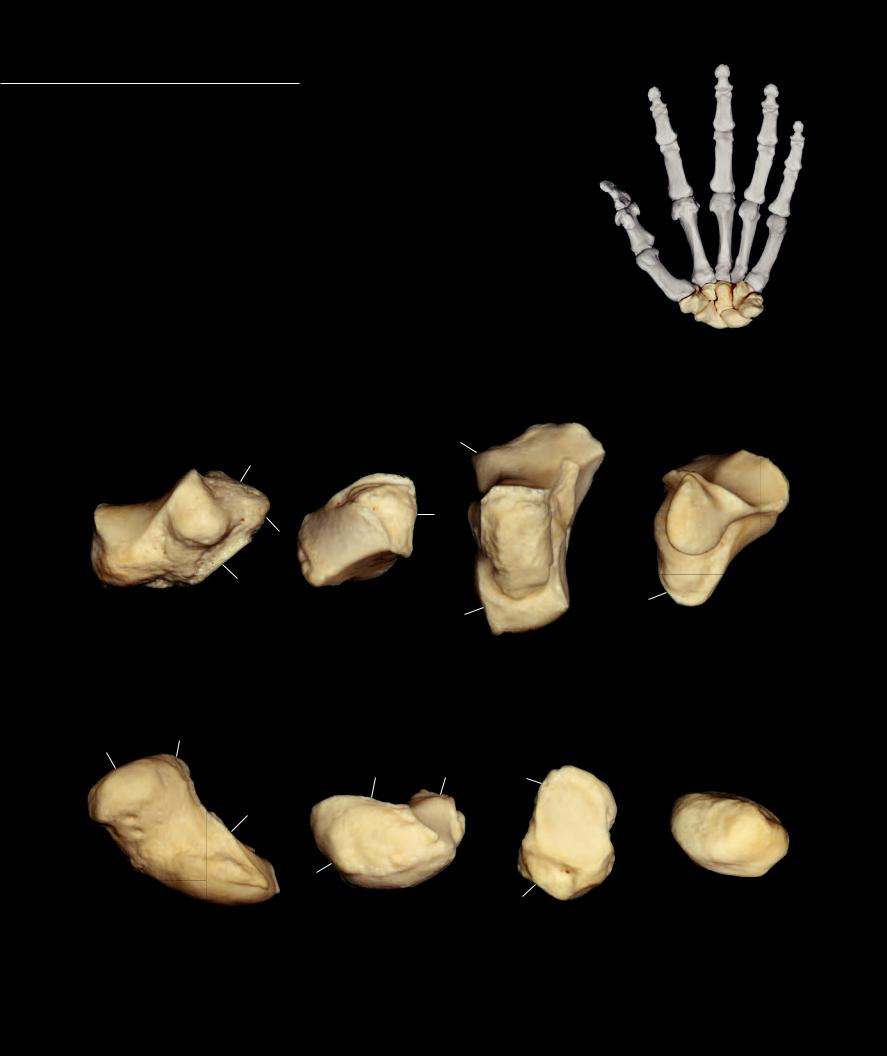
Trapezium |
Scaphoid |
||
1 |
Tubercle of trapezium |
22 |
Scaphoid tubercle |
2 |
Articular surface with scaphoid |
23 |
Articular surface with radius |
3 |
Articular surface with trapezoid |
24 |
Articular surface with trapezium |
4 |
Articular surface with first metacarpal |
25 |
Articular surface with trapezoid |
5 |
Articular surface with second metacarpal |
26 |
Articular surface with capitate |
Trapezoid |
27 |
Articular surface with lunate |
|
6 |
Articular surface with scaphoid |
Lunate |
|
7 |
Articular surface with trapezium |
28 |
Articular surface with radius |
8 |
Articular surface with capitate |
29 |
Articular surface with scaphoid |
9 |
Articular surface with first metacarpal |
30 |
Articular surface with capitate |
Capitate |
31 |
Articular surface with hamate |
|
10 |
Articular surface with scaphoid |
32 |
Articular surface with triquetrum |
11 |
Articular surface with lunate |
Triquetrum |
|
12 |
Articular surface with trapezoid |
33 |
Articular surface with lunate |
13 |
Articular surface with hamate |
34 |
Articular surface with pisiform |
14 |
Articular surface with second metacarpal |
35 |
Articular surface with hamate |
15 |
Articular surface with third metacarpal |
Pisiform |
|
16 |
Articular surface with fourth metacarpal |
36 |
Articular surface with triquetrum |
Hamate
17Hook of hamate or hamulus
18Articular surface with lunate
19Articular surface with triquetrum
20Articular surface with fourth metacarpal
21Articular surface with fifth metacarpal
15
16 |
|
17 |
|
14 |
9 |
|
21 |
20 |
|
|
12 |
|
|
|
5 |
|
|
|
|
|
|
7 |
||
|
|
|
|
|
||||
|
|
|
|
|
|
|
4 |
|
|
|
|
|
|
|
|
|
|
|
|
13 |
|
|
|
|
|
|
|
|
8 |
|
|
|
|
||
|
|
10 |
|
|
|
|
3 |
|
|
|
|
|
|
||||
|
|
|
|
|
|
|
|
|
|
|
11 |
|
|
6 |
|
|
|
|
|
|
|
|
|
|
|
|
19 |
18 |
Left capitate |
Left trapezoid |
|
2 |
|||
Left hamate |
Posterior view, lateral to right |
|
Left trapezium |
|||||
|
|
|
|
|
|
|
||
Posterior view, lateral to right |
Posterior view, lateral to right |
|
|
|
Posterior view, lateral to right |
|||
|
|
|
|
|||||
|
|
31 |
|
|
|
|
35 |
|
|
25 |
24 |
|
32 |
|
|
26 |
|
|
|
|
|
||
|
|
|
|
|
|
36 |
|
|
30 |
|
|
|
|
|
|
|
|
|
|
28 |
29 |
27 |
|
|
|
|
|
|
|
|
33 |
|
|
23 |
|
Left pisiform |
|
|
|
|
|
Left triquetrum |
Left lunate |
|
|
||
Posterior view, lateral to right |
|
|
|||
Posterior view, lateral to right |
Left scaphoid |
|
|||
|
Posterior view, lateral to right |
|
|||
|
|
|
|
||
Posterior view, lateral to right
99