


The axial skeleton is clearly depicted in the photos below. Note that this portion of the skeleton consists of three principal skeletal regions — the cranium, the vertebral column, and the
rib cage. There are 29 cranial bones, 26 vertebral bones, and 25 bones in the rib cage. On the pages that follow, each of the axial skeletal regions and the respective bones will be explored in greater detail.
1
1 |
Cranium |
|
|
2 |
Hyoid bone |
|
|
3 |
Cervical vertebral column |
|
|
4 |
Cervical vertebra 1 - Atlas |
|
|
5 |
Cervical vertebra 2 - Axis |
|
|
6 |
Cervical vertebra 7 |
|
|
7 |
Thoracic vertebral column |
|
|
8 |
Thoracic vertebra 1 |
|
|
9 |
Thoracic vertebra 12 |
2 |
|
10 |
Lumbar vertebral column |
||
11 |
Lumbar vertebra 1 |
|
|
12 |
Lumbar vertebra 5 |
|
|
13 |
Sacrum |
|
|
14 |
Sternum |
6 |
|
15 |
Ribs |
||
|
14
15
9
11
12
13
Axial skeleton
Anterior view
34
1 |
1 |
4 |
4 |
|
|
5 |
5 |
3
2
6
6
8
8
7
9
9
11 |
11 |
10
12
12
13
13
Axial skeleton |
|
Lateral view |
Axial skeleton |
|
|
|
Posterior view |
35

Cranium The cranium is the composite skeleton of the head and is composed of 29 bones. The bones of the cranium range from simple, non-descript plates of bone to the
most intricate bones of the skeleton. The cranial bones have a range of important functions, that include protecting the delicate brain tissue, fi xing the vestibular apparatus of the inner ear in three-dimensional space, maintaining open air passageways for respiration, and acquiring and processing food, to name a few. There are two main subdivisions of the cranium — the neurocranium or brain box is the region that surrounds and encases the brain, and the viscerocranium or facial skeleton is the area contributing to the orbits, nasal cavity, and oral cavity. This page and the facing page, and the four page spreads that follow, depict the fi ve normas, or views, of the cranium in both articulated and disarticulated cranial images. The bones of the skull are labeled on these views, along with key landmarks that can only be labeled on the articulated cranium. Individual landmarks of the bones are labeled on the individual pictures of the cranial bones on the pages that follow. This spread is of the norma facialis or facial aspect of the cranium.
1 Frontal bone
2 Parietal bone
3 Occipital bone
4 Sphenoid bone
5 Temporal bone
6 Ethmoid bone 1
7 Inferior nasal concha
8 Lacrimal bone
9 Nasal bone
10Vomer
11Maxilla
12Palatine bone
13Zygomatic bone
14Mandible
15Bony nasal cavity
16Piriform aperture
17Inferior nasal meatus
18Middle nasal meatu
19Orbit
19 |
|
|
|
|
|
|
|
|
|
|
|
|
12 |
4 |
|
|
|
|
|
|
|
|
9 |
|
8 |
|
|
||
|
|
|
|
|
|
|
|
|
|
|
|
|
||
|
|
|
|
|
|
|
|
|
|
|
|
|
|
|
|
|
|
|
|
|
|
|
|
6 |
18 |
|
|
|
13 |
|
|
|
|
|
|
|
|
|
|
|
|
|||
|
|
|
|
|
|
|
|
|
|
|
|
|||
|
|
|
|
|
|
|
|
|
|
|
|
|
||
|
|
|
|
|
|
|
6 |
|
|
|
|
|
||
7 |
|
|
15 |
|
|
|
|
|
||||||
16 |
|
|
10 |
17 |
|
|
|
|
||||||
|
|
|
|
|
|
|||||||||
|
|
|
|
|
|
|
|
|
|
|
||||
|
|
|
|
|
|
|
|
|
|
|
11 |
|
||
|
|
|
|
|
|
|
|
|
|
|
|
|||
14
Cranium
Anterior view
2
5
36

2
1
3
98
4
4
5
6
10
12
13
7
11
14
Bones of the cranium disarticulated
Anterior view
37

Cranium This page spread depicts the norma lateralis, or lateral aspect of the cranium. In this view both the brain box and facial skeleton are clearly visible and the relative proportions of the two cranial
regions are evident. In the disarticulated view, only those bones that are visible in the lateral aspect are shown.
1 Frontal bone
2 Parietal bone
3 Occipital bone
4 Sphenoid bone
5 Temporal bone
6 Ethmoid bone
7 Lacrimal bone
8 Nasal bone
9 Maxilla
10Zygomatic bone
11Mandible
12Zygomatic arch
13Pterygopalatine fossa
2 |
1 |
|
|
4 |
|
|
6 |
5 |
12 |
7 |
|
|
8
10
3
13 
9
11
Cranium
Lateral view
38

2
1
3
4
6 |
7 |
5
8
10
9
11
Bones of the cranium disarticulated
Lateral view
39

Cranium This page spread depicts the norma occipitalis, or occipital aspect of the cranium. From this posterior view the internal aspects of the bones of the oral and nasal cavities are clearly visible. In the
disarticulated view only those bones that are visible in the occipital aspect of the cranium are depicted.
1 Parietal bone
2 Occipital bone
3 Sphenoid bone
4 Temporal bone
5 Ethmoid bone
6 Inferior nasal concha
7 Vomer
8 Maxilla
9 Palatine bone
10Zygomatic bone
11Mandible
12Choana or posterior nasal aperture
13Inferior orbital fissure
14Bony nasal cavity
15Middle nasal meatus
16Inferior nasal meatus
17Bony palate
18Sutural bone
1
18
2
4
|
|
|
|
|
|
|
3 |
13 |
|
|
|
7 |
5 |
||
|
|
|
|
||||
|
|
|
|
|
|
|
|
12 |
|
|
14 |
15 |
10 |
||
|
|
||||||
|
|
|
|
6 |
|
||
|
|
|
|
|
|
|
|
16 |
|
|
|
|
|
9 |
|
|
|
|
|
|
|
||
|
|
|
|
|
|
||
|
|
|
|
|
|
|
|
17 
8
11
Cranium
Posterior view
40

1
2
4
3
10
5
6
7 |
9 |
8
11
Bones of the cranium disarticulated
Posterior view
41

Cranium This page spread depicts the norma superior, or superior aspect of the cranium. This view clearly depicts the neurocranium or brain box, while the facial skeleton is almost completely hidden from
view. In the disarticulated view only those bones that are visible in the superior aspect of the cranium are depicted.
1 |
Frontal bone |
|
2 |
Parietal bone |
5 |
3 |
Occipital bone |
6 |
4 |
Temporal bone |
|
5 |
Nasal bone |
|
6 |
Maxilla |
|
7 |
Zygomatic bone |
|
7
1
4
2
Cranium
Superior view
42

56
7
1
4
2
3
Bones of the cranium disarticulated
Superior view
43

Cranium This page spread depicts the norma inferior (basalis), or inferior aspect of the cranium. The mandible has been removed to more clearly reveal the basicranium. This view clearly depicts the fl oor
of the brain box, the bony palate forming the roof of the oral cavity, and mandibular tooth row. In the disarticulated view only those bones that are visible in the inferior aspect of the cranium are depicted.
1 |
Occipital bone |
|
|
|
|
2 |
Sphenoid bone |
|
|
|
|
3 |
Temporal bone |
|
|
14 |
|
4 |
Vomer |
|
|
|
|
5 |
Maxilla |
|
|
|
|
6 |
Palatine bone |
|
|
|
|
7 |
Zygomatic bone |
|
|
5 |
|
8 |
Bony palate |
|
|
||
|
|
|
|
||
9 |
Choana or posterior nasal aperture |
8 |
|
7 |
|
|
|||||
10 |
Zygomatic arch |
|
|
||
|
|
|
|
||
11 |
Jugular foramen |
|
|
|
|
12 |
Foramen lacerum |
|
|
|
|
13 |
Greater palatine foramen |
|
|
|
|
14 |
Incisive fossa |
|
|
6 |
|
|
|
|
|
||
|
13 |
|
|
10 |
|
|
|
|
|
|
|
|
|
9 |
|
||
|
|
4 |
|||
|
|
2 |
|||
|
12 |
3 |
|||
11
1
Cranium
Inferior view
44
5
6
7
4
2
3
1
Bones of the cranium disarticulated
Inferior view
45

Cranium This page spread depicts the cranium sectioned in a parasagittal plane through the right side of the nasal cavity just lateral to the bony nasal septum. The section below depicts the lateral wall of
the right nasal cavity, and the section on the opposite page depicts the medial (septal) wall of the right nasal cavity. The osseous sinuses that communicate with the nasal cavity are all visible in these sections.
2
1
18 |
|
|
8 |
3 |
|
19 |
6 |
|
5 |
||
|
||
|
17 |
|
|
23 |
|
20 |
4 |
|
7 |
|
|
24 |
10 |
11 |
|
12
Parasagittal section of the cranium
Medial view of the right side
46

1 |
Frontal bone |
7 |
Inferior nasal concha |
13 |
External table of calvaria |
19 Ethmoidal air cells (sinuses) |
|
2 |
Parietal bone |
8 |
Nasal bone |
14 |
Diploë |
20 |
Maxillary sinus |
3 |
Occipital bone |
9 |
Vomer |
15 |
Internal table of calvaria |
21 |
Incisive canal |
4 |
Sphenoid bone |
10 |
Maxilla |
16 |
Groove for sigmoid sinus |
22 |
Bony nasal septum |
5 |
Temporal bone |
11 |
Palatine bone |
17 |
Sphenoidal sinus |
23 |
Sphenopalatine foramen |
6 |
Ethmoid bone |
12 |
Mandible |
18 |
Frontal sinus |
24 |
Inferior nasal meatus |
14
2
13
15
1
18
3 |
5 |
|
|
|
8 |
|
|
|
|
||||
|
|
17 |
6 |
|||
|
|
|
|
|
|
|
16 |
|
4 |
22 |
|
|
|
|
|
|
|
|
|
|
9
11
10
21
12
Parasagittal section of the cranium
Medial view of the left side
47

Cranium This page spread depicts the cranium sectioned in a horizontal plane through the neurocranium, or brain box, revealing the internal aspects of the fl oor and roof of the sectioned cranial cavity. On this page the fl oor of the neurocranium is visible, while on the opposing page the roof of the neurocranium is visible. The superior portion of the cranium, depicted on the opposite page, is called the calvaria.
1
8
13 
6
4
14 
9
11
7 12
12
5
2
10
15 
3
Cranium with calvaria removed
Superior or internal view of the cranial base
48

1 |
Frontal bone |
5 |
Temporal bone |
9 |
Foramen lacerum |
13 |
Anterior cranial fossa |
2 |
Parietal bone |
6 |
Ethmoid bone |
10 |
Jugular foramen |
14 |
Middle cranial fossa |
3 |
Occipital bone |
7 |
Clivus |
11 |
Petrosphenoidal fissure |
15 |
Posterior cranial fossa |
4 |
Sphenoid bone |
8 |
Foramen caecum |
12 |
Petro-occipital fissure |
16 |
Granular foveolae |
1
16
2
3
Removed calvaria
Inferior or internal view
49

The unpaired frontal bone has a bowl-like shape that consists of two parts, an internally concave
vertical portion termed the squama and a horizontal plate that forms the superior walls of the orbits. The bone has a smooth external surface, while its internal surface consists of impressions made by the meningeal vessels and scattered foramina that transmit diploic vessels. The squamous portion of the bone is thick. It consists of internal and external laminae of compact bone sandwiching a layer of trabecular bone called diploë. Near the anterior, inferior midline the spongy bone is absent between the external and internal laminae and in its place are variably sized spaces — the frontal sinuses. The orbital plate consists of a thin plate of compact bone, which is often so thin that it is translucent. The frontal bone articulates with twelve bones.
1 Squamous part
2 Frontal tuber
3 Glabella
4 Superciliary arch
5 Supra-orbital notch or foramen
6 Frontal notch or foramen
7 Temporal surface
8 Zygomatic process
9 Frontal crest
10Groove for superior sagittal sinus
11Nasal spine
12Orbital surface
13Trochlear spine
14Lacrimal fossa
15Ethmoidal notch
16Frontal sinus
1
2
7
5
46
|
|
3 |
|
|
12 |
|
|
14 |
8 |
||
|
|||
|
|
13 |
|
|
|
11 |
Frontal bone
Anterior view
1
10
9
8
11
Frontal bone
Posterior view
50

1
2
Frontal bone |
|
11 |
|
Superior view, anterior to bottom |
|
|
|
|
5 |
|
|
|
14 |
16 |
|
|
|
|
|
|
|
12 |
8 |
|
|
15 |
|
Frontal bone
Inferior view, anterior to top
1
2
7
4
3
12 8
13
11
Frontal bone
Lateral view, anterior to left
51

The parietal bones are large quadrilateral bones forming the greater part of the roof and sides of the cra-
nium. The external surface of each parietal bone is slightly convex while the internal surface is concave and marked with impressions from meningeal vessels. The inferior border forms a beveled articular surface, while the superior, anterior, and posterior borders form deeply denticulate articular surfaces. The bone consists of inner and outer laminae of compact bone sandwiching a layer of trabecular bone, the diploë. Each parietal bone articulates with five bones.
1 |
Groove for sigmoid sinus |
|
|
2 |
Groove for superior sagittal sinus |
|
|
3 |
Grooves for middle meningeal artery |
4 |
|
4 |
Superior temporal line |
||
5 |
Inferior temporal line |
11 |
|
6 |
Parietal tuber |
||
6 |
|||
7 |
Squamosal border |
||
12 |
|||
8 |
Occipital border |
||
5 |
|||
9 |
Frontal border |
|
|
10 |
Sagittal border |
9 |
|
11 |
Frontal angle |
||
|
|||
12 |
Occipital angle |
8 |
13Sphenoid angle
14Mastoid angle
15Parietal foramen
7
14
13
Left parietal bone
Lateral view, anterior to right
10 2
11
12
8
9
3
14
1
13
Left parietal bone
Medial view, anterior to right
52

11 |
10 |
12 |
|
|
|
15
8
9
6
Left parietal bone |
10 |
11 |
Superior view, anterior to left |
|
|
2 |
|
|
12 |
|
|
8 |
3 |
9 |
|
|
13
14
7
Left parietal bone
Inferior view, anterior to right
10 |
|
10 |
|||
|
|
|
|
|
|
11 |
|
|
|
|
|
|
|
12 |
|||
|
|
4 |
|||
|
|
|
6 |
|
|
|
|
|
|||
|
|
6 |
|
|
|
|
|
|
|
||
|
9 |
8 |
|||
|
|
|
|
|
|
|
|
5 |
|||
|
14 |
13 |
|
|
Left parietal bone |
Left parietal bone |
Posterior view |
Anterior view |
|
53

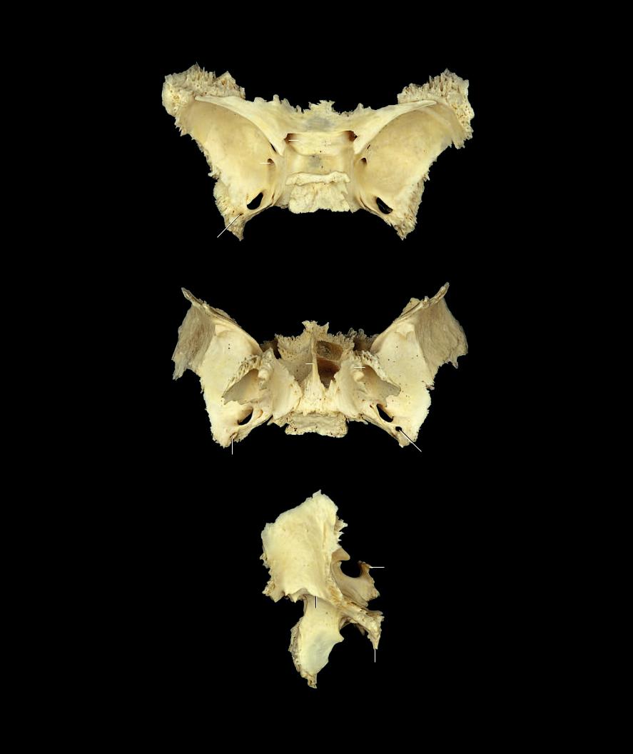
The occipital bone forms the greater part of the posterior and inferior cranium. Viewed from
behind it has an oval to round shape. The bone has four distinct regions. The squamous portion is the internally concave posterosuperior plate and forms the greater part of the bone. The thick quadrilateral basioccipital, or basilar part, contributes to the base of the cranium anterior to the foramen magnum. Lateral to this and converging with the squama are the two condylar parts or exoccipitals. Together the four regions of the bone form the borders to the large circular opening, the foramen magnum, which provides passage for the spinal cord between the cranial vault and the spinal canal. The occipital bone articulates with six bones.
1 Foramen magnum
2 Clivus
3 Pharyngeal tubercle
4 Squamous part
5 Mastoid border
6 Lambdoid border
7 Occipital condyle
8 Condylar canal
9 Hypoglossal canal
10Condylar fossa
11Jugular tubercle
12Jugular notch
13Jugular process
14External occipital protuberance
15Superior nuchal line
16Inferior nuchal line
17Internal occipital protuberance
18Groove for transverse sinus
19Groove for occipital sinus
20Groove for superior sagittal sinus
21Cerebral fossa
22Cerebellar fossa
64
20
|
21 |
18 |
|
17 |
|
19 |
22 |
|
1
8 5
13 |
11 |
|
Occipital bone
Anterior view
54
6
4
15
14
16
5
7
Occipital bone
Posterior view
|
|
|
|
|
4 |
|
|
15 |
14 |
|
|
6 |
|
|
|
16 |
|
|
|
|
18 |
|
|
|
|
|
|
|
|
||
|
|
|
|
|
17 |
|
|
|
|
|
|
22 |
|
|
|
|
1 |
|
|
|
|
|
|
5 |
10 |
|
|
|
|
||
|
|
|
|
|
|
|
|
|
|
|
|
5 |
1 |
|
|
7 |
|
|
|
|
|
|
|
|
|
|
|
|
|
8 |
|
13 |
12 |
|
|
|
|||
|
|
||||||
|
|
|
|
||||
|
|
13 |
|||||
|
|
|
|
|
|||
|
9 |
12 |
11 |
|
|
||
|
|
|
|
||||
|
|
|
3 |
|
2 |
|
|
|
|
|
|
|
|||
|
|
|
|
|
|
|
|
Occipital bone |
Occipital bone |
Inferior view, anterior to bottom |
Superior view, anterior to bottom |
6
4
15
14
16
10 |
2 |
|
5 |
||
|
||
|
13 |
|
|
7 |
Occipital bone 3
Lateral view, anterior to right
55

The temporal bone is a complex bone with fi ve distinct parts. The squamous part of
the bone is the thin lateral plate that contributes to the lateral wall of the cranium. It projects anteriorly as the zygomatic process and forms the mandibular fossa for the temporomandibular joint. The styloid part is represented by the styloid process. This projection of bone arises from the upper elements of the second pharyngeal arch. The petrous part forms the thick pyramidal base of the bone. It begins posterior to the external acoustic meatus as the mastoid process and ends where it forms a junction with the basi-occipital and greater wing of the sphenoid. The name petrous describes its rock-like appearance. This is the thickest part of the temporal bone. It arises from the otic capsules that stabilize the delicate internal ear structures. The mastoid is the posterolateral protuberance of the petrous portion that is easily palpable just posterior to the ear. The tympanic part of the temporal bone is the ring-like plate that forms the walls of the external acoustic meatus. Each temporal bone articulates with five bones.
1 Petrous part
2 Mastoid process
3 Mastoid notch
4 Groove for sigmoid sinus 22
5 Carotid canal
6 Apex of petrous part
7 Musculotubal canal
8 Tegmen tympani
9 Hiatus for greater petrosal nerve
10Hiatus for lesser petrosal nerve
11Trigeminal impression
12Internal acoustic meatus
13Mastoid canaliculus
14Tympanic canaliculus
15 |
Styloid process (broken) |
23 |
24 |
|
|
|
|
19 |
|
|
|||
16 |
Stylomastoid foramen |
|
25 |
|
|
|
17 |
Jugular notch |
|
|
|
|
|
|
|
|
|
|
||
18 |
Tympanic ring |
|
|
18 |
|
27 |
19 |
External acoustic meatus |
|
21 |
|
|
|
|
|
|
|
|||
20 |
Greater tympanic spine |
|
20 |
|
2 |
|
21 |
Lesser tympanic spine |
|
|
|||
22Squamous part
23Zygomatic process
24 |
Mandibular fossa |
Left temporal bone |
|
25 |
Articular tubercle |
||
Lateral view, anterior to left |
|||
26 |
Petrotympanic fissure |
||
|
|||
27 |
Tympanomastoid fissure |
|
|
|
|
22 |
23
1
12
5
4
2
Left temporal bone
Medial view, anterior to right
56

22
|
8 |
|
||
|
10 |
|
|
|
|
1 |
|
|
|
|
|
|
|
|
11 |
|
|
|
9 |
|
||||
6 |
|
23 |
|
|
|
5 |
7 |
24 |
|
||
|
|
Left temporal bone
Anterior view
23
22
6
11
10 
9 1
8
Left temporal bone
Superior view, anterior at top
22
12
23 |
1 |
3
2
Left temporal bone
Posterior view
3
215
13
14 17
18 |
1 |
|
5 |
24
26
25
23
Left temporal bone
Inferior view, anterior at bottom
57

The sphenoid bone is a complex bone that has the spread -winged appearance of a but-
terfl y. Like it name suggests, it is wedged into the center of the cranium where it articulates with twelve neighboring bones and contributes to much of the cranial base. It is divisible into four principal components — the body, greater wings, lesser wings, and pterygoid processes. With the calvaria removed the bone is visible from any view. This bone plays a prominent role at the base of the skull. It supports the brain, serves to protect the optic stalks and capsules, provides passage for many vessels and nerves entering and leaving the skull, and forms a sinus cavity that communicates with the nasal cavity.
1 |
Jugum |
|
|
|
|
|
|
|
2 |
Sella turcica |
|
|
|
|
|
|
|
3 |
Tuberculum sellae |
|
|
|
|
|
|
|
4 |
Hypophysial fossa |
|
|
|
|
|
|
|
5 |
Dorsum sellae |
|
|
|
|
|
|
|
6 |
Posterior clinoid process |
|
|
|
|
|
|
|
7 |
Middle clinoid process |
|
5 |
6 |
|
|||
8 |
Carotid sulcus |
|
|
|||||
|
|
|
|
|
|
|
||
9 |
Sphenoidal crest |
17 |
13 |
|
|
|
|
|
10 |
Sphenoidal rostrum |
|
|
|
|
|
||
11 |
Sphenoidal sinus |
|
1 |
16 |
||||
12 |
Sphenoidal concha |
|
||||||
|
|
|
|
|
|
|
||
13 |
Lesser wing |
|
|
|
|
|
|
|
14 |
Optic canal |
|
11 |
19 |
||||
15 |
Anterior clinoid process |
|
|
12 9 |
|
|
|
|
|
|
|
|
|
|
|||
16 |
Superior orbital fissure |
|
|
|
|
|
|
|
17 |
Greater wing |
|
|
|
|
20 |
||
|
31 |
|
||||||
18 |
Infratemporal crest |
|
|
|
|
|
||
|
|
|
|
|
|
|
||
19 |
Orbital surface |
|
|
|
|
29 |
|
|
20 |
Foramen rotundum |
|
10 |
|
|
|
||
21 |
Foramen ovale |
24 |
|
|
|
25 |
|
|
22 |
Foramen spinosum |
|
|
|
|
|||
|
|
|
|
|
|
|
||
23 |
Spine of sphenoid bone |
|
|
|
|
|
|
|
24 Lateral plate of pterygoid process |
|
30 |
|
|
|
|
|
|
25 Medial plate of pterygoid process |
|
|
|
|
|
|
||
|
|
|
|
26 |
||||
26 |
Pterygoid notch |
|
|
|
|
|||
|
|
Sphenoid bone |
||||||
27 |
Pterygoid fossa |
|
|
|||||
28 |
Scaphoid fossa |
|
|
Anterior view |
||||
29Vaginal process
30Pterygoid hamulus
31Pterygoid canal
|
|
|
15 |
3 |
|
|
||
|
|
|
13 |
|||||
16 |
|
|||||||
|
17 |
|||||||
|
|
|
|
8 |
|
|||
23 |
|
|
|
|
|
|
|
31 |
|
|
|
|
|||||
|
|
28 |
|
|
18 |
|||
|
|
|
|
|||||
|
|
|
|
|
||||
|
|
|
|
|||||
|
|
|
|
|
|
|
|
|
|
|
|
|
29 |
|
|
||
10 27
24 25
30
26
Sphenoid bone
Posterior view
58
13 |
|
|
|
|
|
17 |
|
|
|
|
1 |
|
|
14 |
15 |
||
|
|||||
|
|
|
|
|
|
7 |
3 |
||||
20 |
|
|
|
|
4 |
|
|
|
|
||
|
|
|
|
|
|
6 |
|
5 |
|||
21 |
|
|
|
8 |
|
22
Sphenoid bone
Superior view, anterior at top
17 |
|
|
|
|
10 |
|
|
|
|
26 |
|
|||
27 |
|
|||
25 |
||||
29 |
||||
|
|
|
||
23 |
28 |
|||
|
|
|
||
23 Sphenoid bone
Inferior view, anterior at top
17
15
5
2
18
24
18 
24
21
22
6
23
Sphenoid bone
Lateral view, anterior to left
59

The maxillae are large, paired bones that unite to form the upper jaw. They also contribute to the
walls of the nasal cavity, orbit, oral cavity, and maxillary sinus. The maxillary sinus is the hollow central cavity within the large body of the maxilla. Four variable-shaped processes project from the maxillary body. The processes are the posterolateral zygomatic process, the medial projecting palatine process, the arched inferior process called the alveolar, and the superiorly projecting frontal process. Each maxilla articulates with nine bones.
|
|
10 |
|
1 |
Orbital surface |
|
|
2 |
Infra-orbital groove |
|
|
3 |
Infra-orbital foramen |
|
|
4 |
Anterior nasal spine |
|
|
5 |
Canine fossa |
|
|
6 |
Maxillary tuberosity |
|
|
7 |
Lacrimal groove |
|
|
8 |
Maxillary sinus |
|
|
9 |
Greater palatine groove |
3 |
|
10 |
Frontal process |
||
11 |
|||
11 |
Zygomatic process |
||
|
|||
12 |
Palatine process |
|
|
13 |
Incisive canal |
5 |
|
14 |
Alveolar process |
4 |
|
15 |
Interalveolar septum |
|
|
|
|
14 |
|
|
|
15 |
Left maxilla
Anterior view
10
11
6
12
Left maxilla
Posterior view
60

10
7
1
3 11
6
5
4
14
15
Left maxilla
Lateral view, anterior to left
14
10
7
11 |
1 |
12 |
|
2
Left maxilla
Superior view, anterior at top
10
8
9 |
12 |
|
4 |
|||
|
|
|||||
|
14 |
|
|
|
|
13 |
|
|
|
|
|
||
|
|
|||||
|
|
|
|
|
|
|
Left maxilla
Medial view, anterior to right
13 15
14
12
11
Left maxilla
Inferior view, anterior at top
61

The mandible, the largest of the facial bones, forms the lower jaw. The bone has an
arched body with a tooth-bearing alveolar process. Posteriorly each side of the arched body joins the vertically directed rami at the mandibular angle. The superior aspects of the two rami articulate with the temporal bones at the base of the cranium. The mandible is a strong bone composed predominantly of compact bone. It houses the lower tooth row in its alveolar arch. The strong masticatory muscles act on this bone to move it in the temporomandibular joint. Its shape can vary exceedingly with age. If the teeth are lost, bone gets resorbed on the alveolar surface leading to the thinning of the dental arch. The mandible articulates with two bones.
1 |
Body of mandible |
|
|
|
2 |
Mental protuberance |
18 |
15 |
|
3 |
Mental foramen |
|
||
|
|
|||
4 |
Mental tubercle |
|
|
|
5 |
Oblique line |
|
|
|
6 |
Digastric fossa |
|
|
|
7 |
Mental spines |
|
|
|
8 |
Mylohyoid line |
5 |
|
|
9 |
Submandibular fossa |
12 |
||
|
10Alveolar part
11Retromolar triangle
12Ramus of mandible
13Angle of mandible
14Mandibular foramen
15 |
Coronoid process |
|
10 |
|
|
20 |
|||
16 |
Mandibular notch |
13 |
|
|
17 |
Condylar process |
3 |
||
1 |
||||
18 |
Head of mandible |
|||
|
||||
19 |
Pterygoid fovea |
|
|
|
20 |
Masseteric tuberosity |
|
2 |
|
21 |
Pterygoid tuberosity |
|
||
|
4 |
|||
|
|
|
Mandible
Anterior view
18
|
|
|
17 |
|
12 |
|
|
|
|
14 |
|
|
|
|
21 |
|
10 |
|
|
8 |
1 |
9 |
||
|
||||
13 |
|
|||
|
|
|
7 |
6 |
|
Mandible
Posterior view
62

18
19
17
15
11
7
17
4
2
Mandible
Superior view, anterior at bottom
14
21
13
9
8
1
7
18 |
15 |
6
17
16
Mandible
Inferior view, anterior at bottom
|
12 |
|
|
10 |
|
|
3 |
|
20 |
1 |
|
|
||
|
||
13 |
2 |
|
|
4 |
|
Mandible
Lateral view, anterior to right
63

The term ethmoid comes from the Greek term ethmos meaning sieve. Galen called the bone the
sieve-like bone because of the many small foramina that transmit the olfactory nerves to the nasal cavity. This unpaired bone is both complex and delicate and is the central bone of the nasal cavity. Wedged between the two orbits, the bone consists of a median vertical plate, a horizontal plate perforated by many small foramina, and bilateral pneumatic, labyrinthine regions. The labyrinthine regions form most of the medial walls of the orbit and the superior and middle nasal conchae. This bone consists of thin laminae of compact bone surrounding many small air sinuses, which communicate with the nasal cavity. The ethmoid bone articulates with thirteen bones, more articulations than any other cranial bone.
1 Cribriform plate
2 Cribriform foramina
3 Crista galli
4 Perpendicular plate
5 Ethmoidal air cells
6 Orbital plate
7 Superior nasal concha
8 Middle nasal concha
9 Ethmoidal bulla
10Uncinate process
11Ethmoidal infundibulum
3
5
6
5 |
5 |
4
8
Ethmoid bone
Anterior view
3
2
1
6
5
3
Ethmoid bone
Superior view, anterior at top
6
10
Ethmoid bone
Lateral view, anterior at right
3
1
7
4 |
8 |
10 |
Ethmoid bone
Posterior view
11
4
9 8 1
7
10
Ethmoid bone
Inferior view, anterior at top
64
The zygomatic bone, originally named by Galen the os zygoma, comes from
the Greek word zygon meaning yoke, after its resemblance to a yoke placed on oxen. This yoke-shaped bone has three distinct surfaces, fi ve borders, and two processes. It is situated anterolateral on the face as the “cheekbone”, and contributes to the lateral and inferior walls of the orbit. It consists of external and internal laminae of compact bone with an inner core of spongy bone. The zygomatic bone articulates with four bones.
1 Orbital surface
2 Temporal surface
3 Lateral surface
4 Temporal process
5 Frontal process
6 Zygomatico-orbital foramen
7 Zygomaticofacial foramen
8 Zygomaticotemporal foramen
5
1
3
Right zygomatic bone
Anterior view
6
1
5
4
Right zygomatic bone
Superior view, anterior to top
5
7
7
3
4
5
2
8
4
Right zygomatic bone
Posterior view
3
4
5
Right zygomatic bone
Inferior view, anterior to top
5
8
2
1
4
Right zygomatic bone |
Right zygomatic bone |
Medial view, anterior to left |
|
Lateral view, anterior to right |
|
65

The palatine bone is a delicate and intricate bone that forms the shape of the letter L. It sits deep
in the posterior facial region where it contributes to the roof of the mouth, fl oor of the orbit, fl oor and lateral walls of the nasal cavity, and to the pterygopalatine fossa. It has a strong horizontal plate with a delicate vertical lamina that projects superiorly. The palatine bone articulates with six bones.
1 Perpendicular plate
2 Sphenopalatine notch
3 Greater palatine groove
4 Pyramidal process
5 Orbital process
6 Lesser palatine foramina
7 Posterior nasal spine
8 Conchal crest
9 Horizontal plate
66
9
5
7
4
Left palatine bone
Superior view, anterior at top
25
1
9
4
Left palatine bone
Anterior view, lateral at right
5 2
1
4
Left palatine bone
Lateral view, anterior at left
4
7
9
3
Left palatine bone
Inferior view, anterior at bottom
52
1
|
3 |
7 |
4 |
9 |
|
|
|
Left palatine bone
Posterior view, lateral at left
5
1
8
9
4
Left palatine bone
Medial view, anterior at right

The vomer is a fl at, triangular bone that resembles a plow. It has a fl at, median, vertical blade-like process
with transverse posterosuperior projections resembling the handles of the plow. This is a small, thin, unpaired bone that sits in the median plane. It is wider at its superoposterior base and it tapers toward its antero-inferior apex. It forms the inferior portion of the bony nasal septum. Its surfaces face laterally and form the lower, medial wall of the nasal cavities. The vomer articulates with six bones and one cartilage.
1 Ala of vomer
2 Vomerine groove
3 Vomerine crest of choana
4 Cuneiform part
1 |
|
3 |
|
2
|
|
4 |
1 |
|
|
3 |
Vomer |
Vomer |
|
Posterior view |
Anterior view
4
1 |
1 |
|
2
Vomer
Lateral view, anterior at left
Vomer |
Vomer |
Superior view, anterior at bottom |
Inferior view, anterior at bottom |
67

The paired nasal bones are small, rectangular bones with a subtle bow-like shape. They form the bridge of the nose
upon which a pair of eye glasses rest. The external surface of the bones provides attachment for the procerus and nasalis muscles, two thin muscles of facial expression. Each nasal bone articulates with four bones.
1 Ethmoidal groove
2 Nasal foramina
3 Superior border
4 Inferior border
5 Lateral border
6 Medial border
3
2
56
4
Left nasal bone
Anterior view, lateral at left
3
5
4
Left nasal bone
Lateral view, anterior at right
1
3
Left nasal bone
Superior view, anterior at bottom
3
1
65
4
Left nasal bone
Posterior view, lateral at right
3
6
4
Left nasal bone
Medial view, anterior at left
4
Left nasal bone
Inferior view, anterior at bottom
68