

This page intentionally left blank

ATLAS OF
HUMAN ANATOMY
Mark Nielsen
University of Utah
Shawn Miller
University of Utah
JOHN WILEY & SONS, INC.
Vice President & Executive Publisher |
Kaye Pace |
Acquisitions Editor |
Bonnie Roesch |
Project Editor |
Lorraina Raccuia |
Production Manager |
Dorothy Sinclair |
Senior Production Editor |
Anna Melhorn |
Marketing Manager |
Clay Stone |
Creative Director |
Harry Nolan |
Senior Designer |
Madelyn Lesure |
Media Editor |
Linda Muriello |
Cover Photo Credit |
Mark Nielsen |
This book was set in Minion Pro by Mark Nielsen & Aptara®, Inc. and printed and bound by World Color USA Dubuque. The cover was printed by World Color USA Dubuque.
Copyright © 2011 John Wiley & Sons, Inc. All rights reserved. No part of this publication may be reproduced, stored in a retrieval system or transmitted in any form or by any means, electronic, mechanical, photocopying, recording, scanning or otherwise, except as permitted under Sections 107 or 108 of the 1976 United States Copyright Act, without either the prior written permission of the Publisher, or authorization through payment of the appropriate per-copy fee to the Copyright Clearance Center, Inc. 222 Rosewood Drive, Danvers, MA 01923, website www.copyright.com. Requests to the Publisher for permission should be addressed to the Permissions Department, John Wiley & Sons, Inc., 111 River Street, Hoboken, NJ 07030-5774, (201)748-6011, fax (201)748-6008, website http://www.wiley.com/go/permissions.
Founded in 1807, John Wiley & Sons, Inc. has been a valued source of knowledge and understanding for more than 200 years, helping people around the world meet their needs and fulfill their aspirations. Our company is built on a foundation of principles that include responsibility to the communities we serve and where we live and work. In 2008, we launched a Corporate Citizenship Initiative, a global effort to address the environmental, social, economic, and ethical challenges we face in our business. Among the issues we are addressing are carbon impact, paper specifications and procurement, ethical conduct within our business and among our vendors, and community and charitable support. For more information, please visit our website: www.wiley.com/go/citizenship.
Evaluation copies are provided to qualified academics and professionals for review purposes only, for use in their courses during the next academic year. These copies are licensed and may not be sold or transferred to a third party. Upon completion of the review period, please return the evaluation copy to Wiley. Return instructions and a free of charge return shipping label are available at www.wiley.com/go/returnlabel. Outside of the United States, please contact your local representative.
ISBN-13: 978-0470-50145-0
Printed in the United States of America
10 9 8 7 6 5 4 3 2 1

Preface
Anatomy is a visual science, and in no other subject does the age-old saying ring so true — “a picture is worth a thousand words.” With this in mind we created this book to teach anatomy with the real thing — photographs of cadaver dissections and the bones of the skeleton, and micrographs of the body’s tissues. We believe that every word that has ever been written about anatomy is the result of someone describing what they observed in a dissection (or as is the case of many authors today, the words are paraphrased from somebody else’s knowledge and writings about dissection). In this book we provide you with the images of real anatomy, with the hope that this will help you better visualize the words of anatomy.
We often hear that photographs can never clarify and teach anatomy as well as art. While it is true that the artist has much more creative license than the dissector, it is also true that a lot of anatomical art does not always accurately depict what is actually observed by a dissector; or for that matter, a surgeon in a clinical setting. We believe that good dissection and photography can be instructive, especially when creatively coupled with teaching concepts. With this in mind, another objective of this book is to present images that teach, and not just showcase a plethora of anatomy. Each dissection was made with an instructive purpose and reference images are used to highlight and focus on the patterns or concepts depicted by the dissections. There are many simple patterns of design that organize and clarify the structure of the vertebrate body. We attempt to show these patterns in our presentation of anatomical structure throughout the chapters of this book. The few words that accompany the images in the book draw attention to the patterns and the basic structure-function relationships of the dissections and micrographs.
It has also been our goal to create a book that will benefit students at all levels of anatomy education. The chapters are constructed with a systematic approach to anatomy to meet the needs of the typical undergraduate anatomy course. Each chapter illustrates the concepts and features of a body system and depicts those features with clear dissections and reference images of the dissections. On the other hand, because it is dissection based the book is also an excellent reference for the medical student, physical therapy student, or other graduate student who is studying cadaver anatomy from a regional approach. Even the layperson who wants to learn more about their amazing body can benefit from the beautiful anatomy images throughout the book. Students can continue their exploration of anatomy using Real Anatomy, 3-D imaging software that enables students to dissect through layers of the real human body.
To learn more about Real Anatomy, visit http://www.wiley.com/college/sc/realanatomy
In conclusion we would like to thank a few individuals for their help with the dissections that were photographed for this book. Good dissection is a time consuming task that requires a strong knowledge of anatomy, skill and dexterity, and above all a lot of patience. Nathan Mortensen played a major role in helping with the dissections throughout the pages of this book. Also, the following individuals each contributed one or two dissections, and we want to thank them for their contribution: Richard Homer, Torrence Meyer, Jordan Barker, Jon Groot, and John Dimitropoulos. We also want to thank Alexa Doig who took a few of the cadaver photographs.
We hope this book expands your vista of the amazing machine we call the human body. We would love to have any feedback you have on how we might improve the book for future editions.
Mark Nielsen, University of Utah marknielsen@bioscience.utah.edu Shawn Miller, University of Utah smiller@biology.utah.edu
iii
Content
Chapter 1 Introduction . . . . . . . . . . . . . . . . . . . . . . . . . . . . . . . . . . . . . . . . . . . 1
Chapter 2 Histology . . . . . . . . . . . . . . . . . . . . . . . . . . . . . . . . . . . . . . . . . . . . . 3
Chapter 3 Integument . . . . . . . . . . . . . . . . . . . . . . . . . . . . . . . . . . . . . . . . . . 17
Chapter 4 Skeletal System . . . . . . . . . . . . . . . . . . . . . . . . . . . . . . . . . . . . . . . 25
Chapter 5 Axial Skeleton . . . . . . . . . . . . . . . . . . . . . . . . . . . . . . . . . . . . . . . . 33
Chapter 6 Appendicular Skeleton . . . . . . . . . . . . . . . . . . . . . . . . . . . . . . . . . 83
Chapter 7 Articular System . . . . . . . . . . . . . . . . . . . . . . . . . . . . . . . . . . . . . 123
Chapter 8 Muscular System . . . . . . . . . . . . . . . . . . . . . . . . . . . . . . . . . . . . . 139
Chapter 9 Head Muscles . . . . . . . . . . . . . . . . . . . . . . . . . . . . . . . . . . . . . . . . 143
Chapter 10 Trunk Muscles . . . . . . . . . . . . . . . . . . . . . . . . . . . . . . . . . . . . . . . 157
Chapter 11 Upper Limb Muscles . . . . . . . . . . . . . . . . . . . . . . . . . . . . . . . . . . 175
Chapter 12 Lower Limb Muscles . . . . . . . . . . . . . . . . . . . . . . . . . . . . . . . . . . 195
Chapter 13 Peripheral Nervous System . . . . . . . . . . . . . . . . . . . . . . . . . . . . . 211
Chapter 14 Central Nervous System . . . . . . . . . . . . . . . . . . . . . . . . . . . . . . . 231
Chapter 15 Endocrine System . . . . . . . . . . . . . . . . . . . . . . . . . . . . . . . . . . . . 249
Chapter 16 Cardiovascular System . . . . . . . . . . . . . . . . . . . . . . . . . . . . . . . . 261
Chapter 17 Respiratory System . . . . . . . . . . . . . . . . . . . . . . . . . . . . . . . . . . . 289
Chapter 18 Digestive System . . . . . . . . . . . . . . . . . . . . . . . . . . . . . . . . . . . . . 299
Chapter 19 Urinary System . . . . . . . . . . . . . . . . . . . . . . . . . . . . . . . . . . . . . . 313
Chapter 20 Reproductive System . . . . . . . . . . . . . . . . . . . . . . . . . . . . . . . . . . 321
iv
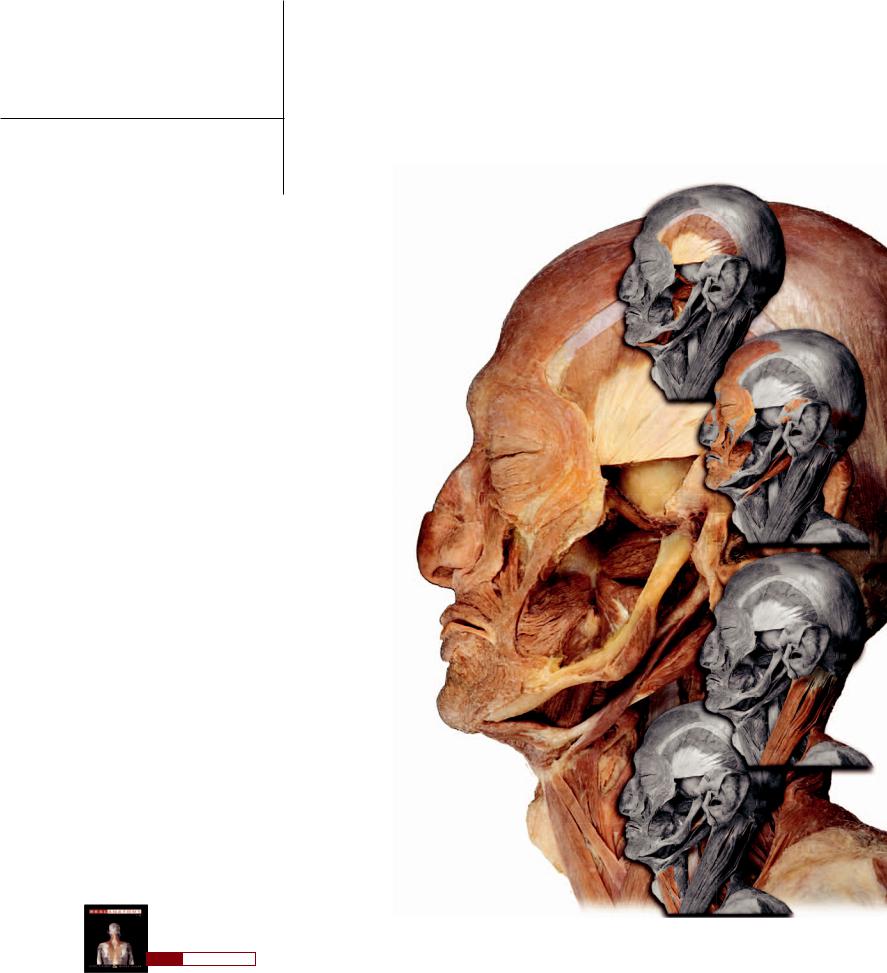
1 Introduction
Human anatomy is the science that deals with the structure and design of the human body. A knowledge of anatomy is not only impor-
tant for the anatomist, but is an essential tool for all the professionals who deal with the human body in any of a variety of ways. Furthermore, everyone can benefit from a knowledge of anatomy because it is what we are, and understanding our bodies can be invaluable.
Anatomy is an ancient science. The principal methods anatomists used, and still use, to reveal what is known about anatomy are dissection and microscopy. Dissection involves the cutting apart of
a body to reveal its gross structure. This was the first technique used to discover the structure of the body and is still the best way to truly understand the design and relationship of anatomical detail. The best drawings, photos, and virtual images can never reveal what the dissector experiences during a dissection. The advent of the microscope expanded anatomical knowledge by revealing microscopic perspectives that were not available
to the unaided eye. This understanding of microscopic structure opened the door to an increased knowledge of the functional aspects of anatomy.
In this atlas we attempt to teach the elegant structure and design of the human body using the tools and methods of the anatomist — dissection and microscopy.
While there are numerous excellent visual resources that depict anatomy, we believe that, with the exception of personal dissection study, excellent photographs based on excellent dissections and microscopy are the truest form of anatomical imagery. Nothing depicts the actual thing as well as the actual thing. Our goal is to create images that teach, and to use that imagery to highlight the patterns and design features of anatomy.
This atlas approaches the body from a systemic perspective; that is, it covers each body system and the organs associated
with that system. Each system is highlighted in the dissection photos. However, the dissections of the systemic anatomy often reveal regional perspectives and relationships, and the structural details of regional anatomy are labeled on every image. Have fun exploring what we think might be the next best thing to dissection.
Find more information about anatomy in
R E A L A N AT O M Y
1
The design features of the Atlas of Human Anatomy are illustrated on this page using a sample page from the book. Each page will begin with a short introduction to the featured anatomy of the page.
This brief narrative will occupy this text space. Below this narrative, the majority of the page will focus on the images of anatomy and the appropriate labels for the images. The design elements used to teach and illustrate the anatomy are highlighted in the boxes below.
Featured Structure |
Descriptive Narrative |
Reference Image |
The page heading will list the |
A brief description of the structure |
The reference image helps to quickly |
anatomical structure or feature that is |
and function of the anatomical |
identify the featured anatomy and see |
the focal point of the page |
structures on the page |
its relationships |
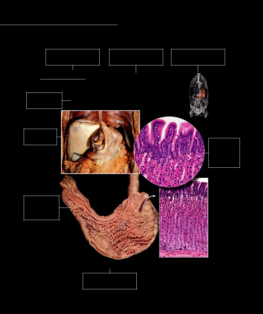
Stomach The stomach is a J-shaped organ of variable size and shape and has the greatest diameter of any part of the gut tube. It occupies the upper left quadrant of the
abdominal cavity, where it is anchored to the posterior abdominal wall by a mesentery. The stomach performs several functions, the most important of which is to store ingested food until it can be emptied into the small intestine at a rate that allows for optimal digestion and absorption.
Structure List |
|
1 |
Stomach |
7 |
Pylorus |
13 |
Surface mucous cell |
|
|
||
Numbered list of all the |
|
2 |
Cardia of stomach |
8 |
Pyloric sphincter |
14 |
Lamina propria |
|
|
||
|
3 |
Fundus of stomach |
9 |
Gastric rugae |
15 |
Mucous neck cell |
|
|
|||
structures visible on the |
|
4 |
Body of stomach |
10 |
Greater curvature |
16 |
Gastric glands |
|
|
||
anatomical images |
|
5 |
Pyloric antrum |
11 |
Lesser curvature |
17 |
Liver |
|
|
||
|
6 |
Pyloric canal |
12 |
Gastric pit |
18 |
Gallbladder |
|
|
|||
|
|
|
|
|
|
|
|
19 |
Spleen |
|
|
|
|
|
|
|
|
|
|
20 |
Greater omentum |
|
|
Dissection Images |
|
|
|
|
|
19 |
|
12 |
14 |
|
|
Beautiful dissections |
|
|
|
|
|
|
|
|
|||
|
|
17 |
|
|
|
|
13 |
|
|||
illustrate the anatomy |
|
|
|
|
|
|
|
|
|||
|
|
|
|
|
1 |
|
|
|
|
||
of the body system |
|
|
|
|
11 |
|
|
|
|
||
|
|
|
|
|
|
|
|
|
|
|
|
|
|
|
|
|
|
|
10 |
|
|
|
|
|
|
|
18 |
|
|
|
|
|
|
|
|
|
|
|
|
|
|
20 |
|
|
|
|
|
|
|
|
|
|
|
|
|
|
15 |
|
|
|
|
|
|
Abdominal dissection revealing stomach |
|
|
|
|
|||
|
|
|
|
|
Anterior view |
|
|
|
|
|
|
|
|
8 |
7 |
8 |
|
|
|
|
|
|
|
Numbered |
|
|
|
|
|
2 |
|
|
|
||
|
|
|
|
|
|
|
|
3 |
|
|
|
Structures |
|
|
|
|
|
|
|
|
|
|
|
Unobtrusive numbered |
|
|
|
|
6 |
|
|
|
|
15 |
|
structures without the |
|
|
|
|
|
|
|
|
|
||
|
|
|
|
|
|
|
|
|
|
||
clutter and distraction of |
|
|
|
|
11 |
|
|
|
|
||
leader lines |
|
|
|
|
|
9 |
|
|
|
|
|
|
|
|
|
5 |
|
4 |
|
|
16 |
|
|
|
|
|
|
|
|
|
|
|
|
16 |
|
|
|
|
|
|
|
|
|
|
|
|
|
|
|
|
|
|
|
|
|
10 |
|
|
|
|
|
|
|
|
Frontal section of stomach |
|
Photomicrograph of stomach mucosa |
||||
|
|
|
|
|
|
|
with callout above |
||||
|
|
|
|
|
|
Anterior view |
|
|
|||
|
|
|
|
|
|
|
|
40x and 100x |
|
||
|
|
|
|
|
|
|
|
|
|
|
|
Captions
Captions describe the image and the view or magnification of the anatomy or histology
Microscope
Images
Crisp histology photomicrographs illustrate the contextual microscopic structure of the anatomy
2
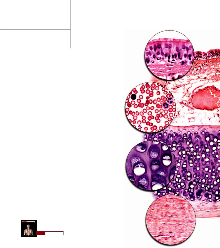
2 Histology
Histology is the study of tissues, and tissues are the building materials of the body. Like the materials we use to make the clothing we wear, tissues are the materials that form the various layers and structures of all the body’s organs. For example, you might wear a light undershirt of cotton beneath a silk long-sleeved shirt and wear a wool sweater over the top of the two shirts. Each layer of clothing is made of a different material, and the material is organized into a unique structure that has its own functional qualities. The same is true of the organs of the body. Each organ consists of distinct struc-
tural layers, and each layer is a specific type of tissue. For example, the stomach has an inner lining of simple columnar epithelium that is in contact with the food we eat and secretes enzymes to help digest the food. This epithelial layer is surrounded by a vascular layer of loose connective tissue that contains the blood vessels that transport the absorbed molecules from the stomach. Smooth muscle tissue surrounds the two inner layers and helps toss and turn the food within the stomach and move it toward the small intestine. The smooth muscle tissue is covered by a slippery, thin layer of simple squamous epithelium that forms the outer surface of the stomach and allows it to
move against neighboring organs while reducing the damaging friction. And just as the layers of clothing have names — undershirt, long-sleeved shirt, sweater—so also do the structural layers of an organ such as the stomach—mucosa, submucosa, muscularis, and serosa.
All the tissues of the body can be organized into four basic tissue catego- ries—epithelial tissue, connective and supporting tissue, muscle tissue, and nervous tissue. Each tissue category has unique structural features that are shared by the tissues of that category. Epithelial tissues are surface tissues that consist of numerous cells tightly packed together. Connective and supporting tissues share the common feature of having relatively few cells that are scattered within a surrounding fibrous extracellular matrix. Muscle tissue consists of elongated cells with specialized protein arrangements that are designed to shorten. Nervous tissue cells are branching, wire-like cells with a great variety of shapes and
lengths. In this chapter you will explore these four tissue categories and the specific tissue types that comprise each category. In the chapters that follow, the
different tissues will be observed in the context of the organs and organ systems they form.
Find more information about histology in
R E A L A N AT O M Y
3