Добавил:
Upload Опубликованный материал нарушает ваши авторские права? Сообщите нам.
Вуз: Предмет: Файл:
Цветной атлас по фармакологии 2005.pdf
Скачиваний:
126
Добавлен:
06.02.2015
Размер:
10.6 Mб
Скачать

330 Therapy of Selected Diseases

Osteoporosis

Osteoporosis is defined as a generalized decrease in bone mass (osteopenia) that equally affects bone matrix and mineral content and is associated with a change in spongiosal architecture. This condition predisposes to collapse of vertebral bodies and bone fractures with trivial trauma (e.g., hip fractures).

Bone substance is subject to continual remodeling. The equilibrium between bone formation and bone resorption is regulated in a complex manner: a remodeling cycle is initiated by osteoblasts when these stimulate uninucleated osteoclast precursor cells to fuse into large multinucleated cells. Stimulation occurs by direct cell-to-cell contact between osteoblasts and osteoclast precursor cells and is mediated by the RANK ligand on the surface of osteoblasts and its receptor on the osteoclasts (or their precursors), as well as cytokines secreted by osteoblasts. These processes are inhibited by estrogens and a protein secreted by osteoblasts (osteoprotegerin). The osteoclast creates an acidic milieu, enabling minerals to be solubilized, and then phagocytoses the organic matrix. Hormones regulate these events.

In hypocalcemia, the parathyroid increases its secretion of parathormone, resulting in enhanced liberation of Ca2+. Calcitonin transfers active osteoclasts into a resting state. Calcitonin given therapeutically relieves pain associated with neoplastic bone metastases and vertebral body collapse. Estrogens diminish bone resorption by (a) inhibiting activation of osteoclasts by osteoblasts and (b) promoting apoptosis of osteoclasts.

Idiopathic osteoporosis cannot be prevented by prophylactic therapy, but its development can be delayed. This requires: a healthy lifestyle with plenty of physical exercise (sports, hiking), daily intake of calcium (1000 mg/day Ca2+) and of vitamin D (1000 IU/day). The same principle holds for postmenopausal osteoporosis. Hormone

replacement therapy in postmenopausal women has not been successful because of an increased incidence of breast cancers, thromboembolism, and other adverse effects (p. 250). Long-term hormone replacement therapy should no longer be carried out.

If osteoporosis has become clinically manifest, attempts can be made at improving the condition by means of drugs, or at least slowing down further deterioration. Besides administration of calcium and vitamin D, the following options are available.

Bisphosphonates (N-containing) structurally mimic endogenous pyrophosphate (see formulae), and like the latter are incorporated into the mineral substance of bone. During phagocytosis of the bone matrix, they are taken up by osteoclasts. There, the N- containing bisphosphonates inhibit prenylation of G-proteins and thus damage the cells. Accordingly, osteoclast activity levels are lowered by alendronate and risedronate, while osteoclast apoptosis is promoted. The result is a reduction in bone resorption and a decreased risk of bone fractures.

Raloxifene exerts an estrogen-like effect on bone, while acting as an estrogen antagonist in the uterus and breast tissue (p. 254). In terms of fracture prophylaxis, its effectiveness appears inferior to that of bisphosphonates. Thromboembolism and edema are reported as adverse effects.

It should also be mentioned that intermittent slight elevation in the plasma concentration of parathormone leads to increased formation of bone substance. The likely explanation is that, under this condition, stimulation of osteoblasts is suf cient to induce synthesis of bone matrix but not strong enough to activate osteoclasts. This strategy is applied therapeutically by administration of a fragment (amino acids 1–34) of recombinant human parathormone (teriparatide, s.c.).


Osteoporosis

331

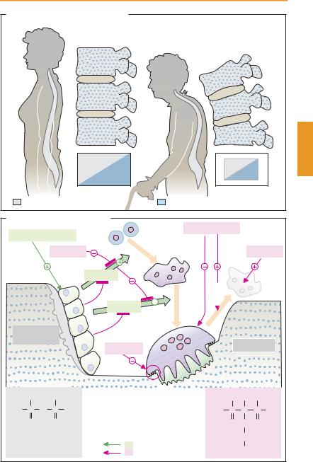
A. Bone: normal state and osteoporosis


Normal state

Osteoporosis


Organic bone matrix

Bone mineral: hydroxyapatite


B. Regulation of bone remodeling


Progenitor

Biphosphonates


Parathyroid hormone

cells


Estrogens

Fusion


Estrogens


RANKL


Apoptosis


OPG


RANKL


Activation


(Pre)Osteo-


OPG


blasts


Osteoclasts


Calcitonin


OH


OH


RANK = receptor activating NF-Κ

-B

OH OH OH


HO

P

O

P

OH

RANKL = RANK-Ligand

HO

P

C

P

OH


O


O


OPG = Osteoprotegerin


O

CH2 O


Pyrophosphate


Bone resorption


(CH2)2


Stimulation

Alendronate


Inhibition


NH2


332 Therapy of Selected Diseases

Rheumatoid Arthritis

Rheumatoid arthritis or chronic polyarthritis (A)isaprogressiveinflammatoryjoint disease that intermittently attacks more and more joints, predominantly those of the fingers and toes. The probable cause of rheumatoid arthritis is a pathological reaction of the immunesystem.Thismalfunctioncanbepromoted or triggered by various conditions, including genetic disposition, age-related wear and tear, hypothermia, and infection. An initialnoxiousstimuluselicitsan inflammation of synovial membranes that, in turn, leads to release of antigens through which the inflammatory process is maintained.

The antigen is taken up by synovial anti- gen-presenting cells; lymphocytes, including T-helper cells (p. 304), are activated and start to proliferate. In the process of interaction between lymphocytes and macrophages, the intensity of inflammation increases. Macrophages release proinflammatory messengers; among these, interleukin-1 and tumor necrosis factor α (TNFα) are important. TNFαis able to elicit a multiplicity of proinflammatory actions (B) that benefit defense against infectious pathogens but are detrimental in rheumatoid arthritis. The cytokines stimulate gene expression for COX- 2; inflammation-promoting prostanoids are produced. The inflammatory reaction increases activity of lymphocytes and macrophages, initiating a vicious circle. Synovial fibroblasts proliferate and release destructive enzymes; the inflamed characteristic pannus tissue develops and destructively invades joint cartilage and subjacent bone. Ultimately, ankylosis (loss of joint motion or bone fusion) with connective tissue scar formation occurs. Concomitant extra-articular disease may be superimposed. The disease process is associated with strong pain and restriction of mobility.

Pharmacotherapy. Acute relief of inflammatory symptoms can be achieved by prostaglandin synthase inhibitors (p. 200; nonse-

lective COX inhibitors or COX-2 inhibitors) and by glucocorticoids (p. 244). The inevitably chronic use of both groups of substances is likely to cause significant adverse effects. Neither can halt the progressive destruction of joints.

Substances that are able to reduce the requirement for nonsteroidal anti-inflam- matory drugs and to slow disease progression are labeled disease-modifying agents. Early use of these drugs is recommended. Their effect develops only after treatment for several weeks. Proliferation of lympho-

cytes can be slowed by

methotrexate

(p. 304) and leflunomide,

which reduces

the availability of pyrimidine nucleotides in lymphocytes (via inhibition of dihydroorotate dehydrogenase). For ciclosporin, see p. 306. In addition, use is made of immune suppressants such as azathioprine and cyclophosphamide. Intralysosomal accumulation and impaired phagocytic function may be involved in the action of chloroquine or hydroxychloroquine, as well as gold compounds (i.m.: aurothioglucose or aurothiomalate; oral: auranofin, less effective). The antibodies, infliximab and adalimumab, as well as the fusion protein etanercept intercept TNF-αmolecules, preventing them from interacting with membrane receptors of target cells. Anakinra is a recombinant analogue of the endogenous interleukin-1 antagonist. The mechanisms of action of D-pen- icillamine and sulfasalazine are unknown. Same of the drugs mentioned possess considerable potential for adverse effects. Sulfasalazine and methotrexate exhibit a relatively favorable risk–benefit ratio. Combination of disease-modifying drugs is possible.

Surgical removal of the inflamed synovial membrane (synovectomy) frequently provides long-term relief. If feasible, this approach is preferred because all pharmacotherapeutic measures entail significant adverse effects.


Rheumatoid Arthritis


333

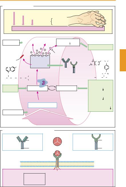
A. Rheumatoid Arthritis


Genetic predisposition


Environmental factors


Precipitating

Infection


causes

Trauma


Immune system: reactive against autologous joint tissue


Sulfasalazine


?

Prostaglandin


synthesis


COX inhibitors

IL-1 receptor


Cox-2


Anakinra


Cytokines, etc.


NH2


N

CH2 N

CH3


IL-1, TNFα


N


N

Cl


Infliximab

H2N

N

N


Methotrexate


NH


Etanercept


H

C


C2H5


N

O

H3C CH CH2 CH2 CH2 N


HOOC

(CH2)2

CH

COOH

Chloroquine


C2H5


ÐMethotrexate


Gold,


?

Macrophages

Lymphocytes

(purine


synthesis

)


chloroquine


ÐLeflunomide


(pyrimidine


synthesis

)


ÐCiclosporin


Antigen (unknown)


(IL-2 synthesis


in T-helper cells )

D-Penicillamine


?


B. Tumor necrosis factor α and inhibitors


Homotrimer


TNF α


Murine


Infliximab


portion (Fab)

TNF α

Etanercept


receptor-


parts

(chimeric


Human


(fusion protein)


IgG antibody)


portion (Fc)


Fc portion


TNF α


receptor


Activation


Vessels:


Tumor cells


Ð Proliferation


Macrophages:


Synovial membrane:


Bone:


Ð Activation


Ð Proliferation


Ð Lysis


Ð Adhesion of


Ð Resorption


Ð Chemotaxis


Ð Pannus formation


blood cells