Добавил:
Upload Опубликованный материал нарушает ваши авторские права? Сообщите нам.
Вуз: Предмет: Файл:
Цветной атлас по фармакологии 2005.pdf
Скачиваний:
126
Добавлен:
06.02.2015
Размер:
10.6 Mб
Скачать

318 Therapy of Selected Diseases

Antianginal Drugs

Antianginal agents derive from three drug groups, the pharmacological properties of which have already been presented in more detail: the organic nitrates (p.124), the calcium antagonists (p.126), and the β-blockers (p. 96).

Organic nitrates (A) increase blood flow, hence O2 supply, because diastolic wall tension (preload) declines as venous return to the heart is diminished. Thus, the nitrates enable myocardial flow resistance to be reduced even in the presence of coronary sclerosis with angina pectoris. In angina due to coronary spasm, arterial dilation overcomes the vasospasm and restores myocardial perfusion to normal. O2 demand falls because of the ensuing decrease in the two variables that determine systolic wall tension (afterload): ventricular filling volume and aortic blood pressure.

Calcium antagonists (B) decrease O2 demand by lowering aortic pressure, one of the components contributing to afterload. The dihydropyridine nifedipine, is devoid of a cardiodepressant effect, but may give rise to reflex tachycardia and an associated increase in O2 demand. The catamphiphilic drugs verapamil and diltiazem are cardiodepressant. Reduced beat frequency and contractility contribute to a reduction in O2 demand; however, AV-block and mechanical insuf ciency can dangerously jeopardize heart function. In coronary spasm, calcium antagonists can induce spasmolysis and improve blood flow.

β-Blockers (C) protect the heart against the O2-wasting effect of sympathetic drive by inhibiting β1-receptor-mediated increases in cardiac rate and speed of contraction.

Uses of antianginal drugs (D). For relief of the acute anginal attack, rapidly absorbed drugs devoid of cardiodepressant activity are preferred. The drug of choice is nitroglycerin (NTG, 0.8–2.4 mg sublingually; onset of ac-

tion within 1–2 minutes; duration of effect ~ 30 minutes). Isosorbide dinitrate (ISDN) can also be used (5–10 mg sublingually); compared with NTG, its action is somewhat delayed in onset but of longer duration. Finally, nifedipine may be useful in chronic stable, or in variant angina (5–20 mg, capsule to be bitten and the content swallowed).

For sustained daytime angina prophylaxis, nitrates are of limited value because “nitrate pauses” of about 12 hours are appropriate if nitrate tolerance is to be avoided. If attacks occur during the day, ISDN, or its metabolite isosorbide mononitrate, may be given in the morning and at noon (e. g., ISDN 40 mg in extended-release capsules). Because of hepatic presystemic elimination, NTG is not suitable for oral administration. Continuous delivery via a transdermal patch would also not seem advisable because of the potential development of tolerance. With molsidomine, there is less risk of a nitrate tolerance; however, its clinical use is restricted owing to its potential carcinogenicity.

The choice between calcium antagonists must take into account the differential effect of nifedipine versus verapamil or diltiazem on cardiac performance (see above). When β-blockers are given, the potential consequences of reducing cardiac contractility (withdrawal of sympathetic drive) must be kept in mind. Since vasodilating β2-receptors are blocked, an increased risk of vasospasm cannot be ruled out. Therefore, monotherapy with β-blockers is recommended only in angina due to coronary sclerosis, but not in variant angina.

Antianginal Drugs

319

A. Effects of nitrates

p

Diastole

Systole

p


Vol


Vol


Resistance

Venous


vessels


capacitance

Preload

Nitrate

Afterload

vessels


tolerance

O2-demand


O2-supply


Vasorelaxation

Relaxation of


coronary spasm


Nitrates, e.g., nitroglycerin (GTN), isosorbide dinitrate (ISDN)

B. Effects of Ca-antagonists

C. Effects of β -blockers

p


Rest

Ca-


antagonists


Sympathetic


Relaxation

system


of


resistance


vessels

β -Blocker


Afterload


Rate


Contraction


velocity


Relaxation of


O2-demand

coronary spasm

Exercise


D. Clinical uses of antianginal drugs


Angina pectoris

Coronary sclerosis


Coronary spasm


Therapy of attack


GTN,

ISDN


Nifedipine


Anginal prophylaxis


Long-acting nitrates


β− Blockers


Ca-antagonists


320 Therapy of Selected Diseases

Acute Coronary Syndrome— Myocardial Infarction

Myocardial infarction (MI) is caused by the acute thrombotic occlusion of a coronary artery. The myocardial region that has been cut off from its blood supply dies within a short time owing to the lack of O2 and glucose. The loss in functional muscle tissue results in reduced cardiac performance. In the infarct border zone, spontaneous pacemaker potentials may develop, leading to fatal ventricular fibrillation. The patient experiences severe pain, a feeling of annihilation, and fear of dying.

MI usually develops after rupture or erosion of an atherosclerotic plaque within a coronary blood vessel. At this site, the clotting cascade is activated and the resultant thrombus occludes the lumen. In all patients under suspicion of MI, immediate therapy has to be initiated by the emergency physician. To relieve the patient from severe pain and anxiety, morphine and a benzodiazepine need to be given. Antiplatelet drugs and heparin are necessary for preventing further formation of thrombi. Nitroglycerin can be used to reduce cardiac load. When blood pressure and heart rate have stabilized, a β-blocker can be administered to lower cardiac O2 consumption and the risk of arrhythmias. Infusion of lidocaine is required to counter the threat of arrhythmias. The chance of survival of the MI patient depends on the interval between the onset of infarction and the start of therapy.

In the hospital, ECG and laboratory tests are performed promptly to determine the subsequent treatment strategy. When cardiomyocytes die, contractile proteins (troponin) or myocardial enzymes (creatine kinase, CK-MB) are liberated and can be detected in blood for diagnostic purposes. Marked elevation of the ST segment in the ECG raises the strong suspicion of a complete occlusion of a coronary artery (STelevation MI, STEMI). In these MI patients, reperfusion of the affected area as early and as completely as

possible may be life-saving. In this situation, removal of the coronary stenosis with a balloon catheter combined with implantation of a stent offers the best chance of survival but can be performed only in specialized cardiac centers. As dilatation of the vascular stenosis by the heart catheter liberates many thrombogenic mediators, platelet aggregation must be prevented by administration of glycoprotein IIb/IIIa receptor antagonists (p.154).

If the MI patient cannot be transported to a cardiac center in time, fibrinolytic treatment of the coronary thrombus is instituted. For this purpose, fibrinolytics (streptokinase or recombinant tissue plasminogen activators) are given intravenously. Fibrinolysis is associated with an increased risk of bleeding; cerebral hemorrhages are of particular concern. A coronary bypass operation is available as a third therapeutic option.

Patients with persistent angina pectoris who lack an elevated ST segment in the ECG but show an elevated blood level of troponin may have a non-STEMI (“NSTEMI”). The most frequent cause is thrombi that have moved from the larger coronary arteries into smaller branches to produce a blockage there. If neither ST segment elevation nor biochemical MI markers can be ascertained, unstable angina pectoris is present, which is initially treated with antiplatelet drugs.

Post-MI management calls for strict adherence to a program of secondary prevention. Cardiac risk factors have to be excluded or modified, for instance, by reduction of overweight, cessation of smoking, optimal control of diabetes mellitus, and physical exercise (a dog that loves to run is an ideal training partner). Supportive pharmacotherapeutic measures include administration of platelet aggregation inhibitors, β-blockers, and ACE inhibitors.


Myocardial Infarction

321

A. Myocardial infarction: pharmacotherapeutic approaches


Acute symptoms:

Acute care measures


severe pain


– Nitroglycerin (reduction of


sense of impending

preand afterload)


doom


– Acetylsalicylic acid


Patient

fear of dying


(if needed i.v.)


(inhibition of platelet


aggregation)


– Morphine (analgesia, sedation)


– Oxygen via nasal tube


Hospitalization


with minimal delay


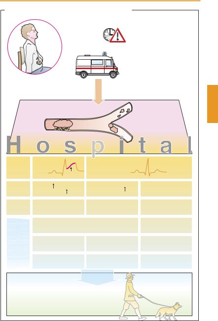
Acute coronary syndrome

Plaque rupture


Angina pectoris > 20 min


Distal


thrombus


Thrombus


ECG

ST-elevation

T

No ST-elevation


S


Laboratory

CK-MB


Troponin-I, -T


Troponin-I, -T


Diagnosis

Myocardial infarction

Myocardial infarction

Unstable


(“STEMI”)


(“NSTEMI”)


angina pectoris


Therapy

General: O2, acetylsalicylic acid,


Acetylsalicylic acid,


heparin, nitrates, β -blocker, morphine


clopidrogel


PTCA (Stent)


PTCA (Stent)


Cardiac


GPIIb/IIIa-Antagonist

GPIIb/IIIa-Antagonist

catheterization


or


fibrinolysis


or


bypass surgery


Secondary prevention


Discharge


possibly:


– Acetylsalicylic acid

– Clopidrogel


– β -Blocker

– Phenprocoumon


– ACE inhibitor

– Statins