- •Preface to the 3rd edition
- •General Pharmacology
- •Systems Pharmacology
- •Therapy of Selected Diseases
- •Subject Index
- •Abbreviations
- •General Pharmacology
- •History of Pharmacology
- •Drug and Active Principle
- •The Aims of Isolating Active Principles
- •European Plants as Sources of Effective Medicines
- •Drug Development
- •Congeneric Drugs and Name Diversity
- •Oral Dosage Forms
- •Drug Administration by Inhalation
- •Dermatological Agents
- •From Application to Distribution in the Body
- •Potential Targets of Drug Action
- •External Barriers of the Body
- •Blood–Tissue Barriers
- •Membrane Permeation
- •Binding to Plasma Proteins
- •The Liver as an Excretory Organ
- •Biotransformation of Drugs
- •Drug Metabolism by Cytochrome P450
- •The Kidney as an Excretory Organ
- •Presystemic Elimination
- •Drug Concentration in the Body as a Function of Time—First Order (Exponential) Rate Processes
- •Time Course of Drug Concentration in Plasma
- •Time Course of Drug Plasma Levels during Repeated Dosing (A)
- •Time Course of Drug Plasma Levels during Irregular Intake (B)
- •Accumulation: Dose, Dose Interval, and Plasma Level Fluctuation (A)
- •Dose–Response Relationship
- •Concentration–Effect Curves (B)
- •Concentration–Binding Curves
- •Types of Binding Forces
- •Agonists—Antagonists
- •Other Forms of Antagonism
- •Enantioselectivity of Drug Action
- •Receptor Types
- •Undesirable Drug Effects, Side Effects
- •Drug Allergy
- •Cutaneous Reactions
- •Drug Toxicity in Pregnancy and Lactation
- •Pharmacogenetics
- •Placebo (A)
- •Systems Pharmacology
- •Sympathetic Nervous System
- •Structure of the Sympathetic Nervous System
- •Adrenergic Synapse
- •Adrenoceptor Subtypes and Catecholamine Actions
- •Smooth Muscle Effects
- •Cardiostimulation
- •Metabolic Effects
- •Structure–Activity Relationships of Sympathomimetics
- •Indirect Sympathomimetics
- •Types of
- •Antiadrenergics
- •Parasympathetic Nervous System
- •Cholinergic Synapse
- •Parasympathomimetics
- •Parasympatholytics
- •Actions of Nicotine
- •Localization of Nicotinic ACh Receptors
- •Effects of Nicotine on Body Function
- •Aids for Smoking Cessation
- •Consequences of Tobacco Smoking
- •Dopamine
- •Histamine Effects and Their Pharmacological Properties
- •Serotonin
- •Vasodilators—Overview
- •Organic Nitrates
- •Calcium Antagonists
- •ACE Inhibitors
- •Drugs Used to Influence Smooth Muscle Organs
- •Cardiac Drugs
- •Cardiac Glycosides
- •Antiarrhythmic Drugs
- •Drugs for the Treatment of Anemias
- •Iron Compounds
- •Prophylaxis and Therapy of Thromboses
- •Possibilities for Interference (B)
- •Heparin (A)
- •Hirudin and Derivatives (B)
- •Fibrinolytics
- •Intra-arterial Thrombus Formation (A)
- •Formation, Activation, and Aggregation of Platelets (B)
- •Inhibitors of Platelet Aggregation (A)
- •Presystemic Effect of ASA
- •Plasma Volume Expanders
- •Lipid-lowering Agents
- •Diuretics—An Overview
- •NaCl Reabsorption in the Kidney (A)
- •Aquaporins (AQP)
- •Osmotic Diuretics (B)
- •Diuretics of the Sulfonamide Type
- •Potassium-sparing Diuretics (A)
- •Vasopressin and Derivatives (B)
- •Drugs for Gastric and Duodenal Ulcers
- •Laxatives
- •Antidiarrheal Agents
- •Drugs Affecting Motor Function
- •Muscle Relaxants
- •Nondepolarizing Muscle Relaxants
- •Depolarizing Muscle Relaxants
- •Antiparkinsonian Drugs
- •Antiepileptics
- •Pain Mechanisms and Pathways
- •Eicosanoids
- •Antipyretic Analgesics
- •Nonsteroidal Anti-inflammatory Drugs (NSAIDs)
- •Cyclooxygenase (COX) Inhibitors
- •Local Anesthetics
- •Opioid Analgesics—Morphine Type
- •General Anesthesia and General Anesthetic Drugs
- •Inhalational Anesthetics
- •Injectable Anesthetics
- •Sedatives, Hypnotics
- •Benzodiazepines
- •Pharmacokinetics of Benzodiazepines
- •Therapy of Depressive Illness
- •Mania
- •Therapy of Schizophrenia
- •Psychotomimetics (Psychedelics, Hallucinogens)
- •Hypothalamic and Hypophyseal Hormones
- •Thyroid Hormone Therapy
- •Glucocorticoid Therapy
- •Follicular Growth and Ovulation, Estrogen and Progestin Production
- •Oral Contraceptives
- •Antiestrogen and Antiprogestin Active Principles
- •Aromatase Inhibitors
- •Insulin Formulations
- •Treatment of Insulin-dependent Diabetes Mellitus
- •Treatment of Maturity-Onset (Type II) Diabetes Mellitus
- •Oral Antidiabetics
- •Drugs for Maintaining Calcium Homeostasis
- •Drugs for Treating Bacterial Infections
- •Inhibitors of Cell Wall Synthesis
- •Inhibitors of Tetrahydrofolate Synthesis
- •Inhibitors of DNA Function
- •Inhibitors of Protein Synthesis
- •Drugs for Treating Mycobacterial Infections
- •Drugs Used in the Treatment of Fungal Infections
- •Chemotherapy of Viral Infections
- •Drugs for the Treatment of AIDS
- •Drugs for Treating Endoparasitic and Ectoparasitic Infestations
- •Antimalarials
- •Other Tropical Diseases
- •Chemotherapy of Malignant Tumors
- •Targeting of Antineoplastic Drug Action (A)
- •Mechanisms of Resistance to Cytostatics (B)
- •Inhibition of Immune Responses
- •Antidotes and Treatment of Poisonings
- •Therapy of Selected Diseases
- •Hypertension
- •Angina Pectoris
- •Antianginal Drugs
- •Acute Coronary Syndrome— Myocardial Infarction
- •Congestive Heart Failure
- •Hypotension
- •Gout
- •Obesity—Sequelae and Therapeutic Approaches
- •Osteoporosis
- •Rheumatoid Arthritis
- •Migraine
- •Common Cold
- •Atopy and Antiallergic Therapy
- •Bronchial Asthma
- •Emesis
- •Alcohol Abuse
- •Local Treatment of Glaucoma
- •Further Reading
- •Further Reading
- •Picture Credits
- •Drug Indexes

38 Drug Elimination
Drug Metabolism by Cytochrome P450
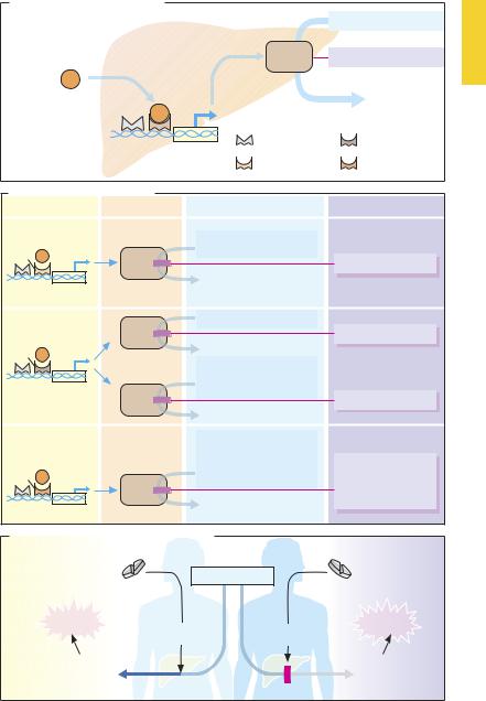
Cytochrome P450 enzyme. A major part of phase I reactions is catalyzed by hemoproteins, the so-called cytochrome P450 (CYP) enzymes (A). To date about 40 genes for cytochrome P450 proteins have been identified in the human; among these, the protein families CYP1, CYP2, and CYP3 are important in drug metabolism (B). The bulk of CYP enzymes are located in the liver and the intestinal wall, which explains why these organs are responsible for the major part of drug metabolism.
Substrates, inhibitors, and inducers. Cytochromes are enzymes with broad substrate specificities. Accordingly, pharmaceuticals of diverse chemical structure can be metabolized by a given enzyme protein. When several drugs are metabolized by the same isozyme, clinically important interactions may result. In these, substrates (drugs metabolized by CYP) can be distinguished from inhibitors (drugs that are bound to CYP with high af nity, interfere with the breakdown of substrates, and are themselves metabolized slowly) (A). The amount of hepatic CYP enzymes is a major determinant of metabolic capacity. An increase in enzyme concentration usually leads to accelerated drug metabolism. Numerous endogenous and exogenous substances, such as drugs, can augment the expression of CYP enzymes andthus actas CYP inducers (p.32). Manyof these inducers activate specific transcription factors inthenucleusof hepatocytes, leading to activation of mRNA synthesis and subsequent production of CYP isozyme protein. Some inducers also increase the expression of P-glycoprotein transporters; as a result, enhanced metabolism by CYP and increased membrane transport by P-glycoprotein can act in concert to render a drug ineffective.
The table in (B) provides an overview of different CYP isozymes along with their substrates, inhibitors, and inducers. Obviously,
when a patient is to be exposed to polypharmaceutic regimens (especially multimorbid subjects), it would be imprudent to start therapywithoutcheckingwhether thedrugs being contemplated include CYP inducers or inhibitors, some of which may dramatically alter pharmacokinetics.
Drug interaction due to CYP induction or inhibition. Life-threatening interactions have been observed in patients taking inducers of CYP3A4 isozyme during treatment with ciclosporin for the prevention of kidney and liver transplant rejection. Intake of rifampin [rifampicin] and also of St. John’s wort preparations (available without prescription) may increase expression of CYP3A4 to such an extent as to lower plasma levels of ciclosporin below the therapeutic range (C). As immunosuppression becomes inadequate, the risk of transplant rejection will be enhanced. In the presence of rifampin, other drugs that are substrates of CYP3A4 may become ineffective. For this reason, theintake of rifampin iscontraindicated in HIV patients being treated with protease inhibitors. As a rule, inhibitors of CYP enzymes elevate plasma levels of drugs that are substrates of the same CYP enzymes; in this manner, they raise the risk of undesirable toxic effects. The antifungal agent ketoconazole enhances the nephrotoxicity of ciclosporin by such a mechanism (C).

Drug Metabolism by Cytochrome P450 |
39 |
A. Cytochrome P450 in the liver |
|
|
|
|
|
|
|
Substrates |
|
|
Protein |
CYP |
Inhibitors |
|
Inducer |
synthesis |
|||
|
|
|||
|
mRNA |
|
|
|
AhR |
CYP gene |
|
||
Transcription |
|
|
|
|
factors |
|
Retinoid-X- |
Constitutive |
|
RXR |
|
receptor |
androstane receptor |
|
|
|
Arylhydrocarbon |
Pregnane-X-receptor |
|
|
|
receptor |
||
|
|
|
||
B. Cytochrome P450 isozymes
Inducers |
Cytochrome |
Substrates |
Inhibitors |
Barbecued meat, |
|
|
|
tobacco smoke, |
|
Clozapine, estradiol, |
|
omeprazole |
|
|
|
|
haloperidol, theophylline |
|
|
AhR |
CYP |
|
|
|
Fluoroquinolone |
||
|
1A2 |
|
|
|
|
|
|
Arylhydrocarbon |
|
|
|
receptor |
|
|
|
Phenobarbital, |
CYP |
Ibuprofen, Losartan |
|
|
|
||
Rifampicin |
|
Isoniazid, Verapamil |
|
2C9 |
|
||
|
|
|
|
CAR |
|
|
|
|
|
Carvedilol, metoprolol, |
|
|
|
tricyclic antidepressants, |
|
Constitutive |
CYP |
neuroleptics, SSRI, codeine |
Quinidine, fluoxetine |
androstane |
2D6 |
|
|
receptor |
|
|
|
|
|
|
|
Rifampicin, carba- |
|
Ciclosporin, tacrolimus, |
|
mazepine, dexa- |
|
|
|
methasone, pheny- |
|
nifedipine, verapamil, statins, |
|
toin, St. John’s wort |
|
estradiol, progesterone, |
HIV protease inhibitors, |
PXR |
|
testosterone, haloperidol |
|
|
amiodarone, macrolides, |
||
CYP |
|
||
|
|
azole antimycotics, |
|
|
3A4 |
|
|
|
|
grapefruit juice |
Pregnane X-receptor
C. Drug interactions and cytochrome P450 |
|
|
|
Rifampicin, |
|
|
Itraconazole |
St. John’s wort |
Ciclosporin |
|
|
|
|
|
|
Transplant |
|
|
Ciclosporin |
rejection |
Induction |
Inhibition |
nephrotoxicity |
|
of CYP3A4 |
of CYP3A4 |
|
Accelerated |
|
|
Delayed |
ciclosporin |
|
|
ciclosporin |
elimination |
|
|
elimination |

40 Drug Elimination
The Kidney as an Excretory Organ
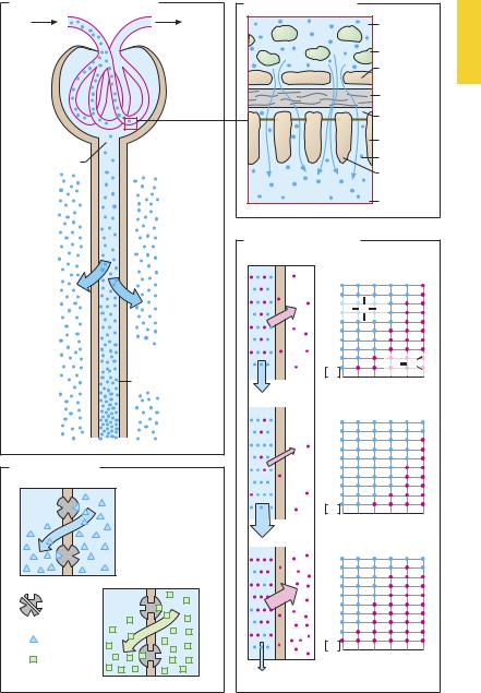
Most drugs are eliminated in urine either chemically unchanged or as metabolites. The kidney permits elimination because the vascular wall structure in the region of the glomerular capillaries (B) allows unimpeded passage into urine of blood solutes having molecular weights(MW) < 5000. Filtration is restricted at MW < 50 000 and ceases at MW > 70 000. With few exceptions, therapeutically used drugs and their metabolites have much smaller molecular weights and can therefore undergo glomerular filtration, i.e., pass from blood into primary urine. Separating the capillary endothelium from the tubular epithelium, the basal membrane contains negatively charged macromolecules and acts as a filtration barrier for high-molecular-weight substances. The relative density of this barrier depends on the electric charge of molecules that attempt to permeate it. In addition, the diaphragmatic slits between podophyte processes play a part in glomerular filtration.
Apart from glomerular filtration (B), drugs present in blood may pass into urine by active secretion (C). Certain cations and anions are secreted by the epithelium of the proximal tubules into the tubular fluid via special energy-consuming transport systems. These transport systems have a limited capacity. When several substrates are present simultaneously, competition for the carrier may occur (see p.326).
During passage down the renal tubule, primary urinary volume shrinks to about 1%; accordingly, there is a corresponding concentration of filtered drug or drug metabolites (A). The resulting concentration gradient between urine and interstitial fluid is preserved in the case of drugs incapable of permeating the tubular epithelium. However, with lipophilic drugs the concentration gradient will favor reabsorption of the filtered molecules. In this case, reabsorption is not based on an active process but results instead from passive diffusion. Accordingly,
for protonated substances, the extent of reabsorption is dependent upon urinary pH or the degree of dissociation. The degree of dissociation varies as a function of the urinary pH and the pKa, which represents the pH value at which half of the substance exists in protonated (or unprotonated) form. This relationship is illustrated graphically (D) with the example of a protonated amine having a pKa of 7. In this case, at urinary pH 7, 50% of the amine will be present in the protonated, hydrophilic, membrane-imperme- ant form (blue dots), whereas the other half, representing the uncharged amine (red dots), can leave the tubular lumen in accordance with the resulting concentration gradient. If the pKa of an amine is higher (pKa = 7.5) or lower (pKa = 6.5), a correspondingly smaller or larger proportion of the amine will be present in the uncharged, reabsorbable form. Lowering or raising urinary pH by half a pH unit would result in analogous changes.
The same considerations hold for acidic molecules, with the important difference that alkalinization of the urine (increased pH) will promote the deprotonization of –COOH groups and thus impede reabsorption. Intentional alteration of urinary pH can be used in intoxications with proton-accep- tor substances in order to hasten elimination of the toxin (e.g., alkalinization † phenobarbital; acidification † methamphetamine).

The Kidney as an Excretory Organ |
41 |
A. Filtration and concentration |
|
B. Glomerular filtration |
|
|
|
|
|
||||||
|
|
|
|
|
|
|
|
|
|
|
Blood |
|
|
|
|
|
|
|
|
|
|
|
|
|
Plasma |
|
|
|
|
|
|
|
|
|
|
|
|
|
protein |
|
|
|
|
|
|
|
|
|
|
|
|
|
Endothelium |
||
|
|
|
|
|
|
|
|
|
|
|
Basal |
|
|
|
|
|
|
|
|
|
|
|
|
|
membrane |
||
|
|
|
|
|
|
|
|
|
|
|
Slit |
|
|
|
|
|
|
|
|
|
|
|
|
|
diaphragm |
||
180 l |
|
|
|
|
Glomerular |
|
|
|
|
Epithelium |
|||
|
|
|
|
|
|
|
|
Drug |
|
||||
Primary urine |
|
|
|
filtration |
|
|
|
|
|
||||
|
|
|
|
|
|
|
|
|
|
||||
|
|
|
|
|
of drug |
|
|
|
|
|
Podocyte |
||
|
|
|
|
|
|
|
|
|
|
|
processes |
||
|
|
|
|
|
|
|
|
|
|
|
Primary |
|
|
|
|
|
|
|
|
|
|
|
|
|
urine |
|
|
|
|
|
|
|
|
|
D. Tubular reabsorption |
|
|
|
|
||
|
|
|
|
|
|
|
pH = 7,0 |
pKa of substance |
|
||||
|
|
|
|
|
|
|
|
100 |
|
pKa = 7,0 |
|
|
|
|
|
|
|
|
|
|
|
|
|
|
|
|
|
|
|
|
|
|
|
|
|
R |
N+ H |
|
|
||
|
|
|
|
|
|
|
|
50 |
|
|
|
|
|
|
|
|
|
|
Concentration |
|
|
|
R |
N |
|
||
1,2 l |
|
|
|
|
of drug |
|
|
% |
|
|
|
||
|
|
|
|
in tubule |
|
6 |
6,5 |
7 |
7,5 |
8 |
|||
Final |
|
|
|
|
|
|
|||||||
|
|
|
|
|
|
|
|
||||||
urine |
|
|
|
|
|
|
|
|
|
|
|
|
|
|
|
|
|
|
|
|
|
100 |
|
pKa = 7,5 |
|
|
|
|
|
|
|
|
|
|
|
|
|
|
|
|
|
C. Active secretion |
|
|
|
|
50 |
|
|
|
|
|
|||
|
|
+ |
+ |
|
|
|
|
|
|
|
|
|
|
|
|
|
|
|
|
|
% |
|
|
|
|
|
|
|
|
+ |
+ |
|
|
|
|
|
|
|
|
|
|
|
|
+ |
|
|
|
|
|
6 |
6,5 |
7 |
7,5 |
8 |
|
|
+ |
+ |
|
|
|
|
|
||||||
+ |
+ |
|
|
|
|
|
|
|
|
|
|
||
+ |
+ |
|
|
|
|
|
|
|
|
|
|
||
+ |
+ |
+ |
|
|
|
|
|
|
pKa = 6,5 |
|
|
||
+ |
|
|
|
|
|
|
|
|
|||||
+ |
|
+ |
|
|
|
|
100 |
|
|
|
|||
|
|
|
|
|
|
|
|
|
|
||||
+ |
+ + |
+ |
+ |
|
|
|
|
|
|
|
|
|
|
|
|
|
|
|
|
|
|
|
|
||||
|
Tubular |
|
|
|
|
- |
- |
|
|
|
|
|
|
|
transport |
|
|
|
|
50 |
|
|
|
|
|
||
|
|
|
|
- |
|
|
|
|
|
|
|||
|
system for |
|
|
- |
- |
|
|
|
|
|
|
||
|
|
|
|
|
|
|
|
|
|
||||
+ |
Cations |
|
|
- |
- |
- |
- |
|
|
|
|
|
|
|
|
|
- |
|
|
|
|
|
|
|
|||
|
- |
|
|
- |
% |
|
|
|
|
|
|||
|
|
|
- |
|
- |
|
|
|
|
|
|||
– |
Anions |
|
|
- |
- |
- |
|
6 |
6,5 |
7 |
7,5 |
8 |
|
|
- |
- |
- |
|
|||||||||
|
|
|
- |
- |
|
|
|
|
|
|
|||
|
|
|
|
|
|
|
pH = 7,0 |
|
|
pH of urine |
|
||
