

24 ELECTROANALGESIA, SYSTEMIC
McB Hodgson J, Sheehan HM. Atlas of Intravascular Ultrasound. New York: Raven Press; 1994.
Oh JK, Seward JB, Tajik AJ. The Echo Manual. Philadelphia: Lippincott-Raven; 1999.
Otto CM. Textbook of Clinical Echocardiography. 2nd ed. Philadelphia: W.B. Saunders; 2000.
Weyman AE. Principles and Practice of Echocardiography. 2nd ed. Philadelphia: Lea & Febiger; 1994.
Zagzebski JA. Essentials of Ultrasound Physics. St. Louis, Mosby; 1994.
Hatle L, Angelsen B. Doppler Ultrasound in Cardiology. Philadelphia: Lea and Febiger; 1985.
Marwick TH. Stress Echocardiography: Its role in the diagnosis and evaluation of coronary artery disease. Dordrecht: Kluwer Academic Publishers; 1994.
Freeman WK, Seward JB, Khandheria BK, Tajik AJ. Transesophageal Echocardiography. Boston: Little Brown; 1994.
See also BIOIMPEDANCE IN CARDIOVASCULAR MEDICINE; ULTRASONIC IMAGING.
ECT. See ELECTROCONVULSIVE THERAPY.
EDUCATION, BIOMEDICAL
ENGINEERING. See BIOMEDICAL ENGINEERING EDUCATION.
EDUCATION, COMPUTERS IN. See MEDICAL
EDUCATION, COMPUTERS IN.
EEG. See ELECTROENCEPHALOGRAPHY.
EGG. See ELECTROGASTROGRAM.
ELECTRICAL TREATMENT OF BONE
NONUNION. See BONE UNUNITED FRACTURE, ELECTRICAL TREATMENT OF.
ELECTROANALGESIA, SYSTEMIC
AIME LIMOGE
The Rene´ Descartes
University of Paris
Paris, France
TED STANLEY
Salt Lake City, Utah
INTRODUCTION
Electroanalgesia, electroanesthesia, neurostimulation, neuromodulation, and other physical methods of producing analgesia, anesthesia, and/or decreased sensitivity to painful stimuli are old concepts that are beginning to be revitalized in the recent past. For > 40 years, there has been a revival of electrotherapy in the treatment of pain. Analgesia by electrical current is now based on transcutaneous or percutaneous nerve stimulation, deep stimulation, posterior spinal cords stimulation, and transcutaneous cranial electrical stimulation (1–8). One reason for this has been the increased awareness of spinal and supraspinal opioid analgesic mechanisms, including the precise pathways,
receptors, and neurotransmitters involved in pain perception, recognition, modulation, and blockade. Another reason is the renewed belief that nonpharmacological manipulation of these receptors and transmitters should be possible with electricity since numerous progress have been made in the development of electric current waveforms that result in significant potentiation of the analgesic and hypnotics action of many intravenous and inhaled anesthetics without producing significant side effects (9– 20). Finally, recent successes of transcutaneous electrical nerve stimulation (TENS) in the treatment of pain and transcutaneous cranial electrical stimulation (TCES) as a supplement during anesthesia to obtain postoperative analgesia by potentiating the anesthetic agents used during the intraand postoperative phases. The popularity of electroacupuncture in a variety of pain and pain related areas have focused the attention of investigators and the public on electricity as a beneficial medical therapy. In this article, some of the most recent developments in nerve and brain stimulatory techniques using electrical stimulation to produce analgesia are addressed.
HISTORY
Alteration of pain perception utilizing forms of electrical stimulation dates back to the Greco-Roman period. Electrostimulation to decrease the pain started with the ‘‘electric fish’’ (torpedo marmorata), as 46 years after Jesus Christ, Scribonius Largus, physician to emperor Claudius, recommended the analgesic shock of the Torpille in the treatment of the pain (21,22). Unfortunately, in those days attempts were crude and success was limited for many reasons none-the-least of which was a poor understanding of the fundamentals of electricity. Interest in electroanalgesia was renewed in the seventeenth century when Von Guericke built the first electrostatic generator to apply locally to relieve pain; however, results were still marginal. At the beginning of the twentieth century, Leduc reawakened interest in the idea of producing sleep and local and general anesthesia with low frequency impulsional electrical current. He used unidirectional rectangular intermittent current of 100 Hz with an ON-time of 1 ms and OFF-time of 9 ms with a moderate amperage (0.5–10 mA) on a variety of animals and on himself to evaluate the effects of electricity on the central nervous system (23,24). Electrodes were placed on the forehead and kidney areas and electrostimulation resulted in apnea, cardiac arrhythmias, cardiac arrest, and convulsions in dogs and a ‘‘nightmarelike state’’ in which the subject was aware of pain. Despite these inauspicious beginnings, studies continued. In 1903, Zimmern and Dimier produced postepilectic coma with transcerebral currents, and in 1907, Jardy reported the first cases of surgery in animals with electroanesthesia. Between 1907 and 1910, Leduc and other performed a number of surgical operations on patients with electricity as an anesthetic supplement (1–5).
In the early decades of the twentieth century, electroanalgesia was always associated with intense side effects including muscle contractures, prolonged coma (cerebral shock), cerebral hemorrhage, hyperthermia, cardiac
arrhythmias, and convulsions. Because of these difficulties, interest waned. In 1944, Frostig and Van Harreveld began experimenting with an alternating current from 50 to 60 mA and a variable voltage bitemporally. The advantage of this more complex method of stimulation was less muscular spasm and contraction (4,5). Unfortunately, these approaches still resulted in transient periods of apnea, cardiac arrhythmias, and standstill as well as fecal and urinary soillage. These problems could be reduced, but not eliminated, by decreasing amperage. Numerous other investigators began using many diverse currents without much success. The most interesting results were obtained in 1951 by Denier (25,26) and in 1952 by Du Cailar (4). Denier began experimenting with high frequency (90 kHz) rectified sinusoidal current with a pulse duration of 3 ms (on time) and a resting time of 13 ms (OFF time), knowing that the effects of modulation at a high frequency current are those of the envelope of its waves. Du Cailar introduced the idea of electropharmaceutical anesthesia by utilizing a premedication of mor- phin–lobelin in association with a barbituric induction, along with the electrical current. This idea of electropharmaceutical anesthesia that was taken up again in the Soviet Union in 1957 by Ananev et al. using the current of Leduc combined with a direct current (1–3), and in the United States by Hardy et al. using an alternating sinusoidal current of 700 Hz current of Knutson (27–29), and during the same period by Smith using the current of Ananev (1). But with these currents the experimenters were always bothered by side effects (muscle contractions of the face with trismus, of the body with apnea, etc.) that required the use of curare and for all practical purposes made this approach to anesthesia more complicated that conventional anesthesia.
Other investigators began studying mixtures of pharmaceutical agents, including opioids, barbiturates, and later benzodiazepines and butyrophenones in combination with electric currents to reduce and hopefully eliminate these problems, which were often attributed to ‘‘the initial shock of the electric current’’. Others began studying the shape of the current waveform and its frequency. Sances Jr., in United States, used the current of Ananev associated with white noise (5,30) while Shimoji et al. in Japan, used a medium frequency (10 kHz) monophasic or biphasic current with sinusoidal or rectangular waves (31,32). Many were able to produce impressive analgesia and anesthesia in animals, but significant problems (apnea, hypersialorrhea, muscular contractures, convulsions, cardiac arrhythmias) continued to occur in humans. As a result, from the 1950s until the present time, many investigators focused on appropriate electrode placement. It was Djourno who thought that the principal problem to resolve was to find the ideal position for the electrodes to determine the trajectory of the electric current so as to touch precise zones of the brain. This is why he advocated electrovector anesthesia applied with three electrode pairs (vertexpalate, temporal-temporal, fronto-occipital) (4,33). During this time the Soviets Satchov et al. preferred interferential currents of middle frequencies (4000–4200 Hz) associated with barbiturates transmitted by two pairs of crossed electrodes (left temporal–right retromastoid and right
ELECTROANALGESIA, SYSTEMIC |
25 |
temporal–left retromastoid). Others suggested that the problems can be minimized by using mixtures of sedative, hypnotic, and analgesic drugs, plus low amperage electrical currents to produce the ideal effect.
The result of all this activity is that there is still no general agreement on the importance of electrode placement (although frontal and occipital are probably most popular), waveform, wave frequency, current strength, interference currents, or the role of supplemental pharmacotherapy (4,34–38). What was agreed was that it appeared impossible to reliably produce problem-free ‘‘complete anesthesia’’ in humans using any available electrical generators and associated apparatus. Instead, the most successful approaches to electroanesthesia have used waveforms, frequencies, and currents that produce few, if any, side effects (and result in significant analgesia), but must be supplemented with pharmacological therapies to be a ‘‘complete anesthetic’’. While some may scoff at these modest gains, others remain optimistic because using a variety of neurostimulatory approaches, reproducible and quantifiable analgesia was now possible without pharmaceutical supplementation.
Analgesia and Electroneurostimulation
The advancement of the spinal gate control theory of pain by Melzach and Wall (39,40), the discovery of central nervous system opiate receptors, and the popularity and apparent effectiveness of acupuncture in some forms of pain management have given support to the basis that neurostimulatory techniques can produce analgesia via readily understandable neurophysiological changes rather than mysterious semimetaphysical flows of mysterious energy forces (41,42). It is now clear that electrical stimulation of the brain and peripheral nerves can markedly increase the concentration of some endogenous opiates (b-endorphin, d-sleep producing factor, etc.) in certain areas of the brain and produce various degrees of analgesia. It is proposed that pain relief from electrical stimulation also results from a variety of other mechanisms including alteration in central nervous system concentrations of other neurotransmitters (serotonin, substance P), direct depolarization of peripheral nerves, peripheral nerve fatigue, and more complex nervous interactions (43–46).
Whatever the mechanisms producing analgesia with electrical stimulation, many clinicians are beginning to realize the advantages of these techniques. Neurostimulatory techniques are relatively simple, devices are often portable, their parameters (controls) are easy to understand and manipulate, and application usually requires minimal skills. Moreover, there are few, if any, side effects, addiction is unheard of, if a trial proves unsuccessful little harm is done, the techniques reduce requirements for other analgesics, and usually the stimulation itself is pleasant.
Transcutaneous Electrical Nerve Stimulators
Transcutaneous electrical nerve stimulation, currently called TENS, is the most frequently used device for treatment of acute postoperative and chronic pain of most etiologies. The first portable transcutaneous electrical stimulators were produced in the 1970s with controllable
26 ELECTROANALGESIA, SYSTEMIC
wave forms and modulable patterns of stimulation. The goal was to produce a compact, lightweight, portable miniaturized current generator to provide stimulation by means of skin contacting electrodes, and able to be used as the patient went about normal daily activities. To that end, as well as safety reasons, the devices were battery powered. A plethora of electrical nerve stimulators can be found on the market. Dimensions are approximately the size of a pack of cigarettes and can be worn by the patient by use of straps or belts. These stimulators, that have one or more adjustable electric parameters that provide no ease of operation, deliver biphasic waves of low frequency of 1–250 Hz with current intensity from 50 to 100 mA. These electrical stimulations result in a tingling or vibrating sensation. Patients are able to adjust the dial settings with respect to frequency and intensity of the stimulus.
The stimulation electrodes must permit uniform current density and have a stimulation surface > 4 cm2 in order to avoid cutaneous irritation caused by elevated current densities. The material must be hypoallergenic, soft, and flexible to allow maximal reduction of any discomfort while providing for lengthy stimulation in diverse situations. The impedance at the biologic electrode–skin interface can be minimized by the choice of material as well as the use of a conducting gel. Materials used to make the electrodes can be carbon-based elastomeres as well as malleable metals. Most recent developments use adhe- sive-type ribbons impregnated with silver and are activated by a solvent and provide improved conductibility. For a clinician who is inexperienced in electronics or electroneurophysiology, it is difficult to choose wisely as parameters available for use are created by inventors or producers with absolutely no scientific basis. Analysis of results obtained with the majority of these devices is based on subjectivity of the physician or the patient. The domain is merely empiric. It is a pity that the parameters chosen in the production and use of these devices is by researchers that have not taken advantage of the available scientific works in electrophysiology, notably those of Willer (47,48) on the nociceptive reflex of exercise in humans. A neurostimulator must be selected that will provide proper nerve excitation that is reproducible and durable and that does not cause lesions from burns or electrolysis. Consequently, all those stimulators that deliver direct or polarized current should be used carefully as well as those that deliver a radio frequency (RF) in excess of 800 kHz. One must chose stimulators that deliver a constant biphasic asymmetric current, that is, one that delivers a positive charge that is equal to the negative charge providing an average intensity of zero. To guide the clinician, it must be recalled that current always takes the path of least resistance, and therefore a current of low frequency can only be peripheral the more one increases the frequency. Otherwise, undesirable effects will be produced under electrodes. It is know that a sensation of numbness appears from 70 to 100 Hz and that a motor action appears from 1 to 5 Hz.
Implantable Electrical Nerve Stimulators
Other forms of stimulation consist of implanted neurostimulators, spinal cord stimulation (SCS) (dorsal column
stimulators), and deep brain stimulation (DBS). Peripheral nerve neurostimulation implants are also often used for chronic pain but may be employed for acute ulnar, brachial plexus, or sciatic pain in critically ill patients (8).
There are two types of implantable electrical stimulators: Passive-type stimulator with RF made up of as totally implantable element (receptor) and an external element (transmitter) that supplies the subcutaneous receiver through the skin using an RF modulated wave (500 kHz–2 MHz). Active-type totally implantable stimulator, supplied by two mercury batteries (which lasts for 2–4 years) or a lithium battery, which lasts for 5 or 10 years. These devices enable several parameters to be controlled (amplitude peak, wave width, frequency gradient). The variation of these parameters obviously depends on the patient, the region stimulated and the symptom which it is desired to modify.
ACTUAL CLINICAL NEUROSTIMULATORY TECHNIQUES
Certain precautions must be taken and the patient must be well advised as to the technique, the principles of stimulation, and all desired effects. These techniques should not be used on patients wearing a cardiac pacemaker, pregnant women, or in the vicinity of the carotid sinus. The methods demand the utmost in patience, attention to detail, and perseverance. It must be regularly practiced by medical or paramedical personnel.
The most important application of neurostimulatory techniques in clinical use today is in management of acute postoperative and chronic pain, however, since 1980 numerous terms are used in the articles to describe the diverse techniques for electrical stimulation of nervous system. Certain words do not harmonize with reality, such as TransCranial Electrostimulation Treatment (TCET) or Transcranial Electrostimulation (TE). In reality, the microamperage and low frequency used do not enable penetration of the current into the brain, they correspond to a peripheral electrostimulation, which is a bad variant of Transcutaneous Electrical Nerve Stimulation, now being used for certain painful conditions.
Transcutaneous Electrical Methods
Transcutaneous Electrical Nerve Stimulation (TENS). The purpose of this method is to achieve sensitive stimulation, by a transcutaneous pathway, of the tactile proprioceptive fibers of rapid conduction with minimal response of nociceptive fibers of slow conduction and of efferent motor fibers. Numerous studies have documented that TENS in the early postoperative period reduces pain, and thus the need for narcotic analgesics, and improves pulmonary function as measured by functional residual capacity. TENS is also frequently applied in chronic unremitting pain when other approaches are less effective or ineffective. This method is the simplest technique, and appears to be effective by alleviating the appreciation of pain (6,49).
The points of stimulation and the stimulation adjustments must be multiple and carefully determined before concluding that the effect is negative. Different stimulation points are used by the various authors: One can stimulate
either locally by placing the electrodes in the patient at the level of the painful cutaneous area and more particularly on the trigger point that may be at times some distance from the painful zone (50), or along a nerve pathway ‘‘upstream’’ away from the painful zone to cause parasthesia in the painful area, or an acupuncture point corresponding to the points depicted an acupuncture charts (41,42). The stimulation time is usually 20–30 min and repeated at fixed hourly intervals, and discontinued when the pain is relieved. Whatever method is used, one must avoid the production of harmful stimulations or muscle contractions and the stimulation must be conducted with the patient at rest.
In acute injury states where pain is localized, TENS can produce analgesia in up to 80% of patients (51), but this percentage decreases to 20% effectiveness at the end of a year. In order to obtain this result, this stimulation has the sensation of ‘‘pins and needles’’ in the area of the cutaneous stimulation. This phenomenon appears to be in part similar to a placebo effect estimated at 33% regardless of the type of current employed or the location of applied current. As the affected area increases in size, TENS is less likely to be sufficient and is also less effective in chronic pain, especially if the cause of the pain itself is diffuse.
The mechanism by which TENS suppresses pain is probably related to spinal and/or brain modulation of neurotransmitter and/or opiate or other g-aminobutyric acid (GABA) receptor function. This method works best with peripheral nerve injuries and phantom and stump pains. Transcutaneous nerve stimulators are usually less effective in low back pain or in patients who have had multiple operations. It is often totally unsatisfactory for pain (particularly chronic pain) that does not have a peripheral nerve cause such as pain with a central nervous system etiology or an important psychological component (depression and anxiety) (52–55).
Transcutaneous Acupoint Electrical Stimulation (TAES).
Acupuncture, in its traditional form, depends on the insertion of needles into specific acupuncture points in the body as determined by historical charts. Electrical Acupuncture (EA) or TAES employs Low Frequency (LF) stimuli of 5–200 Hz in the needles inserted at the classical acupuncture points. Occasionally, nontraditional acupuncturists use the needles at or near the painful area. Usually these types of treatments produce mild degrees of analgesia. Electrical acupuncture is essentially as benign as TENS and produces its effects by similar mechanisms (42,53,56). Unfortunaly, EA is more expensive toperform than TENS because it necessitates the presence of an acupuncturist clinician. Thus, it is likely that EA will not become as popular as TENS for treatment of most pain problems.
Transcutaneous Cranial Electrical Stimulation (TCES). This method is a special form of electrical stimulation that employs a stimulator that gives a complex current (specific waveforms and high frequency). It was developed by a French group headed by Limoge (35–38,57–59). The TCES method has been used for analgesia during labor pain and before, during, and after surgery, and has recently been shown to be effective to potentiate the analgesic drugs for
ELECTROANALGESIA, SYSTEMIC |
27 |
major surgery, and cancer pain (60). With TCES two electrodes are placed in back of the ear lobe and behind the mastoid bone and one electrode at intersection of the line of the eyebrowns and the sagittal plane. The resulting analgesia is systemic rather than regional (see the section Electrical Anesthesia for a more complete description of the current).
Neurosurgical Methods
Percutaneous Electrical Nerve Stimulation (PENS). This method consists of an electric stimulation by means of a surgically implanted electrode (subcutaneous) coupled by RF induction to an external stimulator nerve. This surgical technique produces long-term positive results of 70% (61,62). It is possible to carryout this procedure quite simply by temporarily implanting needle electrodes at the acupuncture points or auriculotherapy points. This technique produces results similar to those of classic TENS (63,64).
Spinal Cord Stimulation (SCS). This is a neurosurgical method utilized in cases of failure of simple pharmacological or physical treatment where the percutaneous test was positive. As with PENS an RF stimulator is implanted, which this time is connected to electrodes at a level with the posterior spinal cord. The electrodes are actually placed in the epidural space, to provide a percutaneous pathway, under local anesthesia and radiological control. It is often difficult to obtain good electrode position and electrodes can easily become displaced. This technique is reserved for desperate cases as the results are of long term. Approximately 30% are discouraging results (8).
Deep Brain Stimulation (DBS). This method is a complicated and awkward procedure bringing to mind stereotaxis
(8). It consists of implanting electrodes at the level of the Ventral Postero-Lateral (VPL) nucleus of the thalamus, which is in relation to afferent posterior cords at the level of PeriAcqueductal Grey Matter (PAGM) or at the level of the PeriVentricular Grey Matter (PVGM), where endorphin and serotonin neurons are found at the motor cortex, which is the start of the pyramidal fascia (10–13). Results obtained are encouraging in cases of consecutive pains at the deafferentation (72%), but of no value in case of pains of nociception. Deep brain stimulation is employed in patients when pain is severe, when other approaches have failed, and when there is a desire to avoid a ‘‘drugged existence’’ and life expectancy is at best a few months. It is often an approach to patients with metastatic cancer. This method is less successful when pain originates from the central nervous system (secondary to stroke, trauma, quadriplegia). The DBS-stimulating probes are usually targeted for the periaqueductal gray matter when pain is deep seated, or for the sensory thalamus or medial lemniscus when is superficial.
Electrical Anesthesia
As mentioned previously, it has never been nor is not now possible to produce, ‘‘complete anesthesia’’ with electricity alone in humans without producing serious side effects. On
28 ELECTROANALGESIA, SYSTEMIC
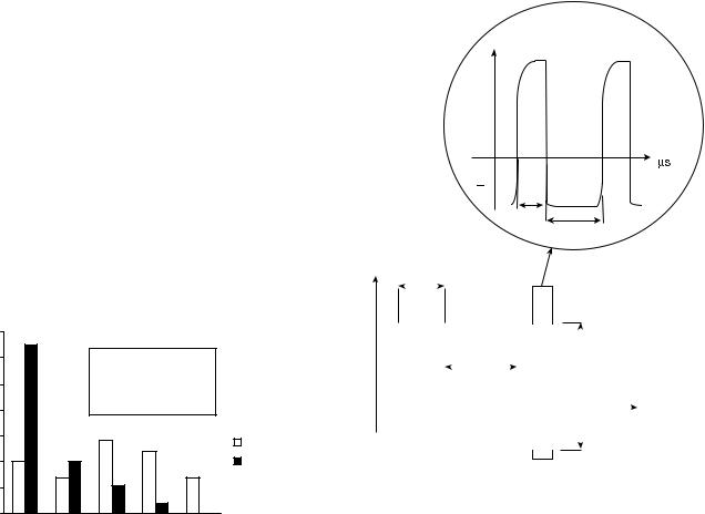
the other hand, work by numerous investigators has demonstrated that one or more methods of electropharmaceutical anesthesia (anesthesia consisting of a combination of an electric current with anesthetic agents) is not only possible, but also desirable because of the lack of side effects and reduced requirements for neurodepressants. During past years, progress in chemical anesthesia has been so successful that the objective was not to replace classical anesthesia, but to more precisely confirm studies performed on animals, potentiation of anesthetic drugs by Transcutaneous Cranial Electrical Stimulation (TCES) to obtain postoperative electromedicinal analgesia, to the end that toxicity induced by chemical drugs could be dramatically reduced. The use of TCES is not without considerable supporting data, as from 1972 to 2000, many clinical trials involving TCES had been carried out on patients under electromedicinal anesthesia and provide > 20 specific references (7). During those clinical trials, anesthetists noticed that complaints of patients operated under TCES were less numerous in the recovery room than complaints of patients operated with chemical anesthesia. It seems that a state of indifference and reduction of painful sensation persisted in TCES-treated patients. These observations were scientifically confirmed in a study (59) in which 100 patients operated under electroanesthesia (EA) was compared to another 100 patients submitted to narco-neurolept-analgesia, a classical anesthesia (CA): the head nurses were ordered to administered 15 mg (i.m.) of pentazocine in case of patient complaints. It is worth noting that the first 16 postoperative hours, the average intake of pentazocine for the patients of the EA group was 8.1 mg/ patient, whereas it was 29.7 mg/patient (3.67 time higher) for the patients of the CA group. This difference between groups is highly statistically significant (p < 0.001) (Fig. 1).
This residual and prolonged analgesia is surely one of the most important advantages of TCES, but few clinicians benefit from its advantages at the present time. The most likely reason that few clinicians benefit from TCES is that it is not yet approved for use in the United States, Canada, and many countries in Europe by the respective regulatory agencies. Recent research carried on in numerous laboratories has increased our knowledge of the neurobiological
Number of |
|
|
|
|
|
patients |
|
|
|
|
|
70 |
|
|
|
|
|
60 |
|
average per head (mg) |
|
||
|
|
CA: 8,1 (12.69 s.e.) |
|
||
50 |
|
EA: 29.7 (19.88 s.e.) |
|
||
40 |
|
(χ2 test) P<0.001 |
|
||
|
|
|
|
|
|
30 |
|
|
|
|
I(CA) n=100 |
|
|
|
|
|
|
20 |
|
|
|
|
(EA) n=100 |
10 |
|
|
|
|
|
0 |
|
|
|
|
|
0 |
15 |
30 |
45 |
60 |
Pentazocin (mg) |
Figure 1. Comparison of two groups receiving pentazocin during the first 16 h after surgery.
effects of these currents, and allowed the establishment of serious protocols dedicated to new clinical applications (7).
Nature of the Limoge’s Current. Limoge et al. demonstrated that complex currents of their design are capable of producing profound analgesia without provoking initial shock, pain, or unpleasant sensations, burns, other cutaneous damage, muscular contractures, cerebral damage or convulsions, and respiratory or circulatory depression (58). The Limoge current consists of high frequency (HF) biphasic asymmetrical wave trains composed of modulated high frequency (166 kHz) pulse trains, regularly interrupted with a repetition cycle of 100 Hz (7,57). These wave trains are composed of successive impulsional waves of a particular shape: one positive impulse of high intensity and short duration (2 ms), followed by a negative impulse of weak intensity and long duration (4 ms) adjusted in such a way that the positive surface is equal to the negative surface. The average intensity of this current equals 0 mA. The use of such a negative phase makes it possible to eliminate all risk of burns. The ‘‘on-time’’ of the low frequency (LF) wave trains is 4 ms, followed by a 6 ms ‘‘OFF-time’’ (Fig. 2).
|
High frequency |
I |
166 kHz |
+
0
2
4
I |
|
Magnification (× 1000) |
|
|
|
4
|
|
|
|
|
|
|
|
|
|
|
|
|
|
|
|
|
|
|
|
|
|
+ |
|
6 |
|
|
|
280 mA |
||||
0 |
|
|
|
|
|
|
|
|
|
|
|
|
|
|
|
Off |
|
|
|
ms |
|
|
|
|
||||||||
|
|
|
|
|
|
|
|
|
|
|
|
|
|
|
|
|
|
|
|
|
|
On
Low frequency 100 Hz
Figure 2. The Limoge waveform pattern: a modulated HF (166 kHz) pulse trains (top) regularly interrupted with a repetition cycle of 100 Hz. Concerning the high frequency, note the exponential ascent and acute fall of the waveform, and also note the area of the positive deflection is equal to that of negative deflection.
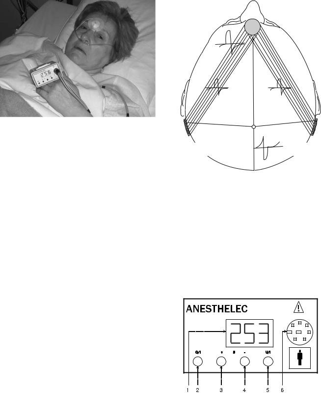
Figure 3. Application of the device on a patient during post operative period. See the placement of the frontal electrode.
This type of current was gradually developed over 20 years through numerous human clinical studies. The shape and cyclic ration of the HF waves are felt to be of utmost importance in the production of analgesia. Various shapes of waves have been tested (triangular, rectangular, exponential). Clinical impressions suggest that the most profound analgesia occurs with HF waveforms having an exponential ascent and acute fall. The most effective cyclic ratios are 2:5 with LF waves and 1:3 with HF waves with peak-to-peak intensity between 250 and 300 mA and peak- to-peak voltage between 30 and 40 V.
Electrodes. Three electrodes are used. One frontal electrode is placed between the eyebrows and two posterior electrodes are placed behind the mastoid process on each side of the occiput (Fig. 3). It is hoped that the intracerebral electric field thus obtained spreads on each side of the median line and it thus successful in stimulating opioid receptors surrounding the third and fourth ventricles and the paraventricular areas of the brain. In addition, some of the electric current spreads over the scalp, thus provoking peripheral electrostimulation (Fig. 4). The use of HF biphasic current permits employment of self-sticking electrodes made of silver (active diameter 30 mm), without risk of burns and without unpleasant sensations under electrodes.
Transcutaneous Cranial Electrical Stimulators Using Limoge Currents. Until now three types of devices only give Limoge currents: two American devices called Foster Biotechnology Neurostimulation Device (FBND) and ElectroAnalgesia Stimulation Equipment (EASE) and one French device called Anesthelec (Fig. 5). The electrical stimulator must abide by general safety rules. The use of electrosurgical units during TCES requires excellent electrical isolation of the generator to avoid any risk of return of the current or skin burns under electrodes, a fault in the electrosurgical unit. These portable devices of type LF with isolated output are composed of one HF oscillator, one oscillator with LF relaxation with internal power supply generating HF (166 kHz), and LF (100 Hz) currents for
ELECTROANALGESIA, SYSTEMIC |
29 |
Frontal
electrode
|
|
Electric |
|
HF.waveform |
|
|
|
field |
Electric |
field |
line |
|
||
|
Median |
|
|
|
|
|
|
Point 0 |
|
Reversal line |
|
Retromastoid |
Retromastoid |
|
electrode |
electrode |
|
Figure 4. Location of the electrodes and shape of wave on the scalp. The center of the frontal electrode is situated at the intersection of the line of the eyebrowns and the sagittal plane. The center of the two retromastoid electrodes is localized in the retromastoid fossa. On the scalp the amplitude of HF waves diminish in measurement as the point O (occipital line) is approached, and behind that point there is an inversion of the wave form. The lines joining the frontal electrode to retromastoid electrodes represent the projected distribution of Limoge currents through the brain with action at the level of the periacqueductal gray matter and the limbic system.
therapeutic use and one delay circuit, to stop output when its level is too high. The battery pack must be protected against short circuit as well as polarity inversion and detachable as it is rechargeable. The patient cable must
Figure 5. Front view of the Anesthelec generator box 1 !3 digits displayer: Current intensity (from 000 to 300 mA); 2 !Pushing button On/Off; 3 !Pushing button for increment of the output current intensity; 4 !Pushing button for decrement of the output current intensity; 5 !Pushing button to select display (intensity of voltage); 6 !Connector for the three electrodes.

30 ELECTROANALGESIA, SYSTEMIC
be an interlocking type preventing accidental disconnection and the functioning of the device must be simplified with detectors to measure the intensity of the current and the voltage applied to the patient to confirm proper contact between skin and electrodes. Concerning electromagnetic compatibility, the device must be autonomous with no possible direct or indirect link mains supplies. Mini box and manipulation components must be made in isolated material and the patient cables must be shrouded and the applied elements must be protected against overvoltage.
Clinical Usage of TCES
The TCES method being used with increasing frequency in France, and many other european countries, in Russia, in Mexico, and in Venezuela. It is not yet approved for use in the United States, but is being evaluated both in patients and in volunteers. Numerous studies have demonstrated that TCES is particularly effective in urologic, thoracic, and gastrointestinal surgery, but is not limited to these types of operative procedures. Patients receiving TCES require less nitrous oxide (N2O) (30–40% less) to prevent movement in response to a pain stimulus. This method potentiates both the amnestic and analgesic effects of N2O and prolongs residual postanesthetic analgesia at sites of trauma. The mechanism of analgesia resulting from TCES during administration of N2O is unknown. Volunteers getting TCES without N2O for 1 h are not sleepy or amnesic, but do report a warm and tingling sensation all over their body, and are objectively analgesic to many forms of painful stimulation (14–16). Similar results have been obtained with some TENS units in patients with chronic pain and after operation in patients with acute postoperative pain (54,55). As mentioned previously, some have suggested that receptor sites situated in the central gray area of the brain, the spinal cord, and other areas in the central nervous system regulate the effects of painful stimulation, analgesia, and the perception of somatic pain. Electrical stimulation of these receptor sites has been shown to result in relief from pain and can be antagonized by narcotic antagonists. Furthermore, the analgesic actions of TENS can be reversed with antagonists like naloxone (9,10). This suggests that TENS and TCES may be producing analgesia by stimulating increased production and/or release of the body’s endogenous analgesics, the endorphins, enkephalins, serotonin and/or other neurotransmitters and neuromodulators (5,11–13,43– 46,63–65).
To separate facts from empiricism and anecdotal information for several years, teams of researchers and clinicians attempted to show in animals and in humans what are the neurobiological mechanisms brought into pay by the TCES with currents of Limoge. For that reason, a study was conducted in France on rats on TCES potentiation of halothane-induced anesthesia and the role of endogenous opioid peptides was addressed (19). Carried out in double blind for 10 h prior to tracheotomy and the inhalation of halothane, the TCES provoked in the stimulated rats (TCES group, n ¼ 10), a significant decrease (p < 0.001) in the Minimum Alveolar Concentration of Halothane (MACH) in comparison with the nonstimulated rats (con-
MACH (vol %)
1,2 |
|
|
|
|
|
|
|
|
|
|
|
|
|
|
|
|
|
|
|
|
|
|
|
|
|
|
|
|
|
|
|
|
|
|
|
|
|
|
|
|
|
|
|
|
|
|
|
|
|
|
|
||
1,0 |
|
|
|
|
|
|
|
*** |
|
|
|
|
|
|
|
|
|
|
|
|
|
|
||||
0,8 |
|
|
|
|
|
|
|
|
|
|
|
|
|
|
|
|
|
|
|
|
|
|||||
0,6 |
|
|
|
|
|
|
|
|
|
|
|
|
|
|
|
|
|
|
|
|
|
|
|
|
|
|
0,4 |
|
|
|
|
|
|
|
|
|
|
|
|
|
|
|
|
|
|
|
|
|
|
|
|
|
|
0,2 |
|
|
|
|
|
|
|
|
|
|
|
|
|
|
|
|
|
|
|
|
|
|
|
|
|
|
0,0 |
|
|
|
|
|
|
|
|
|
|
|
|
|
|
|
|
|
|
|
|
|
|
|
|
|
|
|
Control |
TCES |
Control |
|
TCES |
|||||||||||||||||||||
|
|
|
||||||||||||||||||||||||
|
|
|
|
|
|
|
|
|
|
|
|
|
+ naloxone |
+ naloxone |
||||||||||||
Figure 6. Effects of TCES on halothane requirements in rats.
indicate significant difference between TCES and CONTROL groups (ANOVA, p < 0.001).
trol group, n ¼ 10). This effect was completely inhibited by a subcutaneous injection of 2 mg/kg of naloxone (antagonist of morphine), which restored the MACH to its initial value in the TCES group without affecting the control group (Fig. 6). Moreover, TCES potentiation of halothane-induced anesthesia was dramatically increased by inhibition of enkephalin degradation. Thus the decrease of the MACH is associated with the potentiation the analgesic action of enkephalins released in the cellular space by TCES. These results demonstrate the direct involvement of endogenous opioid peptides on therapeutic effects of TCES.
In addition, a double-blind study carried out during labor and delivery on parturients to provide evidence of a mode of action of TCES on maternal plasma secretion of b-endorphins (66). To evaluate the rate of b-endorphins, blood samples were drawn from two groups of voluntary women in parturition ( a TCES group, n ¼ 23, and a control group, n ¼ 17) at four precise stages: at the moment the electric generator was attached, after 1 h of the current application, at the time of complete dilatation, and finally after the delivery. The dosages were achieved by the radioimmuno enzymatic method. The plasmatic rate of b-endor- phins was identical in the beginning for the two groups as those described in the literature, but this rate was progressively augmented in a significant fashion during the course of the labor from the first hour (p < 0.05) for the TCES group (Table 1).
It is more interesting to know the rate of endorphins produced in the cerebral structures known for their abundance of opiate receptors, more so than in the plasma. The exploration of the effects of TCES on brain opioid peptides was conducted at the Vishnevski Institute in Moscow by dosing endorphins in the cerebral spinal fluid (CSF) before cardiac surgery and after 30 min of TCES. The dosage showed that TCES augmented significantly (p < 0.01) the rate of b-endorphins in the CSF when compared to the control group and the effects of TCES reversed by naloxone (49,67,68).
These studies can partially explain the mode of action of TCES with currents of Limoge in the brain and permit not only rectification of protocols for clinical trials already carried out, but also provide better indication for utilization of TCES.
For all clinical applications, it must be keep in mind that the currents of Limoge provoke endogenous neurosecretions (7), which are not immediate, they require a certain

|
|
|
|
ELECTROANALGESIA, SYSTEMIC |
31 |
|||
Table 1. Evaluation of b-Endorphin During Labor and Delivery on Parturientsa |
|
|
|
|
|
|||
|
|
|
b-Endorphin Plasmatic Rate, pg mL 1 |
|
|
|||
|
Medication |
Labor Time |
Installation |
After 1 h |
Dilatation |
Delivery |
|
|
|
|
|
|
|
|
|
|
|
Control (n ¼ 17) |
Peridural: 1 patient |
< 1 h 30 min: 4 patients |
123 ( 12) |
127 ( 10) |
|
|
|
|
|
Morphine: 11 patients |
> 1 h 30 min: 13 patients |
124 |
160 |
|
|
||
TCES (n ¼ 23) |
None: 5 patients |
|
|
|
|
|
|
|
Peridural: 2 patient |
< 1 h 30 min: 11 patients |
133 ( 11) N.S.b |
167 ( 12)c |
186 |
182 |
|
|
|
|
Morphine: 3 patients |
> 1 h 30 min: 12 patients |
|
|
||||
|
None: 18 patients |
|
|
|
|
|
|
|
aResults of b-endorphin rates are expressed as mean s.e.m. (when available).
bN.S. indicates no difference between b-endorphin rates of control and TCES groups when measured at the installation of labor.
cIndicates significant difference between b-endorphin rates of control and TCES groups (t-test, p < 0.05) when measured 1 h after the installation of labor.
amount of time for their induction, and then are maintained all along the stimulation application. In consequence, the utilization of this technique in classical anesthesia is not the best indication except for major interventions of long duration. One must also remember that during > 10,000 major surgical interventions carried out under classical anesthesia combined with TCES it has been proven that the Limoge currents has a potentiation effect on opioid and non-opioid analgesics, morphinomimetics, psychotropes, and psycholeptics, (14–20) and this potentiation allows a decrease in drug doses, and therefore a decrease in toxicity. But one must admit objectively that, during past years, progress in chemical anesthesia has been so successful that the TCES will not replace classical anesthesia. The potentiation of drugs nevertheless by TCES can open new perspectives in the treatment of pain whether postoperative or chronic. To be precise the potentiation of opioid analgesia by TCES under specific conditions, was demonstrated by Stinus et al. (17). The authors showed that potentiation was a function of (a) the intensity of the stimulation, (b) the opioid dose administered, (c) the duration of TCES applied preceding opioid administration, and (d) the position and the polarity of the electrodes. This experimental approach was of prime importance as it allowed determination of the most efficient parameters, studied the therapeutic effects of TCES in humans, and increased our knowledge of the effects of TCES on neurobiological substrates.
Taking account of animal experimentation and clinical trials, one must know that to be successful in clinical applications, a correct basal protocol for TCES use should be followed. The main parameters are, the correct placement of the electrodes, starting electrostimulation no < 2 h
prior to the introduction of drugs and continuation of TCES delivery during the pharmacokinetic action of drugs.
Abolition of Postoperative Pain (Fig. 3). Patients operated under TCES associated with pharmaceutical anesthesia complain strikingly less often about pain than those operated with a classical anesthesia. The TCES method induces a postoperative analgesia for an average of 16 h. A doubleblind study has been made during per and postoperative period on 39 patients (TCES group n ¼ 20 and control group n ¼ 19) undergoing an abdominal surgery (20). Upon arrival in the recovery room, patients were given a computerized, patient-controlled analgesia (PCA) device to deliver IV buprenorphine (50 mg boluses, 30 min lock-out) during the first four postoperative hours. The recorded variables included postoperative requirements, pain scores with pain visual analogue scale (VAS) (from 0 ¼ no pain to 10 ¼ worst), sedation, (from 0 ¼ not arousable to 4 ¼ awake) awake) and were collected hourly from the first to the sixth postoperative hour by a blinded investigator. There was a highly significant reduction of cumulative buprenorphine requirements in the TCES group compared with the control group (2.36 0.19 vs. 3.43 0.29 mg kg 1 h 1; p < 0.01) (Table 2). At each postoperative hour, patients required less buprenorphine in the TCES group. These results indicate that TCES reduces narcotic requirements for postoperative analgesia. TCES may have potential to facilitate early postoperative analgesia in patients undergoing major surgery.Therefore this technique allows a maximal restriction of pharmaceutical contribution.
Obstetric Electroanalgesia (66,69). In order to test the analgesic efficacy of TCES with Limoge currents during
Table 2. Buprenorphine Consumptiona
Postoperative hours (H) |
TCES |
Control |
||
|
|
|
|
|
H1 |
1.35 0.15 |
1.57 |
0.13 |
|
H2 |
0.90 0.16 |
1.21 |
0.18b |
|
H3 |
0.60 0.15 |
1.10 |
0.16b |
|
H4 |
0.60 0.18 |
1.00 |
0.15 |
|
Total dose (mg kg 1 h 1) |
2.36 0.19 |
3.43 0.29c |
||
aData are expressed as mean SEM. bp < 0.05.
cp < 0.01.
32 ELECTROANALGESIA, SYSTEMIC
labor and delivery, a double-blind study was performed with ‘‘anesthelec’’ on 20 cases for whom analgesia was necessary (TCES group I, current ‘‘on’’, n ¼ 10, and control group II, current ‘‘off ’’, n ¼ 10). Labor and delivery were carried out by a medical team different from those using the anesthelec. The results showed that TCES, with or without nitrous oxide inhalation, decreases by 80% the number of epidural analgesia or general anesthesia that would otherwise have been unavoidable. To define the effects of TCES, maternal and fetal parameters of 50 deliveries carried out under TCES were compared with 50 deliveries carried out under epidural analgesia (70).
TCES was used only if analgesia was required. These clinical trials were a retrospective comparison between two similar nonpaired series. Despite the fact that analgesia obtained with TCES was less powerful than with epidural analgesia, this method showed many advantages: total safety for the child and the mother, easy utilization, shorter labor time, decreased number of instrumental extractions and potentially reduced costs. Good acceptance and satisfaction for the mother should stimulate a rapid evolution and acceptance of this new method.
The TCES method should be applied following the first contractions. Analgesia is established after 40 min of stimulation. A diminution of pain is achieved that is comparable to that obtained after an injection (IV) of morphine (but it is less profound than with epidural analgesia), a decrease in vigilance with euphoria is obtained without inducing sleep, but allowing compensatory rest between contractions. The pupils are enlarged. Stimulation is applied throughout the birthing procedure and residual analgesia persists for several hours following delivery. Results are best if the expectant mother participates in a preparatory course for the birthing experience or if she uses musicotherapy in conjunction with TCES. If analgesia is insufficient it is possible to have patients breath nitrous oxide and oxygen (50:50) or to administer an epidural analgesia for the remainder of procedure. Thus obstetrical analgesia utilizing the currents of Limoge allows a reduction of labor time in all primapares (p < 0.001) and is without risk to the mother or child. Mothers in labor appreciate this simple, nonmedicinal, nonpainful technique that allows them to actively participate in the delivery.
Electropharmaceutical Anesthesia in Long Duration Microsurgery. For major operations and those of long duration the results are most encouraging as TCES permits a reduction of anxiolytics and neuroleptics by 45% and reduction of morphinomimetics by 90% and demonstrates the possibilities of drug potentiation to prolong analgesia while at the same time providing a less depressive general anesthetic (7,58,59,68). Early results have improved thanks to animal research and revision of protocols more particularly (17–19). In 1972, it was not know to begin electrostimulation three hours prior to medicinal induction (4).
Potentiation of Morphine Analgesia for Patients with Chronic Pain and Associate Problems (71). For all neurophysiological applications, a basic protocol must be followed. This protocol is as follows: If the patient is being treated pharmacologically, for the first time, never stop the che-
mical medication but diminish the dosage each day until a threshold dose is obtained according to the particular pathology and the patient. Begin TCES at least 1 h before medication whether it be on awakening in the morning or 2 h prior to going to bed. (There is no contraindication in maintenance of stimulation all-night long.)
If the patient is not being treated chemically, the effect of the current is best if there is a ‘‘starter dosage’’ of medicine. It is therefore recommended that a weak medicinal dose be prescribed according to the pathology and begin the TCES 1 h before the patient takes the dose, and continue stimulation during the time of pharmacocinetic action of the medicine.
This protocol will permit treatment of cancer patients at home whenever possible under medical supervision: This is a less traumatizing course of action than having the patients come into hospital every day. In the beginning, one must maintain the standard pain medication therapy and the patient should be connected to the Limoge Current generator for 12 h (during the night, if possible); the potentiometer is turned clockwise to a reading of 35 V and 250–300 mA, peak to peak. After this first treatment phase, the patient can use the machine for 3 h whenever they feels the need. The analgesic effect of TCES may not appear until the third day of treatment. Then TCES is initiated upon awakening. After 1 h, standard pain medication is given and TCES therapy is continued for another hour. Three hours before bedtime, TCES is again administered for 2 h, then standard pain medication is given and TCES therapy continued for another hour. The patient should enjoy restful sleep. After 8 days, the standard pain medication therapeutic dose should be decreased gradually, but not totally terminated. After this status has been achieved, patients may use the machine whenever they feel the need, for 3 h preferably with the reduced dose of the standard pain medication. The minimal therapeutic dose of the pain medication, however, may have to be adjusted upward somewhat due to individual differences in some patients.
CONCLUSION
All numerous and previous clinical trials have demonstrated that TCES reduces narcotic (fentanyl) requirements in patients undergoing urologic operations with pure neuroleptanesthesia (droperidol, diazepam, fentanyl, and air-oxygen) (20,36–38). Use of TCES in a randomized double-blind trial of these patients resulted in a 40% decrease in fentanyl requirements for the entire operation. Unfortunenately, while available TCES units (using currents of 250–300 mA peak to peak, with an average intensity of zero) provide analgesia and amnesia, they do not produce complete anesthesia. Whether any form of TCES or the use of very high frequency (VHF) will provide more analgesia and amnesia, that is, amounts sufficient to result in complete anesthesia without need for pharmaceutical supplementation, without problems has yet to be carefully evaluated but obviously needs to be studied. Considerable research must continue in this area.
Theoretically, lower doses of narcotics or lower concentrations of inhalation anesthetics should result in fewer
alterations in major organ system function during anesthesia. This could mean that anesthesia with TCES produces less physiological insult than more standard anesthetic techniques and results in a shorter postoperative recovery period. It has been observed that TCES plus N2O results in analgesia that persists after stimulation is terminated and N2O is exhaled (7,14,15,20,58–60). This suggests that intraoperative use of TCES might reduce postanesthetic analgesic requirements, and that future clinical trials must be initiated to confirm this suggestion.
The 30.000 plus major interventions realized under TCES in France and in Russia since 1972 and the > 5000 drug withdrawals undertaken in opioid addicted patients at the Medical Center of the University of Bordeaux since 1979 without even the most minor incident permits us to conclude that the currents of LIMOGE are absolutely innocuous and cause no side effects. This simple technique reduced the use of sedative medicaments such as psychotropes or psycholeptics that often lead to ‘‘legal’’ addiction. The TCES is atoxic, reproductible, causes no personality change and is without habituation. Briefly, this technique fits perfectly into the domaine of all aspects of classical and alternative medicine as well as human ecology.
BIBLIOGRAPHY
Cited References
1.Smith RH. Electrical Anesthesia. Springfield (IL): CC Thomas Publ.; 1963.
2.Smith RH, Tatsuno J, Zouhar RL. Electroanesthesia: a review-1966. Anesth Analg 1967;40:109–125.
3.Smith RH. Electroanesthesia Review article. Anesthesiology 1971;34:61–72.
4.Limoge A. An Introduction to Electroanesthesia. Baltimore: University Park Press; 1975. p 1–121.
5.Sances Jr A, Larson SJ. Electroanesthesia. New York: Academic Press; 1975. p 1–367.
6.Shealy CN, Maurer D. Transcutaneous nerve stimulation for control pain. Surg Neurol 1974;2:45–47.
7.Limoge A, Robert C, Stanley TH. Transcutaneous cranial electrical stimulation (TCES): a review 1998. Neurosci Biobehav Rev 1999;23:529–538.
8.White PF, Li S, Chiu JW. Electroanalgesia: Its role in acute and chronic pain management. Anesth Analg 2001;92:505–513.
9.Adams JE. Naloxone reversal of analgesia produced by brain stimulation in the human. Pain 1976;2:161–166.
10.Hosofrichi Y, Adams JE, Linchitz R. Pain relief by electrical stimulation of the central gray matter and its reversal by naloxone. Science 1977;197:183–186.
11.Snyder SH, Goodman RR. Multiple neurotransmitter receptors. Neurochemistry 1980;35:5–15.
12.Snyder SH. Brain peptides as neurotransmitters. Science 1980;209:976–983.
13.Pasternack GW. Opiate enkephalin and endorphin analgesia: Relations to a single subpopulation of opiate receptors. Neurology 1981;31:1311–1315.
14.Stanley TH, et al. Transcutaneous cranial electrical stimulation increases the potency of nitrous oxide in humans. Anesthesiology 1982;57:293–297.
15.Stanley TH, et al. Transcutaneous cranial electrical stimulation decreases narcotic requirements during neuroleptanesthesia and operation in man. Anesthol Analg 1982;61: 863–866.
ELECTROANALGESIA, SYSTEMIC |
33 |
16.Bourke DL, et al. TENS reduces halothane requirements during hand surgery. Anesthesiology 1982;61:769–772.
17.Stinus L, et al. Transcranial electrical stimulation with high frequency intermittent current (Limoge’s) potentiates opiate-induced analgesia: blind studies. Pain 1990;42:351– 363.
18.Auriacombe M, et al. Transcutaneous electrical stimulation with Limoge current potentiates morphine analgesia and attenuates opiate abstinence syndrome. Biol Psychiat 1990;28:650–656.
19.Mantz J, et al. Transcutaneous cranial electrical stimulation with Limoge’s currents decreases halothane requirements in rats: evidence for involvement of endogenous opioids. Anesthesiology 1992;76:253–260.
20.Mignon A, et al. Transcutaneous cranial electrical stimulation (Limoge’s currents) decreases bupremorphine analgesic requirements after abdominal surgery. Anesth Analg 1996;83:771–775.
21.Scribonius L. Compositiones medicae. Padua: Frambottus; 1655. Chapts. 11 and 162.
22.Kane K, Taub A. A history of local electrical anesthesia. Pain 1975;1:125–138.
23.Leduc S. Production du sommeil et de l’anesthe´sie ge´ne´rale et locale par les courants e´lectriques. C R Acad Sci Paris 1902;135:199–200.
24.Leduc S. L’e´lectrisation ce´re´brale. Arch Electr Med 1903; 11:403–410.
25.Denier A. Electro-anesthe´sie. Anesth Analg Re´an 1951;8(1): 47–48.
26.Denier A. Anesthe´sie e´lectrique. EMC 36550 A 10 1958;4:1–8.
27.Knutson RC. Experiments in electronarcosis. A preliminary study, Anesthesiology 1954;15:551–558.
28.Knutson RC, et al. The use of electric current as an anesthetic agent. Anesthesiology 1956;17:815–825.
29.Knutson RC, Tichy FY, Reitman J. The use of electrical current as an anesthetic agent. Anesthesiology 1966;17: 815–825.
30.Cara M, Cara-Beurton M, Debras C, Limoge A, Sances Jr A, Reigel DH. Essai d’anesthe´sie e´lectrique chez l’Homme. Ann Anesth Franc¸ 1972;13:521–528.
31.Shimoji K, et al. Clinical electroanesthesia with several methods of current application. Anesth Analg 1971;50:409– 416.
32.Shimoji K, Higashi H, Kano T. Clinical application of electroanesthesia. In: Limoge A, Cara M, Debras Ch, editors. Electrotherapeutic Sleep and Electroanesthesia. Volume IV, Paris: Masson; 1978. p 96–102.
33.Djourno A, Kayser AD. Anesthe´sie et sommeil e´lectriques. Paris: P.U.F.; 1968.
34.Sachkov VI, Liventsev NM, Kuzin MI, Zhukovsky VD. Experiences with interference currents in clinical surgery. In: Wageneder FM, Schuy S, editors. Electrotherapeutic Sleep and Electroanesthesia. Excerpta Medica Foundation; 1967. p 321–326.
35.Limoge A. The use of rectified high frequency current in electrical anaesthesia. In: Wageneder FM, Schuy St, editors. Electrotherapeutic sleep and electroanaesthesia. Volume I, Amsterdam: Excerpta Medica Foundation; 1967. p 231– 236.
36.Cara M, Debras Ch, Dufour B, Limoge A. Essais d’anesthe´sie e´lectrome´dicamenteuse en chirurgie urologique majeure. Bull Acad Me´d 1972;156:352–359.
37.Debras C, Coeytaux R, Limoge A, Cara M. Electromedicamentous anesthesia in Man. Preliminary results Rev I E S A 1974; 18–19, 57–68.
38.Limoge A, Cara M, Debras C. Electrotherapeutic Sleep and Electroanesthesia. Paris: Masson; 1978.