

17.Epstein E, Epstein E, Jr, editors. Techniques in Skin Surgery. Philadelphia: Lea & Febiger; 1979.
18.Burdick KH. Electrosurgical Apparatus and Their Application in Dermatology. Springfield (IL): Chas C Thomas; 1966.
19.Pearce JA. Electrosurgery. Chichester: Chapman & Hall; 1986.
20.Knecht C, Clark RL, Fletcher OJ. Healing of sharp incisions and electroincisions in dogs. JAVMA 1971;159:1447–1452.
21.Kelly HA, Ward GE. Electrosurgery. Philadelphia: Saunders; 1932.
22.Operating and service instructions for model 770 Electrosectilis, The Birtcher Corporation, El Monte (CA).
23.Operator and service manual, MF-380, Aspen Laboratories Inc., Littleton (CO).
24.Valleylab SSE2-K service manual. Boulder (CO): The Valleylab Corporation;
25.User’s guide, Force 2 electrosurgical generator. The Valleylab Corporation, Boulder (CO).
26.Medical electrical equipment part 1: General requirements for safety. IEC/EN 60601-1, International Electrotechnical Commission; 1988.
27.Medical electrical equipment part 2-2: Particular requirements for safety—High frequency surgical equipment. IEC/ EN 60601-2-2, International Electrotechnical Commission; 1999.
28.Electrosurgical units. Health Devices. Jun–Jul 1973;2:n8–9.
29.Henriques FC. Studies of thermal injury V: The predictability and the significance of thermally induced rate processes leading to irreversible epidermal injury. Arch Pathol 1947;43:489–502.
30.Panescu D, Fleischman SD, Whayne JG, Swanson DK. Contiguous Lesions by Radiofrequency Multielectrode Ablation. Proc IEEE-Eng Med Biol Soc 17th Annu Meet. 1995; 17, n1.
31.Pearce JA, Thomsen S. Numerical Models of RF Ablation in Myocardium. Proc IEEE-Eng Med Biol Soc 17th Annu Meet; vol. 17, n1, 1995; p 269–270.
32.Lemole GM, Anderson RR, DeCoste S. Preliminary evaluation of collagen as a component in the thermally-induced ‘weld’. Proc SPIE 1991;1422:116–122.
33.Kopchock GE, et al. CO2 and argon laser vascular welding: acute histologic an thermodynamic comparison. Lasers Surg. Med. 1988;8(6):584–8.
34.Schober R, et al. Laser-induced alteration of collagen substructure allows microsurgical tissue welding. Science 1986;232:1421–11.
35.Collagen Volume I Biochemistry. Nimni ME, editors. Boca Raton (FL): CRC Press; 1988.
36.Pearce JA, Thomsen S, Vijverberg H, McMurray T. Kinetic rate coefficients of birefringence changes in rat skin heated in vitro. Proc SPIE 1993;1876:180–186.
37.Chen SS, Wright NT, Humphrey JD. Heat-induced changes in the mechanics of a collagenous tissue: isothermal, isotonic shrinkage. Trans ASME J Biomech Eng 1998;120:382–388.
38.Chen SS, Wright NT, Humphrey JD. Phenomenological evolution equations for heat-induced shrinkage of a collagenous tissue. IEEE Trans Biomed Eng 1998;BME-45:1234–1240.
39.Honig WM. The mechanism of cutting in electrosurgery. IEEE Trans Bio-Med Eng 1975;BME-22:58–62.
40.Geddes LA, Tacker WA, Cabler PA. A new electrical hazard associated with the electrocautery. Biophys Bioengr Med Instrum 1975;9n2:112–113.
41.Hungerbuhler RF, et al. Ventricular fibrillation associated with the use of electrocautery: a case report. JAMA 21 Oct 1974;230n3:432–435.
42.Orland HJ. Cardiac pacemaker induced ventricular fibrillation during surgical diathermy. Anesth Analg Nov 1975;3n4; 321–326.
ENDOSCOPES 177
43.Titel JH, et al. Fibrillation resulting from pacemaker electrodes and electrocautery during surgery. Anesthesiol Jul– Aug 1968;29:845–846.
44.Batra YK, et al. Effect of coagulating and cutting current on a demand pacemaker during transurethral resection of the prostate: a case report. Can Anesthes Soc J Jan 1978;25n1: 65–66.
45.Fein RL. Transurethral electrocautery procedures in patients with cardiac pacemakers. JAMA 2 Oct 1967;202: 101–103.
46.Greene LF. Transurethral operations employing high frequency electrical currents in patients with demand cardiac pacemakers. J Urol Sept 1972;108:446–448.
47.Krull EA, et al. Effects of electrosurgery on cardiac pacemakers. J Derm Surg Oct 1975;1n3:43–45.
48.Wiley JD, Webster JG. Analysis and control of the current distribution under circular dispersive electrodes. IEEE Trans BioMed Eng May 1982;BME-29n5:381–384.
49.Choi BJ, Kim J, Welch AJ, Pearce JA. Dynamic impedance measurements during radio-frequency heating of cornea. IEEE Trans Biomed Eng 2002;49n12:1610–1616.
See also CRYOSURGERY; ION-SENSITIVE FIELD EFFECT TRANSISTORS.
EMERGENCY MEDICAL CARE. See
CARDIOPULMONARY RESUSCITATION.
EMG. See ELECTROMYOGRAPHY.
ENDOSCOPES
BRETT A. HOOPER
Arete´ Associates
Arlington, Virginia
INTRODUCTION
The word endoscope is derived from two Greek words, endo meaning ‘‘inside’’ and scope meaning ‘‘to view’’. The term endoscopy is defined as, ‘‘using an instrument (endoscope) to visually examine the interior of a hollow organ or cavity of the body.’’ In this second edition of the Encyclopedia of Medical Devices and Instrumentation, we will revisit the excellent background provided in the first edition, and then move on from the conventional ‘‘view’’ of endoscopes and endoscopy to a more global ‘‘view’’ of how light can be delivered inside the body and the myriad light-tissue interactions that can be used for both diagnostic and therapeutic procedures using endoscopes. We will update the medical uses of endoscopy presented in the first edition, and then look at the medical specialties that have new capabilities in endoscopy since the first edition; cardiology and neurosurgery. This will be by no means an exhaustive list, but instead a sampling of the many capabilities now available using endoscopes. We will also present new delivery devices (fibers, waveguides, etc.) that have been introduced since the optical fiber to allow a broader range of wavelengths and more applications that deliver light inside the body.
178 ENDOSCOPES
HISTORY
Pre-1800s
Endoscopy had its beginnings in antiquity with the inspection of bodily openings using the speculum, a spoon-shaped instrument for spreading open the mouth, anus, and vagina. Another instrument of fundamental importance to endoscopy is the catheter, as it has been used to evacuate overfilled bladders for more than 3000 years. Catheters and a rectoscope were used by Hippocrates II (460–377 BC), where inspection of the oral cavity and of the pharynx was routine, including operations on tonsils, uvula, and nasal polyps. This trend continued for the better part of two millennia.
1800–1900
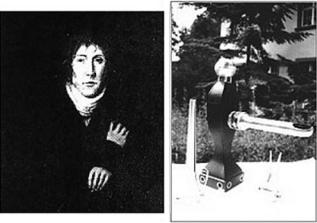
In 1804, Phillip Bozzini came upon the scene with the Lichtleiter, and attempts were made to view into the living body through the endoscope’s narrow opening (1). It took almost 100 years to achieve this goal. These prototype endoscopes consisted of hollow tubes through which light of a candle, and even sunlight, was projected in order to visualize the inside of a bodily opening. The Lichtleiter, the light conductor, consists of two parts: (1) the light container with the optical part; and (2) the mechanical part, which consists of the viewing tubes fitted to accommodate the anatomical accesses of the organs to be examined. The apparatus is shaped like a vase, is 35 cm tall, made of hollow lead, and covered with paper and leather. On its front is a large, round opening divided into two parts by a vertical partition. In one half, a wax candle is placed and held by springs such that the flame is always in the same position. Concave mirrors are placed behind the candle and reflect the candle light through the one-half of the tube onto the object to be examined. The image of the object is then reflected back through the other half of the tube to the eye of the observer. Depending on the width of the cavity to be examined, different specula were used. These specula consisted of leaves that could be spread open by use of a screw device in order to expand the channels. An example is shown in Fig. 1.
Figure 1. Phillip Bozzini’s Lichtleiter endoscope.
In 1828, the physicist C. H. Pfaff mentioned that platinum wires could be made incandescent through electric current. The glowing platinum wire has a desirable side effect; its white-hot heat illuminates the cavity of a surgical field so brightly that it provided the first internal light source. In 1845, the Viennese dentist Moritz Heider was the first to combine the illumination and tissue heating capabilities of the platinum wires when he cauterized a dental pulp with this method. However, the simultaneous heat that is produced by the wire is so intense that it was difficult to attach to the tip of small endoscopes. The term endoscopy was first used in the medical literature in 1865 by two French physicians, Segals and Desmoreaux. In 1873, Kussmaul, successfully passed a hollow tube into the stomach of a sword-swallower. Observation was limited because these long stiff hollow tubes were poorly lit by sun, candlelight, and mirrors. It took the advent of X rays to prove the swallowing of a sword was not a trick (2). Later that year, Gustave Trouve´, an instrument maker from Paris, introduced the polyscope at the World Exhibition in Vienna. His polyscope took many forms; rectoscope, gastroscope, laryngoscope, and cystoscope for looking into the rectum, stomach, larynx, and urinary tract, respectively. Trouve´ was responsible for the idea of using electric current successfully for endoscopic illumination by placing the light source at the tip of the instrument, and the first to accomplish internal illumination. He was also the first to utilize a double prism for two observers by splitting the field with a Lutz prism by 908, and incorporated a Galilean lens system, which magnified 2.5-fold, but did not enlarge the visual field. In 1876, Maximillian Nitze started his work on the urethroscope, and by the fall of 1877 he had instruments for illumination of the urethra, bladder, and larynx that used the platinum wire illumination method. Nitze also reported the first cystoscopy of the urinary bladder in 1877. As early as 1879, Nitze had his instrument patented for bladder, urethra, stomach, and esophagus in both Europe and the United States. The use of fiber optics was foreshadowed by Dr. Roth and Professor Reuss of Vienna in 1888 when they used bent glass rods to illuminate body cavities. With the invention of the light bulb by Thomas Edison, small light sources could be attached to the tip of the endoscopes without the need for cooling. The mignon lamp with a relatively small battery to operate it, led the next resurgence in endoscopy.
1900–Present
It was not until the early 1900s, however, that endoscopy became more than a curiosity. With the advancement of more sophisticated equipment and the discoveries of electrically related technologies, illumination capabilities were greatly improved. New and better quality lenses, prisms, and mirrors dramatically enhanced the potential applications of endoscopy and opened up new avenues of application. During this period, Elner is credited in 1910 with the first report of a technically advanced gastroscope (3) to view the stomach, and only 2 years later, Sussmann reported the first partially flexible gastroscope, which utilized screw systems and levers to contort the scope’s shaft
(4). The diagnostic merit of endoscopy was beginning to be

realized, as an atlas of endoscopic pathologies was being developed (5). Methods for saving images of the scene under investigation ranged from the early metal halide photographic process to 35 mm film cameras. Cameras were attached on view ports to allow simultaneous documentation of the view that the endoscopist had. Only within the last few decades has the use of digital photography been applied to the field of endoscopy, with an enhancement in diagnostic potential because of digital signal processing capabilities. In spite of the technical advances, problems with heat produced at the tip, blind spots in the field of view, and organ perforation limited the widespread use of endoscopes.
The most significant technological development began in the 1950s with the introduction of fiber optics. These thin glass or plastic optical fibers (on the order of 1 mm) allowed for ‘‘cold light’’ illumination at the tip, controlled flexibility to remove blind spots, and with quality image transmission opened up photographic capabilities, thus improving previous limitations. The use of fiber optics also allowed for the incorporation of ancillary channels for the passage of air, water, suction, and implementation of biopsy forceps or instruments for therapeutic procedures. Obviously, the most significant capabilities fiber optics has brought to endoscopes are their small size, flexibility, and ability to deliver laser light. Lasers are now used in both diagnostic and therapeutic procedures. In fact, the overwhelming advantage of using laser light is that it can act as both a diagnostic and therapeutic light source, and often in the same endoscope. Let us now look briefly at the theory behind efficient propagation of light in an optical fiber.
LIGHT DELIVERY WITH OPTICAL FIBERS
Efficient light delivery is made possible in solid-core optical fibers by use of total internal reflection. Total internal reflection (TIR) is achieved at the interface between two media when the incident medium has a refractive index larger than the transmitting medium, ni > nt, and the angle of incidence is greater than the critical angle. The critical angle is defined as the angle of incidence i where the transmitted angle t goes to 908, and with no transmittance there is total internal reflection. For a given interface where ni and nt are known, the critical angle can be calculated from Snell’s law as (6)
ucritical ¼ sin 1ðnt sin ut=niÞ |
ð1Þ |
For the optical fiber, TIR is achieved by designing the core refractive index ni to be greater than the cladding refractive index nt, and by focusing the input light so that it propagates along the axis of the fiber thereby maintaining a large angle with respect to the interface between core and cladding (see Fig. 2). Apart from choosing a fiber material that has close to lossless transmission at the wavelength of choice, one needs to focus the light beam so that the diameter of the fiber core is at least three times the radius of the input light beam and the cone of convergent focused light of angle 2 is less than the acceptance cone of the fiber core. The acceptance half-angle of the fiber is related to the
ENDOSCOPES 179
|
|
|
|
n0 |
nt |
θ t |
|
θi |
ni |
||
|
θ 
Figure 2. Light coupling into an optical fiber with core refractive index ni and cladding refractive index nt, and the associated incident angles (u and ui), refracted angles, and their ray trajectories. The numerical aperture (NA) is a measure of the acceptance angle u of a fiber.
NA and the refractive indexes by
NA ¼ n0 sin u ¼ ðn2i n2t Þ1=2 ð2Þ
Numerical apertures for solid-core fibers range from 0.2 to 1 for fibers in air, where n0 ¼ 1.0, and are a measure of the light gathering capability of a fiber. An NA of 1 indicates that essentially all light incident at the fiber, even light near a 908 angle of incidence, can be launched into the fiber. In practicality, the NA approaches 1 and focusing and transverse mode distribution then play an important role in efficient light coupling. This is the case for unclad singlecrystal sapphire fibers where the NA approaches one. For hollow-core waveguides (HWGs) the same rules apply for efficient coupling, that is, core diameter at least three times the radius of the input light beam and the convergence angle of the focused light u less than the acceptance angle of the waveguide. Waveguiding is accomplished by coating the inside of the HWG with a metallic or dielectric coating depending on the desired wavelength. Fibers are available in diameters from several micrometers, for single-mode transmission, to 1 mm, for multimode fibers. A propagating mode in a fiber is a defined path in which the light travels. Light propagates on a single path in a single-mode fiber or on many paths in a multimode fiber. The mode depends on geometry, the refractive index profile of the fiber, and the wavelength of the light. In the next section, we will look at the many different kinds of ‘‘optical fibers’’ that have become available since the previous edition and the myriad wavelengths that are now available for diagnostic and therapeutic procedures through endoscopes.
NEW ‘‘OPTICAL FIBER’’ DEVICES
There are now available to the endoscopist an array of different light delivery devices that can be incorporated into an endoscope. Conventional silica glass fibers transmit wavelengths from the visible (400 nm) to the mid-infrared (IR) (2.5 mm). New solid-core fibers are available for the transmission of IR wavelengths; these include germaniumoxide glass, fluoride glass, sapphire (out to 5 mm), and silver halide fibers (out to 30 mm) (7–9). In addition to the conventional solid-core optical fiber, there are now fibers available that have a hollow core and fibers that are intentionally ‘‘holey’’, that is to say they have a solid core with an array of periodic holes that run the length of the fiber. The hollow waveguides come in two flavors, but each efficiently transmits IR wavelengths.
The first hollow waveguide is a small silica tube that is metal coated on the inside and efficiently transmits IR light
180 ENDOSCOPES
from 2 to 10 mm. These HWGs can be optimized for transmission of 2.94 mm Er:YAG laser light or 10.6 mm CO2 laser light, and can therefore handle high laser power for therapeutic procedures (Polymicro Technologies, LLC). The second HWG is known as a photonic bandgap fiber (PBG) and is made from a multilayer thin-film process, rolled in a tube, and then heat drawn into the shape of a fiber. These PBG fibers are designed to transmit IR wavelengths, but only over a narrow band of 1–2 mm, for example, 9–11 mm for delivery of CO2 laser light (Omniguide Communications, Inc.). The ‘‘holey’’ fibers are silica fibers that have been very carefully etched to create holes in a periodic pattern about the center of the core that span the length of the fiber. The pattern of these holes is chosen to propagate light very efficiently at a particular band of wavelengths, hence these fibers can be designed for a particular application; to-date largely in the visible and near-IR.
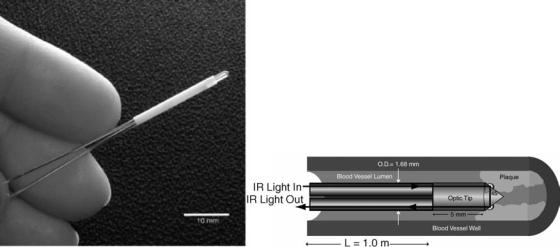
With each of these new fibers there are trade-offs in flexibility, transmission bandwidth, and ability to handle high power laser light. There are also differing fiber needs for diagnostic and therapeutic application. Diagnostic imaging fibers, for example, require a large NA for high light gathering capability. Comparatively, a therapeutic fiber endoscope may require a small NA to confine the delivery of the high power laser light to a specific region. This can also be achieved by use of evanescent waves launched at the interface between a high refractive-index optic and the tissue. Evanescent waves are different from the freely propagating light that typically exits fibers in that it is a surface wave. This surface wave only penetrates into the tissue on the order of the wavelength of the light. For typical wavelengths in the visible and IR, this amounts to light penetration depths on the order of microns, appropriate for very precise delivery of light into tissue. Both diagnostic and therapeutic applications have been demonstrated by coupling a high refractive-index conic tip (sapphire and zinc sulfide) to a HWG (see Fig. 3) (10). The
diagnostic capability allows Fourier transform infrared (FTIR) spectroscopy to be performed on living (In vivo) tissue, where, for example, fatty tissues can be distinguished from normal intimal aorta tissue. Through the same HWG catheter, tissue ablation of atherosclerotic plaque has proven the therapeutic capabilities. These diagnostic and therapeutic applications can potentially take advantage of evanescent waves, HWGs, and midinfrared FT spectroscopy in the 2–10 mm wavelength range (10).
A hybrid optical fiber consisting of a germanium trunk fiber and a low OH silica tip has shown the ability to transmit up to 180 mJ of Er:YAG power for applications requiring contact tissue ablation through a flexible endoscope (11). This pulse energy is more than sufficient for ablation of a variety of hard and soft tissues.
Next, we will look at the potential influence these new light delivery systems may have in endoscopy.
MEDICAL APPLICATIONS USING ENDOSCOPY
Endoscopy has had a major impact on the fields of medicine and surgery. It is largely responsible for the field of minimally invasive surgery. The ability to send diagnostic and therapeutic light into the body via minimally invasive procedures has reduced patient discomfort, pain, and length of hospital stay; and in some cases has dramatically changed the length of stay after a procedure from weeks to days. Optical fibers have had a profound effect on endoscopy, and in doing so, dramatically changed medicine and surgery. These small, flexible light pipes allowed physicians to direct light into the body where it was not thought possible, and even to direct laser light to perform microsurgeries in regions previously too delicate or intricate to access.
In this section, we will examine the state of endoscopy in arthroscopy, bronchoscopy, cardiology, cystoscopy, fetoscopy,
Figure 3. An example of a HWG endoscope. Both diagnostic and therapeutic applications have been demonstrated by coupling a high refractive-index conic tip (sapphire and zinc sulfide) to a HWG. In this geometry with two waveguides, diagnostic spectrometer light can be coupled into the tip in contact with tissue via one waveguide and sent back to a detector via the other waveguide.
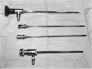
Figure 4. Arthroscope and internal components used to view the interior of knee joint space.
gastrointestinal endoscopy, laparoscopy, neurosurgery, and otolaryngology.
Arthroscopy
Arthroscopy had its birth in 1918 when Takagi modified a pediatric cystoscope and viewed the interior of the knee of a cadaver (12). Today its use is widespread in orthopedic surgery with major emphasis on the knee joint. Arthroscopy has had a major impact on the way knee surgery is performed and with positive outcomes. The surgery is minimally invasive often with only two small incisions; one for an endoscope to visualize the interior of the knee, and another to pass microsurgical instruments for treatment. Presently, endoscopes have the ability to view offaxis at a range of angles from 0 to 1808, with varying field- of-view (see Fig. 4). The larger the field-of-view the more distorted the image, as in a fish-eye lens. A 458 off-axis endoscope, for example, with a 908 field-of-view can be rotated to visualize the entire forward-looking hemisphere.
The most common procedure is arthrotomy, which is simply the surgical exploration of a joint. Possible indications include inspection of the interior of the knee or to perform a synovial biopsy. Synovia are clear viscous fluids that lubricate the linings of joints and the sheaths of tendons. Other indications include drainage of a hematoma or abscess, removal of a loose body or repair of a damaged structure, such as a meniscus or a torn anterior cruciate ligament, and the excision of an inflamed synovium (13,14).
Future directions in orthopedic surgery will see endoscopes put to use in smaller joints as endoscopes miniaturize and become more flexible. The ultimate limit on the diameter of these devices will likely be 10s of micrometers, as single-mode fibers are typically 5–10 mm in diameter. These dimensions will allow for endoscopy in virtually every joint in the body, including very small, delicate joints. Work in the shoulder and metacarpal-phalanges (hand– finger) joints is already increasing because of these new small flexible fiber optic endoscopes.
ENDOSCOPES 181
Bronchoscopy
Bronchoscopy is used to visualize the bronchial tree and lungs. Since its inception in the early 1900s bronchoscopy has been performed with rigid bronchoscopes, with much wider application and acceptance following the introduction of flexible fiber optics in 1968. An advantage of the equipment available is its portability, allowing procedures to be done at a patient’s bedside, if necessary. With the introduction of fiber optics, in 1966, the first fiber optic bronchoscope was constructed, based on specifications and characteristics that were proposed by Ikeda. He demonstrated the instrument’s use and application and named it the bronchofiberscope. Development over the last several decades has seen the use of fiberoptic endoscopes in the application of fiberoptic airway endoscopy in anesthesia and critical care. These endoscopes have improved the safe management of the airway and conduct of tracheal and bronchial intubation. Fiber optic endoscopy has been particularly helpful in the conduct of tracheal and bronchial intubation in the pediatric population.
Bronchoscopy is an integral part in diagnosis and treatment of pulmonary disease (15,16). Bronchoscopic biopsy of lung masses has a diagnostic yield of 70–95%, saving the patient the higher risk associated with a thoracotomy. Pulmonary infection is a major cause of morbidity and mortality, especially in immuno-compromised patients, and bronchoscopy allows quick access to secretions and tissue for diagnosis. Those patients with airway and/or mucous plugs can quickly be relieved of them using the bronchoscope. Another diagnostic use of the bronchoscope is pulmonary alveolar lavage, where sterile saline is instilled into the lung then aspirated out and the cells in the lavage fluid inspected for evidence of sarcoidosis, allergic alveolitis, for example. Lavage is also of therapeutic value in pulmonary alveolar proteinosis.
Bronchoscopy is usually well tolerated by the patient with complications much less than 1% for even minor complications. Laser use has also allowed for significant relief of symptoms in cancer patients.
Cardiology
Early developments in minimally invasive cardiac surgery included the cardiac catheter (1929), the intra-aortic balloon pump (1961), and balloon angioplasty (1968). Endoscopy in cardiology has largely focused on using intravascular catheters to inspect the inside of blood vessels and more recently the inside of the heart itself. Catheters are thin flexible tubes inserted into a part of the body to inject or drain away fluid, or to keep a passage open. Catheters are similar to endoscopes, and they also have many diagnostic and surgical applications.
For diagnostic purposes, angiography uses X rays in concert with radio-opaque dyes (fluoroscopy) to look for blockages in vessels, usually the coronary arteries that supply oxygenated blood to the heart. A catheter is introduced into the femoral artery and sent up the aorta and into the coronary arteries to assess blood flow to the heart. The catheter releases the dye and real-time X-ray fluoroscopy tracks the dye as it is pumped through the coronary artery. Angiography in concert with intravascular
182 ENDOSCOPES
Figure 5. Image of a blood vessel with a stent using IVUS. (Courtesy of LightLab Imaging.)
ultrasound (IVUS) is the currently accepted diagnostic in cardiology (17–21). IVUS emits acoustic energy out the tip of a catheter and listens for echoes to image the inside of coronary arteries (see Fig. 5). An IVUS image is a crosssectional view of the blood vessel, and complements the X-ray fluoroscopy image. The IVUS has been used to assess atherosclerotic plaques in coronary arteries and has been very successful at guiding the placement of stents. Stents are expandable metal mesh cages in the shape of cylinders that act as scaffolding to open obstructions in vessels caused by atherosclerosis.
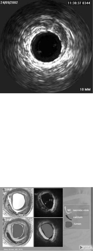
Another technique showing promise in endovascular imaging uses light. The light-driven technology, optical coherence tomography (OCT), is successful at detecting fatty plaques, including those that are vulnerable to rupture. Figure 6 compares OCT images of coronary artery to
Figure 6. Images of coronary artery comparing OCT with conventional histology. (Courtesy of LightLab Imaging.)
conventional histology (LightLab Imaging, Inc.). Visible are the necrotic core of an atherosclerotic plaque and the thin cap at the intimal surface of the vessel. Near-IR light has been used diagnostically for differentiating between oxygenated and deoxygenated blood in myocardium (22– 24). The NIR light in the 2–2.5 mm wavelength range has also been used to characterize myocardial tissue (25). In this wavelength range, the absorption due to water is declining through a local minimum, while there are absorption peaks in fat at 2.3 and 2.4 mm that show distinct spectral signatures. It has also been suggested that IR light might be used to identify atherosclerotic plaques (26–29), including plaques that are at high risk of rupture or thrombosis (30). Arai et al. showed that fibrofatty plaques have characteristic absorbances at mid-IR wavelengths of 3.4 and 5.75 mm that are significantly greater than normal aorta tissue (29). Peaks at these wavelengths, and potentially other subtleties in the absorption spectra [derived from multivariate statistical analysis (MSA)], can be targeted for developing a diagnostic profile similar to that described by Lewis and co-workers for IR and Raman spectroscopic imaging (31,32). These mid-IR frequencies may also be optimized for selective therapy via precise laser surgery (10,33,34).
Surgical laser techniques have become routine over the past two decades in a number of medical specialties such as ophthalmology and dermatology. However, in cardiology initial enthusiasm for fiber optic catheter ablation of atherosclerotic plaque (laser angioplasty) waned in the face of unpredictable vascular perforations, restenosis, and thrombosis (35,36). Therapeutically, IR light has found application in transmyocardial revascularization (TMR) through several partial myocardial perforations (37). Ideally, lasers can ablate tissue with exquisite precision and nearly no damage to the surrounding tissue. This precision requires extremely shallow optical penetration, such that only a microscopic zone near the tissue surface is affected. This has been accomplished by using short laser pulses at wavelengths that are strongly absorbed by proteins (193 nm in the UV) (38) or water (3 mm in the IR) (39).
Therapeutic techniques using catheters recently have targeted the atherosclerotic plaque that is deposited in the vessel wall as we age. The deposition of atherosclerotic plaque in coronary arteries is important for two reasons; the arteries are small (1–3 mm), and they supply blood to the heart muscle itself, so any reduction or blockage of oxygen-supplying blood to the heart shows up symptomatically as angina or worse, a heart attack. Catheters equipped with lasers and inflatable balloons have been used to open these blockages by ablating the plaque or compressing the plaque back into the vessel wall, respectively. Problems with balloon angioplasty have been vessel wall perforation, thrombosis (blood clot formation), and restenosis (reobstruction of the vessel due to an immune response). Recently, balloon angioplasty was augmented by the use of stents, as mentioned above. These expandable cages are opened via a balloon catheter in the vessel lumen and act as a mechanical scaffold to hold the plaque obstruction in place on the vessel wall. Stenting, however, still suffered from restenosis, because of the mechanical injury induced by the stent. Stents coated with small amounts of

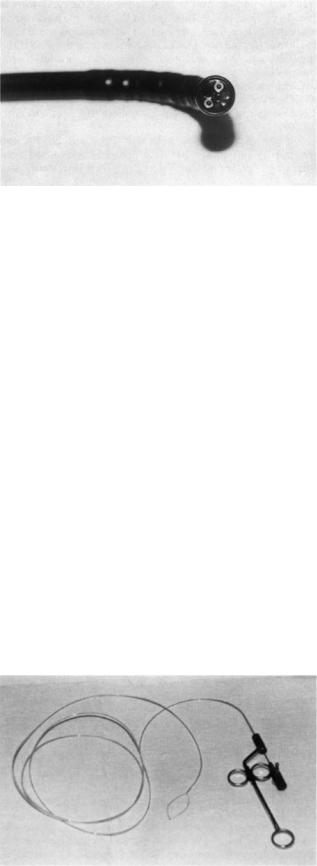
Figure 7. Cystoscope and accessories used for viewing the urinary tract in the medical specialty urology.
drugs (drug-eluding stents) have met with some success, in that they are able to retard the regrowth of intimal hyperplasia responsible for the restenosis.
Cystoscopy
Endoscopy is an integral part of the practice of urology. There are many procedures that look inside the urinary tract using a cystoscope, with typical access through the ureter (see Fig. 7). Common cystoscopy procedures include ureteral catheterization, fulguration and or resection of bladder tumor(s), direct vision internal urethrotomy, insertion of stent, removal of bladder stone, and kidney stone fragmentation (40,41).
A wide variety of therapeutic accessories are available for endoscopic treatment, including snares and baskets for stone removal, and electrohydraulic lithotripsy and laser delivery for fragmenting large stones (42,43). These procedures are minimally invasive and, therefore, can be accomplished with a much lower rate of morbidity and mortality than would be achieved in an open surgical procedure.
Laser incision of urethral, bladder neck, and urethral strictures and fragmentation of kidney stones is being investigated using flexible endoscopes made from germanium fibers to transport Er:YAG laser light. Bladder neck strictures are defined as a narrowing or stenosis of the bladder neck that may result in recalcitrant scarring and urinary incontinence. A significant number of patients undergoing surgery for benign or malignant prostate cancer suffer from bladder neck strictures, and there is no simple and effective minimally invasive treatment. The Er:YAG laser can ablate soft tissue 20–30 times better than a Ho:YAG laser (2.12 mm), which is the laser of choice in urology. The absorption coefficient is many orders of magnitude different for these two lasers,10,000 cm 1 for Er:YAG versus 400 cm 1 for the Ho:YAG. This translates to a 1/e depth of optical penetration of 1 versus 25 mm for these two lasers. Water is the dominant absorptive chromophore in the tissue in this mid-IR region of the spectrum. Hence, the Er:YAG is better suited to procedures where precision is required in laser ablation of soft tissue.
ENDOSCOPES 183
Fetoscopy
Fetoscopy allows for the direct visualization of a fetus in the womb. It also allows for the collection of fetal blood samples and fetal skin sampling, for diagnosis of certain hemoglobinopathies and congenital skin diseases, respectively (44–46).
The instrument is a small-diameter (1–2 mm) needlescope with typical entry through the abdominal wall under local anesthesia and guided by ultrasound. Optimal viewing is from 18 to 22-weeks gestation when the amniotic fluid clarity is greatest. Once the instrument is introduced, a small-gauge needle is typically used to obtain blood samples or small biopsy forceps can be used for skin biopsy.
Complete fetal visualization is not usually achieved. The procedure has some risk with fetal loss 10% and prematurity another 10%. The future of this procedure is not clear, as it is not currently in general use, despite the safe and relatively simple prenatal diagnosis it offers. It has been replaced in many circumstances by ultrasound for the fetal visualization aspect, but is still valuable for blood and skin sampling.
Gastrointestinal Endoscopy
The techniques of fiberoptic gastrointestinal (GI) endoscopy were developed in the 1960s, but surgeons were slow to embrace the techniques. These procedures were developed by gastroenterologists who became skilled practitioners and teachers of the art (see Fig. 8). Gradually, GI surgeons adopted these procedures, and in 1980 the American Board of Surgery mandated that endoscopic training be a part of the curriculum in general surgical training. Endoscopy has since become an integral part of surgical education and practice (47). The GI endoscopy is used in the esophagus, stomach, small bowel and liver, biliary, colon, and in pediatric endoscopy. There are numerous accessories available for the GI endoscope. They include biopsy forceps, graspers, cautery tools, and wire snares (see Figs. 9 and 10).
Figure 8. Upper intestinal panendoscope for the adult patient.
184 ENDOSCOPES
Figure 9. An end view of the distal tip of a panendoscope illustrating the accessory channels and illumination ports.
In the esophagus, endoscopy is used in the dilation of benign esophageal strictures, balloon dilatation of Achalasia, management of foreign bodies and bezoars of the upper GI tract, and endoscopic laser therapy of GI neoplasms (48– 50). Endoscopy of benign esophageal strictures is a common procedure performed by gastroenterologists to treat esophageal narrowing and relieve dysphagia. Achalasia is a motility disorder of the esophagus characterized by symptoms of progressive dysphagia for both solids and liquids with (1) aperistalsis in the body of the esophagus,
(2) high lower esophageal sphincter (LES) pressure, and (3) failure of the LES to relax. Examples of foreign bodies are coins, meat impaction, frequently in the elderly population, sharp and pointed objects, such as, a toothpick, a chicken or fish bone, needles, and hatpins. Bezoars are a hard mass of material often found in the stomach and are divided into three main types: phytobezoars, trichobezoars, and miscellaneous. These bezoars can be managed by endoscopy dependent on size and location, typically by capture and removal rather than endoscopic fragmentation. Since the 1970s, the incidence of esophageal adenocarcinoma has increased more rapidly than any other form of cancer and now represents the majority of esophageal neoplasms in the West (51). Esophagectomy is considered the gold standard for the treatment of high grade dysplasia in Barrett’s esophagus (BE) and for noninvasive adenocarcinoma (ACA) of the distal esophagus (52). Barrett’s esopha-
Figure 10. A polyp wire snare device with retraction handle.
gus is the replacement of native squamous mucosa by specialized intestinal metaplasia and is known to be the major risk factor for the development of adenocarcinoma. A recent study of 45 patients supports the use of endoscopic surveillance in patients who have undergone ‘‘curative’’ esophagectomy for Barrett’s dysplasia or localized cancer (53–55). OCT imaging has recently shown the ability to image Barrett’s esophagus through a small fiber endoscope. The GI neoplasm treatment is also particularly amenable using neodymium-YAG laser palliative treatment of malignancies of the esophagus and gastroesophageal junction as first described by Fleischer in 1982.
For the stomach, endoscopic therapy for benign strictures of the gastric outlet is but one procedure. These strictures of the gastric outlet are similar to the esophageal stricture mentioned above. They are most frequently caused by peptic ulcer disease in the region of the pylorus, although chronic ingestion of nonsteroidal anti-inflammatory drugs is a frequent cause as well. Other endoscopic procedures in the stomach include sclerotherapy of esophageal varices (abnormally swollen or knotted vessels, especially veins), percutaneous endoscopic gastrostomy and jejunostomy, injection therapy for upper GI hemorrhage, and thermal coagulation therapy for upper GI bleeding.
In the small bowel and liver, enteroscopy is an endoscopic procedure to directly inspect the small bowel mucosa and take biopsies by enteroscopy from selected sites in the jejunum and proximal ileum. Until the recent development of the enteroscope, these parts of the small bowel were not possible to endoscopically evaluate. Enteroscopy is in its technological infancy. It presently allows for access to the most proximal aspects of the small bowel to obtain tissue at bleeding lesions. However, it cannot be utilized to adequately visualize the entire mucosa.
Endoscopy in the biliary system is used to perform sphincterotomies, papillotomies and stent insertions, to manage large bile duct stones and malignant biliary strictures, biliary and pancreatic manometry, and endoscopic retrograde cholangiopancreatography. The use of endoscopes all but changed the surgical technique for gallbladder removal and moved its outcome from a major inpatient surgery to a minimally invasive surgery with only small incisions for the endoscope.
In the colon, nonoperative and interventional management of hemorrhoids is performed with endoscopes. Other procedures include dilatation of colonic strictures, approaches to the difficult polyp and difficult colonic intubation, and clinical approaches of anorectal manometry. Additionally, colonoscopy is used for investigating irritable bowel syndrome, Crohn’s disease, and ulcerative colitis (47,56).
Pediatric endoscopy has been used to perform gastroscopy, colonoscopy, and endoscopic retrograde cholangiopancreatography. Advances have largely involved the application of techniques used in adults to the pediatric patient; this was made possible with the introduction of smaller fiberoptic endoscopes.
And finally general endoscopy has been used as a- surveillance program for premalignant lesions, to assess outpatient endoscopy, and endosonography and echo probes.
Laparoscopy
Laparoscopy or peritoneoscopy is an important diagnostic procedure that allows direct visualization of the surface of many intra-abdominal organs, as well as allowing the performance of guided biopsies and minimally invasive therapy. Landmarks in laparoscopic surgery include the first laparoscopic appendectomy (1983) and the first laparoscopic cholescystectomy (1987). The first laparoscope was an ordinary proctoscope with illumination coming from an electric headlight worn by the endoscopist. Since then, with the advent of newer technologies, a fiber optic cable was added to the rigid telescope making flexible laparoscopes available. Today, despite the advent of various noninvasive scanning technologies, laparoscopy is still clinically useful for visualizing and biopsying intra-abdom- inal tumors, particularly those that involve the liver, peritoneum, and pelvic organs (57,58).
The laparoscopic procedure begins with the introduction of a trocar into the abdomen at the inferior umbilical crease for insufflation of carbon dioxide gas. A trocar is a sharply pointed steel rod sheathed with a tight-fitting cylindrical tube (cannula), used together to drain or extract fluid from a body cavity. The whole instrument is inserted then the trocar is removed, leaving the cannula in place. The gas acts as a cushion to permit the safe entry of sharp instruments into the peritoneal cavity and enable a better view. Common procedures include laparoscopic cholecystectomy and laparoscopic appendectomy, laparoscopic removal of the gallbladder and appendix, respectively. Laparoscopic cholecystectomy has all but replaced the conventional surgery for removal of the gallbladder. What used to involve opening the abdominal cavity for gallbladder removal and a 5–7 day stay at the hospital, has been transformed to a minimally invasive procedure with only a few days in the hospital. Lastly, laparoscopic sterilization and abortion are also performed with endoscopes.
Neuroendoscopy
Fiber optics is particularly well suited for the field of neuroendoscopy for both diagnostic and therapeutic procedures in the inner brain, because of size, flexibility, visualization, and laser delivery. We briefly review a case study that highlights some of the advantages of fiber optic endoscopes for minimally invasive surgery in the inner brain.
A recent case report on laser-assisted endoscopic third ventriculostomy (ETV) for obstructive hydrocephalus shows the use of diagnostic and therapeutic endoscopy in neurosurgery (59). Under stereotactic and endoscopic guidance, multiple perforations in the ventricular floor using a 1.32 mm neodymium–yttrium–aluminum–garnet (Nd:YAG) or an aluminum–gallium–arsenide (AlGaAs) 0.805 mm diode laser and removal of intervening coagulated tissue ensued with a 4–6 mm opening between third ventricle and basilar cisterns. These perforations allow for the cerebrospinal fluid (CSF) to be diverted so that a permanent communication can be made between the third cerebral ventricle and arachnoid cisterns of the cranial base. In a series of 40 consecutive cases, 79% of the patients had a favorable outcome. This compares well with a recent
ENDOSCOPES 185
series summarizing > 100 patients and long-term followup with success rates ranging from 50 to 84%.
When the 1.32 mm Nd:YAG laser is used, the high absorption in water requires that the fiber be placed in contact with the ventricular floor. Conversely, the high power diode laser’s dispersion-dominant properties can lead to damage to neural structures around the ventricular cavity. Therefore, the 0.805 mm diode laser was used in a contact mode, but only after carbonization of the fiber tip so that thermal increase of the fiber tip allowing ventricular floor perforation was due to absorption of the laser energy by the carbon layer only and not by direct laser–tissue interaction. The 1.32 mm Nd:YAG laser was found to have higher efficiency for coagulation and perforation than the 0.805 mm diode laser, and would appear to be the better choice for neuroendoscopic use in this procedure. The endoscope allows for visualization and treatment of a very difficult part of the brain to access, and the use of lasers in endoscopes is advantageous in cases of distorted anatomy and microstructures and may reduce technical failures.
In another case of endoscopic-delivered laser light for therapeutic purpose, an IR free-electron laser (FEL) was used to ablate (cut) a suspected meningioma brain tumor at Vanderbilt’s Keck FEL Center (60). A HWG catheter was used to deliver 6.45 mm IR FEL light to a benign neural tumor in the first human surgery to use a FEL. The 6.45 mm FEL light is a candidate for soft tissue surgery because of its ability to ablate (cut) soft tissue with a minimum of thermal damage to the surrounding tissue; on the order of micrometers of damage. This is obviously very important for surgery in the brain where viable, eloquent tissue may be in contact with the tumorous tissue that is being removed.
The FEL is a research center device that generates laser light over a vast portion of the electromagnetic spectrum; to date FELs have generated laser light from the UV (190 nm) to millimeter (61). The FEL is beneficial in identifying wavelengths, particularly in the IR where there are no other laser sources, for selective laser-tissue interaction.
Otolaryngology
Early use of endoscopes for ear, nose, and throat often focused on the interior of the ear. The benefit of endoscopes for diagnosis and therapy had been recognized early on with the advent of the laser and fiber optics (62–65). Recent investigations at the University of Ulm on the use of an Er:YAG laser with a germanium-oxide fiber delivery system has focused on tympanoplasty and stapedotomy (middle ear surgery) (66). The Er:YAG laser was found to be optimum for operating on the eardrum along the ossicles as far as the footplate without carbonization, and with sharpedged, 0.2-mm-diameter canals ‘‘drilled’’ through the bone. Using this technique, children with mucotympanon could have their eardrums reopened in the doctor’s office without the need for drain tubes.
An endoscope suitable for quantitatively examining the larynx (vocal chords) uses a green laser and a double reflecting mirror (67). The device can be clipped onto the shaft of a commercial rigid laryngoscope. The double reflecting mirror sends out two beams parallel to one
186 ENDOSCOPES
another that allows for quantitative morphometry of laryngeal structures such as, vocal cords, glottis, lesions, and polyps.
The miniaturization and flexibility of fiber optics has allowed endoscopes to be applied in the small and delicate organ of the ear with much success. A case in point for the unique capabilities that the fiber optic endoscope has that can be applied to many fields of medicine in a very productive manner.
Future Directions
One pressing issue for ‘‘reusable’’ endoscopes is the ability to guarantee a clean, sterile device for more than one procedure. With the advent of the World Wide Web (WWW), many web sites are available to gain information on endoscopes, as well as the procedures they are used in. The U.S. Food and Drug Administration (FDA) has a site at their Center for Devices and Radiological Health (CDRH) that monitors medical devices and their performance in approved procedures. The FDA has also created guidelines for cleaning these devices.
The results of an FDA-sponsored survey, Future Trends in Medical Device Technology: Results of an Expert Survey in 1998, expressed a strong view that endoscopy and minimally invasive procedures would experience significant new developments during the next 5 and 10 year periods leading to new clinical products (68). The 15 participants included physicians, engineers, healthcare policymakers and payers, manufacturers, futurists and technology analysts. In interviews and group discussions, survey participants expressed an expectation of continuing advancements in endoscopic procedures including fiber optic laser surgery and optical diagnosis, and a range of
miniaturized devices. Clinically, most participants expected an emphasis on minimally invasive cardiovascular surgery and minimally invasive neurosurgery; two new fields we introduced in this edition. Also predicted were continuing advances in noninvasive medical imaging, including a trend to image-guided procedures. Most profound expectations were for developments in functional and multimodality imaging. Finally, participants observed that longer term trends might ultimately lead to noninvasive technologies. These technologies would direct electromagnetic or ultrasonic energy, not material devices, transdermally to internal body structures and organs for therapeutic interventions.
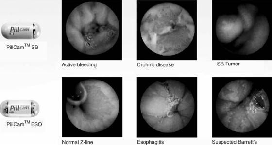
Perhaps a stepping-stone on this path is the PillCam (Given Imaging), a swallowable 11 26-mm capsule with cameras on both ends and a flashing light source, used to image the entire GI tract from the esophagus to the colon. The PillCam capsule has a field of view of 1408, and enables detection of objects as small as 0.1 mm in the esophagus and 0.5 mm in the small bowel. Figure 11 shows the PillCams used for small bowel (SB) and esophagus (ESO) procedures and samples of the images obtained. Shown are examples of active bleeding, Crohn’s disease, and tumor in the small bowel; and normal Z-line, esophagitis, and suspected Barrett’s in the esophagus. Patient exam time is 20 min for an esophageal procedure and 8 h for a small bowel procedure. As the PillCam passes through the GI tract images are acquired at 2 Hz and the information is transmitted via an array of sensors secured to the patient’s chest and abdomen and passed to a data recorder worn around the patient’s waist. The PillCam generates57,000 images in a normal 8 h procedure, while the patient is allowed to carry on their normal activity. An obvious enhancement of patient comfort.
Figure 11. The PillCam from Given Imaging and sample images from the small bowel and esophagus. Shown are examples of active bleeding, Crohn’s disease, and tumor in the small bowel; and normal Z-line, esophagitis, and suspected Barrett’s in the esophagus. The PillCam is a swallowable 11 26 mm capsule with cameras on both ends and a flashing light source, used to image the entire GI tract from the esophagus to the colon.
AESOP (formerly Computer Motion and now Intuitive Surgical) was the first robot, FDA approved in 1994, to maneuver a tiny endoscopic video camera inside a patient according to voice commands provided by the surgeon. This advance marked a major development in closed chest and port-access bypass techniques allowing surgeons direct and precise control of their operative field of view. In 1999, one-third of all minimally invasive procedures used AESOP to control endoscopes.
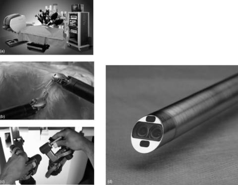
The da Vinci Surgical System (Intuitive Surgical) provides surgeons the flexibility of traditional open surgery while operating through tiny ports by integrating robotic technology with surgeon skill. The da Vinci consists of a surgeon console, a patient-side cart, instruments and image processing equipment (see Fig. 12). The surgeon operates while seated at a console viewing a 3D image of the surgical field. The surgeon’s fingers grasp the master controls below the display with hands and wrists naturally positioned relative to their eyes, and the surgeon’s hand, wrist, and finger movements are translated into precise, real-time movements of endoscopic surgical instruments inside the patient. The patient-side cart provides up to four robotic arms, three instrument arms, and one endoscope arm that execute the surgeon’s commands. The laparoscopic arms pivot at the 1 cm operating ports and are designed with seven degrees of motion that mimic the dexterity of the human hand and wrist. Operating images are enhanced, refined, and optimized using image synchronizers, high intensity illuminators, and camera control units to provide enhanced 3D images of the operative field via a dual-lens three-chip digital camera endoscope. The FDA has cleared da Vinci for use in general laparoscopic surgery, thoracoscopic (chest) surgery, laparoscopic radical
ENDOSCOPES 187
Figure 12. The da Vinci Surgical System consists of (a) a surgeon console, patient-side cart, instruments, and image processing equipment. The surgeon operates while seated at a console viewing a 3D image of the surgical field. The surgeon’s fingers (b) grasp master controls below the display that translate the surgeon’s hand, wrist, and finger movements into precise, real-time movements of the surgical instruments (c) inside the patient. The patient-side cart provides the robotic arms, with two or three instrument arms and one endoscope arm, that execute the surgeon’s commands. The laparoscopic arms pivot at the 1 cm operating ports eliminating the use of the patient’s body wall for leverage and minimizing tissue damage. The instruments are designed with seven degrees of motion that mimic the dexterity of the human hand and wrist. Operating images are enhanced, refined, and optimized for 3D viewing through the (d) 3D endoscopic camera system. (Courtesy of Intuitive Surgical, Inc.)
prostatectomies, and thoracoscopically assisted cardiotomy procedures. Additionally, the da Vinci System is also presently involved in a cardiac clinical trial in the United States for totally endoscopic coronary artery bypass graft surgery. This technology will likely find application in vascular, orthopedic, spinal, neurologic, and other surgical disciplines that will certainly enhance minimally invasive surgery.
Minimally invasive technologies that enhance the present state of endoscopy will continue. The expectation that microelectromechanical systems (MEMS) technology will add a plethora of miniaturized devices to the armament of the endoscopist is well founded, as it is an extension of the impact that fiber optics had on the field of endoscopy. The MEMS will likely add the ability to have light source and detector at the tip of the endoscope, instead of piping the light into and out of the endoscope. Many other functions including ‘‘lab on a chip’’ MEMS technology may allow for tissue biopsies to be performed in situ. This miniaturization will likely lead to more capabilities for endoscopes, as well as, the ability to access previous inaccessible venues in the body. Endoscopy is poised to continue its substantial contribution to minimally invasive procedures in medicine and surgery. This will pave the way for the likely future of noninvasive procedures in surgery and medicine.
BIBLIOGRAPHY
Cited References
1.Matthias Reuter, Rainer Engel, Hans Reuter. History of Endoscopy. Stuttgart: Max Nitze Museum Publications; 1999.
188ENDOSCOPES
2.Wolf RFE, Krikke AP. The X-files of sword swallowing. Available at www.ecr.org/Conferences/ECR1999/sciprg/abs/ p010189.htm.
3.Elner HD. Ein gastroskop. Klin Wochenschr 1910;3:593.
4.Sussmann M. Zur Diptrik des gastroskop. Ther Gegenw 1912;53:115.
5.Schindler R, Lehrbuch U. Atlas D Gastroskop, Munich: Lehmann 1923.
6.Hecht E. Optics. 4th ed. New York: Addison-Wesley; 2002.
7.Harrington JA. A review of IR transmitting, hollow waveguides. Fiber Integr Opt 2000;19:211–227.
8.Rave E, Ephrat P, Goldberg M, Kedmi E, Katzir A. Silver halide photonic crystal fibers for the middle infrared. Appl Opt 2004;43(11):2236–2241.
9.Mackanos MA, Jansen ED, Shaw BL, Sanghera JS, Aggarwal I, Katzir A. Delivery of midinfrared (6 to 7-mm) laser radiation in a liquid environment using infrared-transmitting optical fibers. J Biomed Opt 2003;8(4):583–593.
10.Hooper BA, Maheshwari A, Curry AC, Alter TM. A Catheter for Diagnosis and Therapy using IR Evanescent Waves. Appl Opt 2003;42:3205–3214.
11.Chaney CA, Yang Y, Fried NM. Hybrid germanium/silica optical fibers for endoscopic delivery of erbium:YAG laser radiation. Lasers Surg Med 2004;34:5–11.
12.Altman RD, Kates J. Arthroscopy of the knee. Semin Arthritis Rheum 1983;13:188–199.
13.Drez D, Jr. Arthroscopic evaluation of the injured athelete’s knee. Clin Sports Med 1985;4:275–278.
14.Altman RD, Gray R. Diagnostic and Therapeutic Uses of the Arthroscope in Rheumatoid Arthritis and Osteoarthritis. New York: American Journal of Medicine; 1983. p 50–55.
15.Phillon DP Collins, JV. Current status of fiberoptic bronchoscopy. Postgrad Med J 1984;60:213–217
16.Mitchell DM, Emerson CJ, Collyer J, Collins JV. Fiberoptic bronchoscopy: Ten years on. Br Med J 1980;2:360–363.
17.Nissen S. Coronary angiography and intravascular ultrasound. Am J Cardiol 2001;87(4A):15A–20A.
18.Nissen SE. Who is at risk for atherosclerotic disease? Lessons from intravascular ultrasound. Am J Med 2002;(Suppl 8A):27S–33S.
19.Tuzcu EM, De Franco AC, Goormastic M, Hobbs RE, Rincon G, Bott-Silverman C, McCarthy P, Stewart R, Mayer E, Nissen SE. Dichotomous pattern of coronary atherosclerosis 1 to 9 years after transplantation: Insights from systematic intravascular ultrasound imaging. JACC 1996;27(4):839– 846.
20.Nadkarni SK, Boughner D, Fenster A. Image-based cardiac gating for three-dimensional intravascular ultrasound imaging. Ultrasound Med Biol 2005;1:53–63.
21.Bourantas CV, Plissiti ME, Fotiadis DI, Protopappas VC, Mpozios GV, Katsouras CS, Kourtis IC, Rees MR, Michalis LK. In vivo validation of a novel semi-automated method for border detection in intravascular ultrasound images. Br J Radiol 2005;78(926):122–129.
22.Jobsis FF. Noninvasive, infrared monitoring of cerebral and myocardial oxygen sufficiency and circulatory parameters. Science 1977;198:1264–1267.
23.Parsons WJ, Rembert JC, Bauman RP, Greenfield Jr JC, Piantadosi CA. Dynamic mechanisms of cardiac oxygenation during brief ischemia and reperfusion. Am J Physiol 1990;259 (Heart Circ. Physiol. 28):H1477–H1485.
24.Parsons WJ, Rembert JC, Bauman RP, Greenfield Jr JC, Duhaylongsod FG, Piantadosi CA. Myocardial oxygenation in dogs during partial and complete coronary artery occlusion. Circ Res 1993;73(3):458–464.
25.Nilsson M, Heinrich D, Olajos J, AnderssonEngels S. Near infrared diffuse reflection and laser-induced fluorescence
spectroscopy for myocardial tissue characterisation. Spectrochim Acta Part A-Mol Biomol Spectrosc 1997;51(11): 1901–1912.
26.Manoharan R, Baraga JJ, Rava RP, Dasari RR, Fitzmaurice M, Feld MS. Biochemical-analysis and mapping of atherosclerotic human artery using FT-IR microspectroscopy. Atherosclerosis 1993;103(2):181–193.
27.Baraga JJ, Feld MS, Rava RP. Detection of atherosclerosis in human artery by midinfrared attenuated total reflectance. Appl Spectrosc 1991;45(4):709–710.
28.Rava RP, Baraga JJ, Feld MS. Near-infrared Fourier-Trans- form Raman spectroscopy of human artery. Spectrochim Acta Part A-Mol Biomol Spectrosc 1991;47(3–4):509–512.
29.Arai T, Mizuno K, Fujikawa A, Nakagawa M, Kikuchi M. Infrared absorption spectra ranging from 2.5 to 10 mm at various layers of human normal abdominal aorta and fibrofatty atheroma in vitro. Laser Surg Med 1990;10:357–362.
30.Casscells W, Hathorn B, David M, Krabach T, Vaughn WK, McAllister HA, Bearman G, Willerson JT. Thermal detection of cellular infiltrates in living atherosclerotic plaques: possible implications for plaque rupture and thrombosis. Lancet 1996;347(9013):1447–1449.
31.Colarusso P, Kidder L, Levin I, Fraser J, Arens J, Lewis EN. Infrared spectroscopic imaging: from planetary to cellular systems. Appl Spectrosc 1998;52(3):106A–119A.
32.Kodali DR, Small DM, Powell J, Krishna K. Infrared microimaging of atherosclerotic arteries. Appl Spectosc 1991; 45:1310–1317.
33.Edwards G, Logan R, Copeland M, Reinisch L, Davidson J, Johnson J, Maciunas R, Mendenhall M, Ossoff R, Tribble J, Werkhaven J, O’Day D. Tissue ablation by a Free-Electron Laser tuned to the Amide II band. Nature (London) 1994;371:416–419.
34.Awazu K, Nagai A, Aizawa K. Selective removal of Cholesterol Esters in an Arteriosclerotic region of blood vessels with a free-Electron Laser. Laser Surg Med 1998;23:233–237.
35.Holmes DR, Bresnahan JF. Interventional cardiology. Cardiol Clin 1991;9:115–134.
36.Linsker R, Srinivasan R, Wynne JJ, Alonso DR. Far-ultra- violet laser ablation of atherosclerotic lesions. Laser Surg Med 1984;4:201–206.
37.Hughes GC, Kypson AP, Yin B, St Louis JD, Biswas SS, Coleman RE, DeGrado TR, Annex BH, Donovan CL, Lanolfo KP, Lowe JE. Induction of angiogenesis following transmyocardial laser revascularization in a model of hibernating myocardium: a comparison of holmium:YAG, carbon dioxide, and excimer lasers. Surgical Forum L 1999;115–117.
38.Puliafito CA, Steinert RF, Deutsch TF, Hillenkamp F, Dehm EJ, Alder CM. Excimer laser ablation of cornea and lens: experimental studies. Ophthalmology 1985;92:741–748.
39.Cummings JP, Walsh Jr. JT Erbium laser ablation—the effect of dynamic optical properties. Appl Phys Lett 1993; 62:1988–1990.
40.Walsh PC, Gittes RF, Perlmutter AD, Stamey TS, editors. Campbell’s Urology. Philadelphia: W. B. Saunders; 1986. p 510–540.
41.Segura JW. Endourology. J Urol 1984;132:1079–1084.
42.Powell PH, Manohar V, Ramsden PD, Hall RR. A flexible cytoscope. Br J Urol 1984;56:622–624.
43.Hoffman JL, Clayman RV. Endoscopic visualization of the supravesical urinary tract: Transurethral ureteropuleloscopy and percutaneous nephroscopy. Semin Urol 1985;3:60–75.
44.Benzie RJ. Amniocentesis, amnioscopy, and fetoscopy. Clin Obstet Gynecol 1980;7:439–460.
45.Rodeck CH, Nicolaides KH. Fetoscopy and fetal tissue sampling. Br Med Bull 1983;39:332–337.
46.Rauskolt R. Fetoscopy. J Perinat Med 1983;11:223–231.