

- •VOLUME 3
- •CONTRIBUTOR LIST
- •PREFACE
- •LIST OF ARTICLES
- •ABBREVIATIONS AND ACRONYMS
- •CONVERSION FACTORS AND UNIT SYMBOLS
- •EDUCATION, COMPUTERS IN.
- •ELECTROANALGESIA, SYSTEMIC
- •ELECTROCARDIOGRAPHY, COMPUTERS IN
- •ELECTROCONVULSIVE THERAPHY
- •ELECTRODES.
- •ELECTROENCEPHALOGRAPHY
- •ELECTROGASTROGRAM
- •ELECTROMAGNETIC FLOWMETER.
- •ELECTROMYOGRAPHY
- •ELECTRON MICROSCOPY.
- •ELECTRONEUROGRAPHY
- •ELECTROPHORESIS
- •ELECTROPHYSIOLOGY
- •ELECTRORETINOGRAPHY
- •ELECTROSHOCK THERAPY.
- •ELECTROSTIMULATION OF SPINAL CORD.
- •ELECTROSURGICAL UNIT (ESU)
- •EMERGENCY MEDICAL CARE.
- •ENDOSCOPES
- •ENGINEERED TISSUE
- •ENVIRONMENTAL CONTROL
- •EQUIPMENT ACQUISITION
- •EQUIPMENT MAINTENANCE, BIOMEDICAL
- •ERGONOMICS.
- •ESOPHAGEAL MANOMETRY
- •EVENT-RELATED POTENTIALS.
- •EVOKED POTENTIALS
- •EXERCISE FITNESS, BIOMECHANICS OF.
- •EXERCISE, THERAPEUTIC.
- •EXERCISE STRESS TESTING
- •EYE MOVEMENT, MEASUREMENT TECHNIQUES FOR
- •FETAL MONITORING
- •FETAL SURGERY.
- •FEVER THERAPY.
- •FIBER OPTICS IN MEDICINE
- •FICK TECHNIQUE.
- •FITNESS TECHNOLOGY.
- •FIXATION OF ORTHOPEDIC PROSTHESES.
- •FLAME ATOMIC EMISSON SPECTROMETRY AND ATOMIC ABSORPTION SPECTROMETRY
- •FLAME PHOTOMETRY.
- •FLOWMETERS
- •FLOWMETERS, RESPIRATORY.
- •FLUORESCENCE MEASUREMENTS
- •FLUORESCENCE MICROSCOPY.
- •FLUORESCENCE SPECTROSCOPY.
- •FLUORIMETRY.
- •FRACTURE, ELECTRICAL TREATMENT OF.
- •FUNCTIONAL ELECTRICAL STIMULATION
- •GAMMA CAMERA.
- •GAMMA KNIFE
- •GAS AND VACUUM SYSTEMS, CENTRALLY PIPED MEDICAL
- •GAS EXCHANGE.
- •GASTROINTESTINAL HEMORRHAGE
- •GEL FILTRATION CHROMATOGRAPHY.
- •GLUCOSE SENSORS
- •HBO THERAPY.
- •HEARING IMPAIRMENT.
- •HEART RATE, FETAL, MONITORING OF.
- •HEART VALVE PROSTHESES
- •HEART VALVE PROSTHESES, IN VITRO FLOW DYNAMICS OF
- •HEART VALVES, PROSTHETIC
- •HEART VIBRATION.
- •HEART, ARTIFICIAL
- •HEART–LUNG MACHINES
- •HEAT AND COLD, THERAPEUTIC
- •HEAVY ION RADIOTHERAPY.
- •HEMODYNAMICS
- •HEMODYNAMIC MONITORING.
- •HIGH FREQUENCY VENTILATION
- •HIP JOINTS, ARTIFICIAL
- •HIP REPLACEMENT, TOTAL.
- •HOLTER MONITORING.
- •HOME HEALTH CARE DEVICES
- •HOSPITAL SAFETY PROGRAM.
- •HUMAN FACTORS IN MEDICAL DEVICES
- •HUMAN SPINE, BIOMECHANICS OF
35.Carrel T, Zingg U, Jenni R, Aeschbacher B, Turina M. Early in vivo experience with the hemodynamic plus St. Jude Medical valve in patients with narrowed aortic annulus. Ann Thorac Surg 1996;61:1418–1422.
36.Vitale N, et al. Clinical evaluation of St. Jude Medical hemodynamic plus versus standard aortic valve prostheses: The Italian multicenter, prospective, randomized study. J Thorac Cardiovas Surg 2001;122(4):691–698.
37.Flameng W, et al. Postoperative hemodynamics of two bileaflet heart valves in the aortic position. J Heart Valve Dis 1997; 6:269–273.
38.Kadir I, Wan IY, Walsh C, Dip-Rad, Wilde P, Byran AJ, Angelini GD. Hemodynamic performance of the 21-mm sorin bicarbon mechanical aortic prosthesis using dobutamine Doppler echocardiography. Ann Thorac Surg 2001; 72:49–53.
39.Hammermeister K, Sethi GK, Henderson WG, Grover FL, Oprian C, Rahimtoola SH. Outcomes 15 years after valve replacement with a mechanical versus a bioprosthetic valve: Final report of the Veterans Affairs randomized trial. J Am Coll Cardiol 2000 Oct; 36(4):1152–1158.
40.Khan S, et al. Twenty-year comparison of tissue and mechanical valve replacement. J Throrac Cardiovas Surg 2001; 122:257–269.
41.Bloomfield P. Choice of prosthetic heart valves: 20-year results of the Edinburgh Heart Valve Trial. J Am Coll Cardiol 2004 Aug 4;44(3):667.
42.Koertke H, Minami K, Boethig D, Breymann T, Seifert D, Wagner O, Atmacha N, Krian A, Ennker J, Taborski U, Klovekorn WP, Moosdorf R, Saggau W, Koerfer R. INR self-management permits lower anticoagulation levels after mechanical heart valve replacement. Circulation 2003; 108(Suppl 1):I175–178
43.On-X Aspirin-Only Study with Selected Isolated Aortic Valve Replacements. Available at http://www.onxvalves. com/Med_Aspirin_Study.asp.
44.Cohn L, Soltesz E. The evolution of mitral valve surgery: 1902-2002. Am Heart Hosp J 2003 Winter; 1(1):40–46.
Reading List
Standards
FDA Document Replacement Heart Valve Guidance-Draft Document, October 14, 1994.
ISO 5840, Cardiovascular Implants—Cardiac valve.
EN prEN12006 Cardiovascular Implants—Cardiac valve.
Pathology
Schoen FJ. Cardiovascular Pathology: Pathology of Heart Valve Substitution With Mechanical and Tissue Prostheses. New York: Churchill Livingston; 2001.
Biological Tissue Properties
Sacks MS, Schoen FJ. Mechanical damage to collagen independent of calcification limits bioprosthetic heart valve durability. Biomed Mater Res 2002;62:359–371.
Billiar KL, Sacks MS. Biaxial mechanical properties of the fresh and glutaraldehyde treated porcine aortic valve: Part I - Experimental results. J Biomechan Eng 2000;122:23–30.
Wells SM, Sacks MS. Effects of fixation pressure on the biaxial mechanical behavior of porcine bioprosthetic heart valves with long-term cyclic loading. Biomaterials 2002;23(11): 2389–2399.
HEART, ARTIFICIAL |
449 |
Sacks MS. The biomechanical effects of fatigue on the porcine bioprosthetic heart valve. Long-term Effects Med Implants 2001;11(3&4):231–247.
Pyrolytic Carbon
More R, Bokros J. Carbon Biomaterials, Encyclopedia of Medical Devices and Instrumentation EMD 023. Heart Valve Prostheses In Vitro Flow Dynamics, Encyclopedia of Medical Devices and Instrumentation.
HEART VIBRATION. See PHONOCARDIOGRAPHY.
HEART, ARTIFICIAL
CONRAD M. ZAPANTA
Penn State College of Medicine
Hershey, Pennsylvania
INTRODUCTION
Artificial hearts are broadly defined as devices that either supplement or replace the native (natural) heart. These devices can be classified into two groups: ventricular assist devices and total artificial hearts. This article will define the clinical need, review the native heart anatomy and function, describe design considerations for ventricular assist devices and total artificial hearts, review selected designs, and recommend areas for future development.
CLINICAL NEED
Cardiovascular disease accounted for 38% of all deaths (almost 1.4 million people) in the United States in 2002
(1). Coronary heart disease (53%) represented the majority of these deaths, followed by stroke (18%), and congestive heart failure (6%). Almost 4.9 million Americans suffer from congestive heart failure, with 550,000 new cases diagnosed each year. Over 80% of men and 70% of women with congestive heart failure under the age of 65 will die within 8 years. In people diagnosed with congestive heart failure, sudden cardiac death occurs at six to nine times the rate of the general population.
One treatment for congestive heart failure is heart transplantation. It is estimated that 40,000 Americans could benefit from a heart transplant each year (1,2). However, only 2100 donor hearts were available each year from 1999 to 2004. The number of donor hearts dropped during this period, from a high of 2316 in 1999 to 1939 in 2004. Over 3300 patients were on the waiting list for a donor heart at any time during this period, with >65% of these patients on the waiting list for >1 year. From 1998 to 2004, 630 patients died each year waiting for transplant.
These numbers clearly demonstrate the clinical need for ventricular assist devices and total artificial hearts that support the patient until transplant (bridge to transplant) or permanently assist or replace the natural heart (destination therapy).
450 HEART, ARTIFICIAL
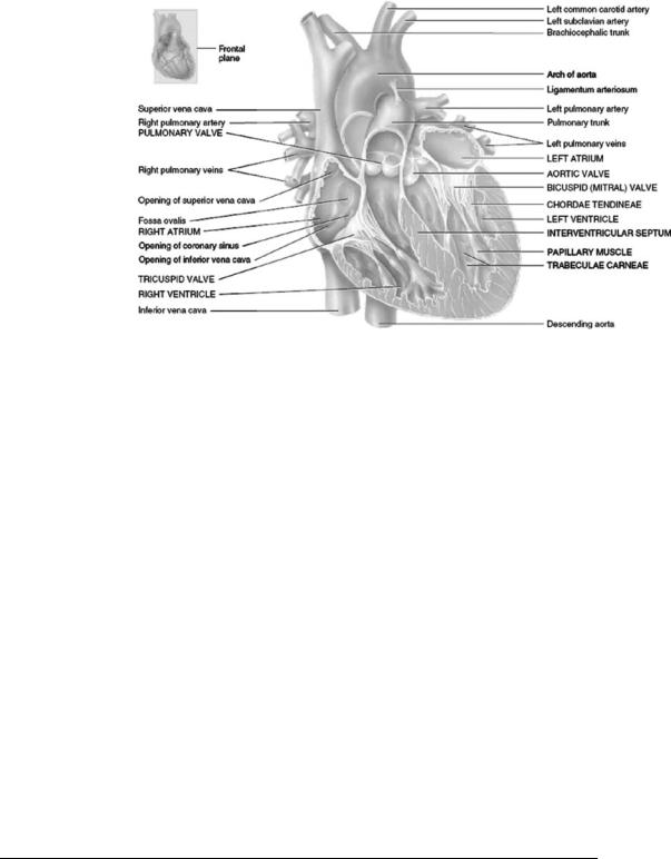
Figure 1. Anatomy of the Native Heart. The heart is composed of two pumps (right and left side) that work simultaneously. The right pump delivers blood to the pulmonary circulation (the lungs), while the left pump delivers blood to the systemic circulation (the body). Each pump consists of an atrium and ventricle. [Reprinted with permission from Gerard J. Tortora and Bryan H. Derrickson, Principles of Anatomy and Physiology, 11th ed., Hoboken (NJ): John Wiley & Sons; 2006.]
NATIVE HEART ANATOMY AND FUNCTION
The anatomy of the native (or natural) heart is shown in Fig. 1. The heart is composed of two pumps (right and left side) that work simultaneously. The right pump delivers blood to the pulmonary circulation (the lungs) while the left pump delivers blood to the systemic circulation (the body). Each pump consists of an atrium and ventricle that make up the heart’s four distinct chambers: right atrium, right ventricle, left atrium, and left ventricle. The atria act as priming chambers for the ventricles. The ventricles pump blood out of the heart to either the pulmonary or systemic circulation. Heart valves located between each atrium and ventricle and at the outlet of each ventricle maintain flow direction during pulsatile flow.
Blood from the systemic circulation enters the right atrium through the superior vena cava (from the head and upper extremities) and inferior vena cava (from the trunk and lower extremities). The blood is then pumped to the right ventricle, which pumps blood to the pulmonary circulation via the pulmonary arteries. Oxygenated blood returns to the left atrium heart from the lungs via the pulmonary vein and is then pumped to the left ventricle. The left ventricle pumps blood to the systemic circulation via the aorta.
Table 1 lists the nominal pressures and flows in the native heart (3). A ventricular assist device or total artificial heart must be able to generate these pressures and flows in order to meet the needs of the recipient.
DESIGN CONSDIRATIONS FOR VENTRICULAR ASSIST DEVICES AND TOTAL ARTIFICIAL HEARTS
Several design considerations must be taken into account when developing a ventricular assist device or total artificial heart. These considerations are detailed below:
1. Size of the Intended Patient: The size of the patient will determine the amount of blood flow required to adequately support the patient. This then determines the size of the ventricular assist device or total artificial heart. For example, a total artificial heart designed for adults would most likely be too large to be implanted within small children. A larger ventricular assist device may be placed externally, while a smaller ventricular assist device could be placed within the native heart. In addition, the size of the patient may dictate the location of some of the components. For example, the power sources may be located either internally (in the abdominal cavity) or externally depending on the size and type of the power source.
2. Pump Performance: A ventricular assist device or total artificial heart can be used to support or replace the native heart. Each of these support modes requires a different cardiac output. For example, a ventricular assist device can provide
Table 1. Nominal Pressures and Flows in the Native (Natural) Heart
Pressures |
|
Left ventricle |
120 mmHg (16.0 kPa) peak systolic normal (into aorta) |
|
10 mmHg (1.33 kPa) mean diastolic (from left atrium) |
Right ventricle |
25 mmHg (3.33 kPa) peak systolic (into pulmonary artery) |
|
5 mmHg (0.667 kPa) mean diastolic (from right atrium) |
Flows |
|
Normal healthy adult at rest: 5 L min 1 |
|
Maximum flow: 25 L min 1 |
|

either a portion of the blood flow required by the patient (partial support) or the entire blood flow (total support). In addition, the decision must be made whether to include a controller that will either passively or actively vary the cardiac output of the ventricular assist device or total artificial heart based on the patient demand.
3. Reliability: The National Institutes of Health (NIH) proposed a reliability goal for ventricular assist devices and total artificial hearts of 80% for a 2 year operation with an 80% confidence level before an artificial heart can begin clinical trials. However, the desired reliability may need to be more demanding for long-term clinical use, such as 95% reliability with 95% confidence for a 5 year operation. The design and components of ventricular assist devices and total artificial hearts must be carefully selected to achieve this reliability goal.
4. Quality of Life: The patient’s quality of life can have a significant impact on the design of a ventricular assist device or total artificial heart. It is important to clearly define what constitutes an acceptable quality of life. For example, if a patient desires to be ambulatory following the implantation of a ventricular assist device or total artificial heart, the power supply must be easily transportable. The environment of the patient (home, work, car, etc.) should also be considered to insure proper function in these different environments. The patient should be able to monitor basic pump operation without the need for a physician or other trained medical personnel. The ventricular assist device or total artificial heart should be designed to clearly provide information through displays and provide alarms to warn the patient of potentially dangerous situations, such as a battery that is running low on power.
VENTRICULAR ASSIST DEVICES
A ventricular assist device (VAD) is designed to assist or replace the function of either the left or right ventricle. These devices are intended to provide either temporary support until a donor heart has been located or the native heart has recovered function, or as a permanent device.
As shown in Table 1, the left ventricle pumps against a higher pressure system than the right ventricle. Therefore, the left ventricle is typically more in need of assistance. Consequently, left ventricular assist devices (LVADs) are more prevalent than right ventricular assist devices (RVADs).
Ventricular assist devices can generate either pulsatile or continuous (nonpulsatile) flow.
Pulsatile
Pulsatile flow ventricular assist devices are composed of a single ventricle that mimics the native ventricle. The ventricle is placed either outside the patient’s body or within the abdominal cavity. There are two types of pulsatile flow ventricular assist devices: pneumatic and electric.
HEART, ARTIFICIAL |
451 |
Figure 2. Penn State/Thoratec Pneumatic Ventricular Assist Device. The ventricle contains a flexible blood sac made of segmented polyurethane that is housed within a rigid polysulfone case. Mechanical heart valves are located in the inlet and outlet positions of the ventricle to control flow direction. Air pulses that are generated by an external drive unit are used to periodically compress the flexible blood sac. (Reprinted with permission from Thoratec Corporation.)
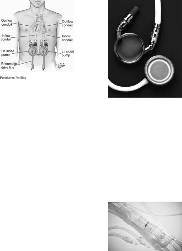
Pneumatic Ventricular Assist Devices. Figure 2 shows a pneumatic ventricular assist device that was originally developed by the Pennsylvania State University and later purchased by Thoratec Corporation (Pleasanton, CA) (4). The ventricle contains a flexible blood sac made of segmented polyurethane that is housed within a rigid polysulfone case. This blood sac is extremely smooth to prevent the formation of clots (or thrombi). Mechanical heart valves are located in the inlet and outlet positions of the ventricle to control flow direction. Air pulses that are generated by an external drive unit are used to periodically compress the flexible blood sac. An automatic control system varies the cardiac output by adjusting the heart rate and the time for ventricular filling in response to an increase in filling pressure.
The device is placed outside the patient on the patient’s abdomen (paracorporeal). The device can be used to assist a single ventricle, or simultaneously with an additional device that assists the both ventricles, as shown in Fig. 3. For the LVAD configuration (right pump in Fig. 3), the inlet cannula is inserted into the apex of the left ventricle and connected to the inlet port of the ventricular assist device. The outflow cannula is attached between the outflow port of the ventricular assist device and the ascending aorta. For the RVAD configuration (left pump in Fig. 3), the inlet cannula is connected to the right atrium and the outlet cannula to the main pulmonary artery. For both types of configurations, the inflow and outflow cannulae pass through the skin below the rib cage. Over 2850 implants have occurred worldwide with the longest duration of 566 days (5). An implantable version of this pump (with a titanium pump casing) was approved by the FDA in August of 2004 (6).
452 HEART, ARTIFICIAL
Figure 3. Implant Location of Penn State/Thoratec Pneumatic Ventricular Assist Device in the RVAD (left) and LVAD (right) Configuration. For the LVAD configuration, the inlet cannula is inserted into the apex of the left ventricle and connected to the inlet port of the ventricular assist device. The outflow cannula is attached between the outflow port of the ventricular assist device and the ascending aorta. For the RVAD configuration, the inlet cannula is connected to the right atrium and the outlet cannula to the main pulmonary artery. For both types of configurations, the inflow and outflow cannulae pass through the skin below the rib cage. (Reprinted with permission from The Cleveland Clinic Foundation.)
Another type of pneumatic ventricular assist device is the Thoratec HeartMate IP (intraperitoneal). The HeartMate IP is an implantable blood pump that is connected to an external drive unit via a percutaenous air drive line (7). The interior of this device is shown in Fig. 4. A unique feature of this device is the use of a textured blood surface in the ventricle that promotes the formation of a cell layer. The cell layer is believed to decrease thrombus formation because the layer mimics the blood contacting surface of a blood vessel. Bioprosthetic heart valves are used to regulate the direction of flow. The HeartMate IP has been implanted in >1300 patients worldwide with the longest duration of 805 days.
Two other types of pneumatic devices include the BVS5000 and AB5000 (both made by ABIOMED, Danvers, MA). Both devices are intended to provide cardiac support as a bridge to transplant or until the native heart recovers. The BVS5000, illustrated in Fig. 5, is an external dualchamber device that can provide support to one or both ventricles as a bridge to transplant (8). The chambers utilize polyurethane valves to regulate the flow direction. More than 6000 implants have been performed worldwide
(9). The AB5000 is a pneumatically driven, paracorporeal device that is similar to a single ventricle of the AbioCor total artificial heart (described in a later section) (10). This device was approved by the FDA in October of 2003 as has been used in > 88 patients. The average duration of support is 15 days with the longest duration of 149 days.
Additional types of pneumatic devices include the Berlin Heart Excor (Berlin Heart AG, Berlin, Germany), the
Figure 4. Interior of Thoratec HeartMate IP Pneumatic Ventricular Assist Device. A unique feature of this device is the use of a textured blood surface in the ventricle that promotes the formation of a cell layer. The cell layer is believed to decrease thrombus formation because the layer mimics the blood contacting surface of a blood vessel. (Reprinted with permission from Thoratec Corporation.)
MEDOS/HIA (Aachen, Germany), and the Toyobo Heart (National Cardiovascular Center, Osaka, Japan). The Berlin Heart Excor is available in a range of sizes (10–80 mL stroke volume) with either tilting disk or polyurethane valves, and has been implanted in > 500 patients (11). The MEDOS/HIA system is also available in a range of sizes and has been implanted in > 200 patients (12). The Toyobo LVAS has been implanted in > 120 patients (13).
ELECTRIC VENTRICULAR ASSIST DEVICES
Electric ventricular assist devices mainly differ from their pneumatic counterparts in their source of power. Electric ventricular assist devices are typically placed within the
Figure 5. ABIOMED BVS 5000 Pneumatic Ventricular Assist Device. The BVS5000 is an external dual-chamber device that can provide support to one or both ventricles as a bridge to transplant
(8). The chambers utilize polyurethane valves to regulate the flow direction. (Reprinted with permission from ABIOMED, Inc.)

HEART, ARTIFICIAL |
453 |
Figure 6. Penn State/Arrow Electric Ventricular Assist Device (LionHeart). The blood pump assembly utilizes a rollerscrew energy converter with a pusher plate. The motion of the pusher plate compresses the blood sac and ejects blood from the ventricle. Mechanical heart valves are used to control the direction of flow into and out of the pump. Energy passes from the external power coil to the subcutaneous (internal) coil by inductive coupling via the transcutaneous energy transmission system (TETS). (Reprinted with permission from Arrow International.)
abdominal cavity. The inlet cannula is inserted into the apex of the native left ventricle and connected to the inlet port of the device. The outlet cannula is attached between the outflow port of the device and the ascending aorta via an end-to-side anastomosis. These types of devices can be used as either bridge-to-transplant or as permanent implants (destination therapy).
Figure 6 illustrates an electric ventricular assist device (LionHeart) developed by the Pennsylvania State University in conjunction with Arrow International (Reading, PA) (14). The blood pump assembly utilizes a rollerscrew energy converter with a pusher plate. The motion of the pusher plate compresses the blood sac and ejects blood from the ventricle. Mechanical heart valves are used to control the direction of flow into and out of the pump. Energy passes from the external power coil to the subcutaneous (internal) coil by inductive coupling via the transcutaneous energy transmission system (TETS). The controller and internal battery supply are also implanted in the abdomen. The internal battery permits operation without the external power coil for 20 min. Air displaced by the blood pump housing enters the polyurethane compliance chamber. Because the air in the compliance chamber can slowly diffuse across the wall of the compliance chamber, the air in the chamber is periodically replenished via the subcutaneous access port. The LionHeart is intended to be used as destination therapy. This device was approved for use in Europe in 2003.
Another type of electric ventricular assist devices is the Novacor LVAS (left ventricular assist system), produced by WorldHeart Corporation (Ottawa, ON). The Novacor LVAS, illustrated in Fig. 7, contains a polyurethane blood sac that is compressed between dual pusher plates (15). The pusher plates are actuated by a solenoid that is coupled to the plates via springs. Bioprosthetic heart valves are utilized to control the direction of flow. A percutaneous power line connects the pump to an external battery pack and controller. The Novacor LVAS has been implanted in
Figure 7. WorldHeart Novacor Electric Ventricular Assist Device. The Novacor contains a polyurethane blood sac that is compressed between dual pusher plates. The pusher plates are actuated by a solenoid that is coupled to the plates via springs. A percutaneous power line connects the pump to an external battery pack and controller. (Reprinted with permission from World Health Corporation, Inc.)
over 1500 patients worldwide. The longest implant duration is > 6 years. No deaths have been attributed to device failure with only 1.4% of the devices needing replacement. The Novacor LVAS is approved as a bridge-to-transplant in the United States and Europe and is in clinical trials for destination therapy in the United States.
The HeartMate XVE (illustrated in Fig. 8) is a derivative to the HeartMate IP (7). The HeartMate XVE uses an electric motor and pusher plate system to pump blood. A percutaneous power line is used to connect the pump to an external battery pack and controller. The HeartMate VE (an earlier version of the XVE) and XVE have been implanted in > 2800 patients worldwide with the longest duration of 1854 days. The HeartMate SNAP-VE was recently approved by the FDA as destination therapy.
The Randomized Evaluation of Mechanical Assistance for the Treatment of Congestive Heart Failure (REMATCH) study examined the clinical utility of ventricular assist devices (16). Patients with end-stage heart failure who were ineligible for cardiac transplantation were split into two groups. The first group (n ¼ 68) received the HeartMate VE LVAS while the second group (n ¼ 61) received optimal medical management. The results showed a reduction of 48% in the risk of death from any cause in the LVAD group versus the medical-therapy group (p ¼ 0.001). The 1 year survival was 52% for the VAD group and 25% for the medical-therapy group (p ¼ 0.002). The 2 year survival

454 HEART, ARTIFICIAL
Figure 8. Thoratec HeartMate XVE Electric Ventricular Assist Device. The HeartMate XVE uses an electric motor and pusher plate system to pump blood. A percutaneous power line is used to connect the pump to an external battery pack and controller. (Reprinted with permission from Thoratec Corporation.)
was 23% for the VAD group and 8% for the medical-therapy group (p ¼ 0.09). Finally, the median survival was 408 days for the VAD group and 150 days for the medical-therapy group. This study clearly showed the clinical utility of ventricular assist devices.
Continuous Flow
Continuous flow ventricular assist devices deliver nonpulsatile flow. Consequently, they do not require heart valves to regulate the direction of blood flow. Continuous flow ventricular assist devices are classified as either centrifugal flow or axial flow pumps based on the direction of the flow as it passes through the pump. These types of pumps are implanted in a similar fashion as their pulsatile counterparts.
Continuous flow assist devices have several potential advantages over pulsatile systems. First, these devices are typically smaller than their pulsatile counterparts and can be used in smaller patients (such as small adults and children). In addition, these pumps have fewer moving parts and are simpler devices than pulsatile systems. These types of devices typically require less energy to operate than the pulsatile pumps.
However, continuous flow pumps have several potential disadvantages. The main disadvantage is that the longterm effects of continuous flow in patients are unknown. Some studies suggest that continuous flow results in lower tissue perfusion (17,18). In addition, these types of devices typically have higher fluid stresses than their pulsatile counterparts, potentially exposing blood components to stress levels that may damage or destroy the cells. However, due to the short residence time of the blood compo-
nents within these pumps, the potential for damage or destruction is reduced (19). Finally, feedback control mechanisms for altering pump speed and flow in response to patient demand are complex and unproven.
Centrifugal Flow Ventricular Assist Device. In a centrifugal flow ventricular assist device, the direction of the outlet port is orthogonal (at a right angle) to the direction of the inlet port. Blood flowing into a centrifugal pump moves onto a spinning impeller. This causes the blood to be propelled away from the impeller due to centrifugal forces. The blood is then channeled to the outlet port by a circular casing (known as the volute) around the impeller. Finally, the blood is discharged through the outlet at a higher pressure than the inlet pressure.
The impeller typically consists of various numbers and geometric configurations of blades, cones, or disks. Typical motor speeds (or rotation rates) for centrifugal flow pumps range vary from 1500 to 5000 rpm (revolutions per minute). This results in flow rates of 2–10 L min 1. Many centrifugal flow pumps utilize electromagnetic impellers that do not make any contact with the interior of the pump when the impeller is spinning. The inlet and outlet ports are connected to the native ventricle and the aorta, respectively, as described previously for pulsatile electric ventricular assist devices.
A major drawback with centrifugal flow pumps is that they are outlet pressure sensitive and may not produce flow if the outflow pressure (the pressure that the pump is working against) becomes greater than the outlet pressure. When this happens, the impeller will continue to spin without producing any flow. In order for the pump to produce flow, either the outflow pressure must be reduced or the impeller speed must be increased (to increase the outlet pressure).
The Bio-Pump (Medtronic BioMedicus, Inc., Minneapolis, MN), shown in Fig. 9, is an extracorporeal, centrifugal flow pump that was originally developed for cardiopulmonary bypass (20). It has been used to provide support for one or both ventricles as a bridge to transplant for short periods
Figure 9. Medtronic BioMedicus Bio-Pump Centrifugal Flow Ventricular Assist Device. The Bio-Pump is an extracorporeal, centrifugal flow pump that was originally developed for cardiopulmonary bypass. It has been used to provide support for one or both ventricles as a bridge to transplant for short periods. (Reprinted with permission from Medtronic, Inc.)
HEART, ARTIFICIAL |
455 |
Figure 10. Thoratec HeartMate III Centrifugal Flow Ventricular Assist Device. The HeartMate III is a centrifugal pump that features a magnetically levitated impeller. (Reprinted with permission from Thoratec Corporation.)
(5 days or less). The pump consists of an acrylic pump head with inlet port and outlet ports placed at right angles to each other. The impeller consists of a stack of parallel cones within a conical acrylic housing. A single magnetic drive unit is coupled with a magnet in the impeller. The pump is driven by an external motor and power console. Two different sizes are available to provide support for both adults and children. Recipients of the Bio-Pump have had mixed results (21). The Sarns/3M Centrifugal system (Terumo, Ann Arbor, MI) is another centrifugal pump that is used primarily for cardiopulmonary bypass (22).
The HeartMate III (Thoratec), shown in Fig. 10, is a centrifugal pump that features a magnetically levitated impeller (23,24). The entire pump is fabricated from titanium. The interior of the pump uses the same type of textured blood contacting surfaces utilized in the HeartMate VE. In addition, the HeartMate III incorporates a TETS that permits it to be fully implantable as a permanent device for destination therapy. The controller is designed to respond automatically to the patient’s needs and to permit both pulsatile and continuous flow. This pump is currently under development. Other centrifugal pumps that utilize a magnetically levitated impeller include the HeartQuest (MedQuest, Salt Lake City, UT) (25) and the Duraheart (Terumo) (26). The Duraheart was first implanted in 2004.
Two centrifugal flow pumps utilize hydrodynamic forces, rather than magnetic levitation, to suspend the impeller: the CorAide (Arrow International) (27) and the VentrAssist (Ventrcor Limited, Chatswood, Australia) (28). The CorAide (shown in Fig. 11) began clinical trials in Europe in 2003 (29), while the VentrAssist (shown in Fig. 12) began clinical trials in Europe in 2004 (30).
Axial Flow Ventricular Assist Devices. An axial flow ventricular assist device is also composed of an impeller spinning in a stationary housing. However, the blood that flows into and out of the device travels in the same direction
Figure 11. Arrow CorAide Centrifugal Flow Ventricular Assist Device. The CorAide utilizes a hydrodynamic bearing, rather than magnetic levitation, to suspend the impeller. (Reprinted with permission from Arrow International, Inc.)
as the axis of rotation of the impeller. The impeller transfers energy to the blood by the propelling, or lifting, action of the vanes on the blood. Stators (stationary flow straighteners) stabilize the blood flow as it enters and exits the impeller. Magnets are embedded within the impeller and are coupled with a rotating magnetic field on the housing. The pumps are typically constructed of titanium.
Axial flow pumps run at speeds of 10,000–20,000 rpm, generating flow rates of up to 10 L min 1. These high motor speeds are not expected to cause excessive hemolysis (damage to blood components) because of the limited exposure of blood within the axial flow pump (19). Like centrifugal pumps, axial flow pumps are also outlet pressure sensitive and may not produce flow in cases when the outflow pressure exceeds the outlet pressure. Mechanical bearings are typically used to support the impeller within the stator.
Figure 12. Ventracor VentrAssist Centrifugal Flow Ventricular Assist Device. The VentrAssist utilizes a hydrodynamic bearing, rather than magnetic levitation, to suspend the impeller. (Reprinted with permission from Ventracor, Inc.)
456 HEART, ARTIFICIAL
Figure 13. MicroMedDebakeyAxialFlowVentricularAssistDevice. The MicroMed is connected to an external controller and power unit. The pump speed is varied manually to meet the needs of the patient. (Reprinted with permission from Micromed Technology, Inc.)
Figure 13 shows the MicroMed Debakey VAD (MicroMed Technology, Houston, TX) axial flow pump. This device operates from 7500 to 12,000 rpm and can provide flows up to 10 L min 1 (31). The flow curves, speed, current and power are displayed in a bedside monitor unit. A pump motor cable along with the flow probe wire exit transcutaneously from the implanted device and connect to the external controller and power unit. The pump speed is varied manually to meet the needs of the patient. The pump can be actuated by two 12 V dc batteries for 4–6 h. This device was approved in Europe in 2001 (32). Clinical trials in the United States began in 2000. Over 280 patients have received the MicroMed Debakey VAD as of January 2005 worldwide. Although this device was originally approved as a bridge to transplant, clinical trials are underway to use the device for destination therapy.
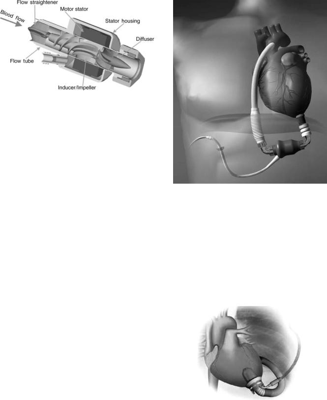
Figure 14 shows the HeartMate II (Thoratec) axial flow ventricular assist device. The rotating impeller is surrounded by a static pump housing with an integral motor (33). The pump’s speed can be controlled either manually or by an automatic controller that relies on an algorithm based on pump speed, the pulsatility of the native heart, and motor current. The HeartMate II is designed to operate between 6000 and 15,000 rpm and deliver as much as 10 L min 1. The initial version of this device is powered through a percutaneous small-diameter electrical cable connected to the system’s external electrical controller. A fully implantable system utilizing a TETS is under development. The first implant HeartMate II implant occurred in 2000 (34). Clinical trials in Europe and the United States are ongoing. This device is intended for both bridge to transplant and destination therapy.
Figure 15 illustrates the Jarvik 2000 (Jarvik Heart, New York). The Jarvik 2000 is intraventricular axial flow pump. The impeller is a neodymium–iron–boron magnet, which is housed inside a welded titanium shell and supported by ceramic bearings (35). A small, percutaneous cable delivers power to the impeller. All of the bloodcontacting surfaces are made of highly polished titanium. The normal operating range for the control system is 8000– 12,000 rpm, which generates an average pump flow rate of 5 L min 1. The pump is placed within the left ventricle with
Figure 14. Thoratec HeartMate II Axial Flow Ventricular Assist Device. The rotating impelleris surroundedbya staticpump housing with an integral motor. The pump’s speed can be controlled either manually or by an automatic controller that relies on an algorithm based on pump speed, the pulsatility of the native heart, and motor current. (Reprinted with permission from Thoratec Corporation.)
a sewing cuff sutured to the ventricle, eliminating the need for an inflow cannula. Over 100 patients have received the Jarvik 2000 as a bridge to transplant or destination therapy, with the longest implant duration of > 4 years (36).
Figure 15. Jarvik 2000 AxialFlowVentricularAssistDevice. Unlike most other axial flow devices, the Jarvik 2000 is intraventricular axial flow pump. The impeller is a neodymium-iron-boron magnet, which is housed inside a welded titanium shell and supported by ceramic bearings. (Reprinted with permission from Jarvik Heart, Inc.)
TOTAL ARTIFICIAL HEARTS
The total artificial heart (TAH) is designed to support both the pulmonary and systemic circulatory systems by replacing the native heart. Two types of artificial hearts have been developed: pneumatic and electric.
Pneumatic Total Artificial Heart
A pneumatic total artificial heart is composed of two ventricles that replace the native left and right ventricle. Each ventricle is of similar design to the Penn State/Thoratec pneumatic ventricular assist device (as described in a previous section) (4). Both ventricles are implanted within the chest. The air pulses are delivered to the ventricles via percutaneous drivelines. An automatic control system varies cardiac output by adjusting the heart rate and the time for ventricular filling in response to an increase in filling pressure.
Pneumatic total artificial hearts are currently used as a bridge to transplant. Several different pneumatic artificial hearts have been used clinically around the world. The only pneumatic TAH approved as a bridge to transplant in the United States is the CardioWest (SynCardia, Tucson, AZ) TAH, illustrated in Fig. 16 (37). The CardioWest (with a stroke volume of 70 mL) is based on the Jarvik-7, which has a stroke volume of 100 mL. A study of 81 recipients of the CardioWest revealed a survival rate to transplant of 79% and a 1 year survival rate of 70%.
Pneumatic total artificial hearts have also been used as a permanent replacement device. The Jarvik-7 pneumatic TAH was permanently implanted in five patients (38).
HEART, ARTIFICIAL |
457 |
Although the longest survivor lived for 620 days, all five patients had hematologic, thromboembolic, and infectious complications. The pneumatic artificial heart is no longer considered for permanent use because of infections associated with the percutaneous pneumatic drive lines and quality of life issues related to the bulky external pneumatic drive units.
Electric Total Artificial Heart
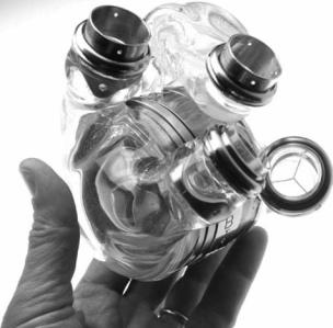
The electric TAH is completely implantable and is designed for permanent use. The Penn State/3M Electric TAH is shown in Fig. 17. The artificial heart is composed of two blood pumps that are of similar design to the Penn State/ Arrow electric ventricular assist device (39). However, the electric TAH uses a single implantable energy converter that alternately drives each ventricle. The implantable controller adjusts the heart rate in response to ventricular filling and maintains left–right balance. The design for this system was completed in 1990 and was the first to incorporate the controller, transcutaneous energy transmission system (TETS), telemetry, and internal power (via rechargeable batteries) into a completely implantable system. The Penn State electric TAH has been successfully implanted in animals for >1 year without thromboembolic complications. In 2000, ABIOMED acquired the rights to the Penn State/3M Electric TAH.
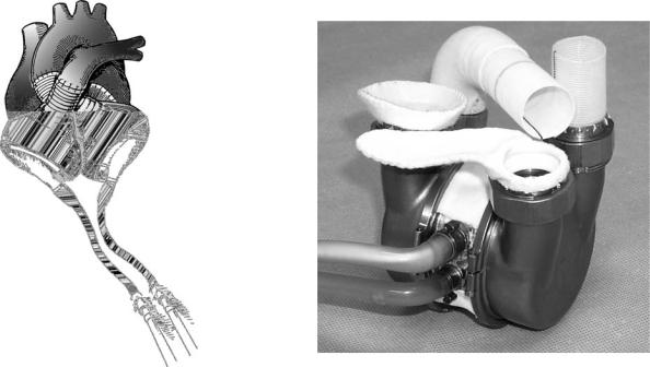
The ABIOMED AbioCor TAH, illustrated in Fig. 18, uses an electrohydraulic energy converter to alternately compress each blood sac (40,41). In addition, the AbioCor uses polymer valves to control flow into and out of each ventricle. The AbioCor is currently undergoing clinical
Figure 16. SynCardia CardioWest Pneumatic Total Artificial Heart. The CardioWest is based on the Jarvik-7 and is the only pneumatic TAH approved as a bridge to transplant in the United States. (Reprinted with permission from SynCardia Systems, Inc.)
Figure 17. Penn State/3M Electric Total Artificial Heart. This artificial heart is composed of two blood pumps that are of similar design to the Penn State/Arrow electric ventricular assist device. However, the electric TAH uses a single implantable energy converter that alternately drives each ventricle.
458 HEART, ARTIFICIAL
Figure 18. ABIOMED AbioCor Electric Total Artificial Heart. The AbioCor TAH uses an electro hydraulic energy converter to alternately compress each blood sac. The AbioCor is currently undergoing clinical trials in the United States. Two patients were discharged from the hospital, with one patient surviving for > 1 year. (Reprinted with permission from ABIOMED, Inc.)
trials in the United States. Fourteen critically ill patients (with an 80% chance of surviving < 30 days) have been implanted. Two patients were discharged from the hospital (one to home), with one patient surviving for >1 year. The causes of death were typically end organ failure and strokes. One pump membrane wore out at 512 days. Smaller, improved totally implantable artificial hearts are currently under development.
FUTURE DIRECTIONS OF RESEARCH
The ventricular assist devices and total artificial hearts presented in this article successfully provide viable cardiac support by either assisting or replacing the native heart. However, there are several areas for future research on artificial hearts. These include the following: Power sources to permit longer intervals between battery changes; Improved control schemes for both pulsatile and nonpulsatile devices that enhance the response of the cardiac assist device to meet physiologic demands; Decrease thromboembolic events associated by modifying the device geometry and/or blood-contacting materials; Determine the long-term effects of continuous, nonpulsatile flow; Decrease incidence of infection by the elimination of all percutaneous lines and creating smaller implantable electronic components; Reduced pump sizes to fit smaller adults, children, and infants; Increased reliability for 5 or more years to 95% (with a 95% confidence level).
Significant progress has been made in the last 20 years. One can only imagine what the next 20 years will bring!
ACKNOWLEDGMENTS
The author would like to acknowledge the support of William S. Pierce, M.D., Gerson Rosenberg, Ph.D., David B. Geselowitz, Ph.D., and the past and present faculty, staff, and graduate students at the Division of Artificial Organs at the Penn State College of Medicine and the Department of Bioengineering at the Pennsylvania State University. The financial support from the National Institutes of Health is also recognized.
BIBLIOGRAPHY
Cited References
1.American Heart Association. Heart Disease and Stroke Statistics—2005 Update. Dallas (TX): American Heart Association; 2005.
2.Organ Procurement and Transplantation Network Data as of May 29, 2005. Available at http://www.optn.org. 2005.
3.Guyton A, Hall J. Textbook of Medical Physiology. Philadelphia: Saunders; 2000.
4.Richenbacher WE, Pierce WS. In: Braunwald HE, editor. Assisted Circulation and the Mechanical Heart Disease: A Textbook of Cardiovascular Medicine, 6th ed. Philadelphia: Saunders; 2001. p 534–547.
5.Thoratec VAD Clinical Results, Available at http://www. thoratec.com. Accessed Nov 2004.
6.Thoratec Corporation Press Release, Aug. 5, 2004 [Online]. Thoratec. Available at http://www.thoratec.com/index.htm. [5/19/2005]. Accessed 2005.
7.HeartMate LVAS Clinical Results, Nov. 2004. Available at http://www.thoratec.com/index.htm. Accessed 2004.
8.Berger EE. ABIOMED’s BVS 5000 biventricular support system. J Heart Lung Transplant 2004;23(5):653.
9.Clinical Information, BVS Clinical Update 2004 [online] ABIOMED. Available at http://www.abiomed.com/clinical information/BVS5000Update.cfm. [5/19/2005]. Accessed 2004.
10.Clinical Information, AB5000 Clinical Update 2004 [online] ABIOMED. Available at http://www.abiomed.com/clinical information/AB5000Update.cfm. [5/19/2005]. Accessed 2004.
11.Berlin Heart AG-The VAD System [online] Berlin Heart. http:// www.berlinheart.com/download/system.pdf. [5/17/2005]. 2003.
12.Reul H. The MEDOS/HIA system: development, results, perspectives. Thorac Cardiovasc Surg 1999;47(Suppl 2):311–315.
13.Takano H, Nakatani T. Ventricular assist systems: experience in Japan with Toyobo pump and Zeon pump. Ann Thorac Surg 1996;61(1):317–322.
14.El-Banayosy A, et al. Preliminary experience with the LionHeart left ventricular assist device in patients with end-stage heart failure. Ann Thorac Surg 2003;75(5):1469–1475.
15.Novacor LVAS-Products-WorldHeart [Online] WorldHeart. Available at http://www.worldheart.com/products/novacor lvas.cfm. [5/17/2005]. Accessed 2005.
16.Rose EA, et al. Long-term mechanical left ventricular assistance for end-stage heart failure. N Engl J Med 2001;345(20): 1435–1443.
17.Baba A, et al. Microcirculation of the bulbar conjunctiva in
the goat implanted with a total artificial heart: effects of pulsatile and nonpulsatile flow. ASAIO J2004;50(4):321–327.
18.Undar A. Myths and truths of pulsatile and nonpulsatile perfusion during acute and chronic cardiac support. Artif Organs 2004;28(5):439–443.
19.Arora D, Behr M, Pasquali M. A tensor-based measure for estimating blood damage. Artif Organs 2004;28(11):1002–1015.