

- •VOLUME 3
- •CONTRIBUTOR LIST
- •PREFACE
- •LIST OF ARTICLES
- •ABBREVIATIONS AND ACRONYMS
- •CONVERSION FACTORS AND UNIT SYMBOLS
- •EDUCATION, COMPUTERS IN.
- •ELECTROANALGESIA, SYSTEMIC
- •ELECTROCARDIOGRAPHY, COMPUTERS IN
- •ELECTROCONVULSIVE THERAPHY
- •ELECTRODES.
- •ELECTROENCEPHALOGRAPHY
- •ELECTROGASTROGRAM
- •ELECTROMAGNETIC FLOWMETER.
- •ELECTROMYOGRAPHY
- •ELECTRON MICROSCOPY.
- •ELECTRONEUROGRAPHY
- •ELECTROPHORESIS
- •ELECTROPHYSIOLOGY
- •ELECTRORETINOGRAPHY
- •ELECTROSHOCK THERAPY.
- •ELECTROSTIMULATION OF SPINAL CORD.
- •ELECTROSURGICAL UNIT (ESU)
- •EMERGENCY MEDICAL CARE.
- •ENDOSCOPES
- •ENGINEERED TISSUE
- •ENVIRONMENTAL CONTROL
- •EQUIPMENT ACQUISITION
- •EQUIPMENT MAINTENANCE, BIOMEDICAL
- •ERGONOMICS.
- •ESOPHAGEAL MANOMETRY
- •EVENT-RELATED POTENTIALS.
- •EVOKED POTENTIALS
- •EXERCISE FITNESS, BIOMECHANICS OF.
- •EXERCISE, THERAPEUTIC.
- •EXERCISE STRESS TESTING
- •EYE MOVEMENT, MEASUREMENT TECHNIQUES FOR
- •FETAL MONITORING
- •FETAL SURGERY.
- •FEVER THERAPY.
- •FIBER OPTICS IN MEDICINE
- •FICK TECHNIQUE.
- •FITNESS TECHNOLOGY.
- •FIXATION OF ORTHOPEDIC PROSTHESES.
- •FLAME ATOMIC EMISSON SPECTROMETRY AND ATOMIC ABSORPTION SPECTROMETRY
- •FLAME PHOTOMETRY.
- •FLOWMETERS
- •FLOWMETERS, RESPIRATORY.
- •FLUORESCENCE MEASUREMENTS
- •FLUORESCENCE MICROSCOPY.
- •FLUORESCENCE SPECTROSCOPY.
- •FLUORIMETRY.
- •FRACTURE, ELECTRICAL TREATMENT OF.
- •FUNCTIONAL ELECTRICAL STIMULATION
- •GAMMA CAMERA.
- •GAMMA KNIFE
- •GAS AND VACUUM SYSTEMS, CENTRALLY PIPED MEDICAL
- •GAS EXCHANGE.
- •GASTROINTESTINAL HEMORRHAGE
- •GEL FILTRATION CHROMATOGRAPHY.
- •GLUCOSE SENSORS
- •HBO THERAPY.
- •HEARING IMPAIRMENT.
- •HEART RATE, FETAL, MONITORING OF.
- •HEART VALVE PROSTHESES
- •HEART VALVE PROSTHESES, IN VITRO FLOW DYNAMICS OF
- •HEART VALVES, PROSTHETIC
- •HEART VIBRATION.
- •HEART, ARTIFICIAL
- •HEART–LUNG MACHINES
- •HEAT AND COLD, THERAPEUTIC
- •HEAVY ION RADIOTHERAPY.
- •HEMODYNAMICS
- •HEMODYNAMIC MONITORING.
- •HIGH FREQUENCY VENTILATION
- •HIP JOINTS, ARTIFICIAL
- •HIP REPLACEMENT, TOTAL.
- •HOLTER MONITORING.
- •HOME HEALTH CARE DEVICES
- •HOSPITAL SAFETY PROGRAM.
- •HUMAN FACTORS IN MEDICAL DEVICES
- •HUMAN SPINE, BIOMECHANICS OF
16.Grace ND. Diagnosis and treatment of gastrointestinal bleeding secondary to portal hypertension. American College of Gastroenterology Practice Parameters Committee. Am J Gastroenterol 1997;92:1081–1091.
17.Bardhan KD, et al. Changing patterns of admissions and operations for duodenal ulcer. Br J Surg 1989;76:230–236.
18.Huang CS, Lichtenstein DR. Nonvariceal upper gastrointestinal bleeding. Gastroenterol Clin N Am 2003;32: 1053–1078.
19.Cook DJ, Guyatt GH, Salena BJ, Laine LA. Endoscopic therapy for acute nonvariceal upper gastrointestinal hemorrhage:a meta-analysis. Gastroenterology 1992;102:139–148.
20.Lau JY, et al. Endoscopic retreatment compared with surgery in patients with recurrent bleeding after initial endoscopic control of bleeding ulcers. N Engl J Med 1999;340: 751–756.
21.Gomes AS, Lois JF, McCoy RD. Angiographic treatment of gastrointestinal hemorrhage: comparison of vasopressin infusion and embolization. Am J Roentgenol 1986;146:1031–1037.
22.Lefkovitz Z, et al. Radiologic diagnosis and treatment of gastrointestinal hemorrhage and ischemia. Med Clin N Am 2002;86:1357–1399.
23.Collins R, Langman M. Treatment with histamine H2 antagonists I acute upper gastrointestinal hemorrhage: implications of randomized trials. N Engl J Med 1985;313: 660–666.
24.Walt RP, et al. Continuous infusion of famotidine for hemorrhage from peptic ulcer. Lancet 1992;340:1058–1062.
25.Javid G, et al. Omeprazole as adjuvant therapy to endoscopic combination injection sclerotherapy for treating bleeding peptic ulcer. Am J Med 2001;111:280–284.
26.Mallory GK, Weiss S. Hemorrhage from lacerations of the cardiac orifice of the stomach due to vomiting. Am J Med Sci 1929;178:506.
27.Onge GS, Bezahler GH. Giant esophageal ulcer associated with cytomegalovirus. Gastroenterology 1982;83:127–130.
28.Schmulewitz N, Fisher DA, Rockey DC. Early colonoscopy for acute lower GI bleeding predicts shorter hospital stay: A retrospective study of experience in a single center. Gastrointest Endosc 2003;58:841–846.
29.Colacchio TA, Forde KA, Patsos TJ, Nunez D. Impact of modern diagnostic methods on the management of rectal bleeding. Am J Surg 1982;143:607–610.
30.Boley SJ, et al. The pathophysiologic basis for the angiographic signs of vascular ectasias of the colon. Radiology 1977;125:615–621.
See also ELECTROGASTROGRAM; ENDOSCOPES.
GEL FILTRATION CHROMATOGRAPHY. See
CHROMATOGRAPHY.
GLUCOSE SENSORS
FARBOD N. RAHAGHI
DAVID A. GOUGH
University of California,
La Jolla, California
INTRODUCTION
Glucose assay is arguably the most common of all medical measurements. Billions of glucose determinations are performed each year by laypeople with diabetes based on
GLUCOSE SENSORS |
393 |
‘‘fingersticking’’ and by healthcare professionals based on blood samples. In fingersticking, sample collection involves the use of a lancet to puncture the skin of the fingertip or forearm to produce a small volume of blood and tissue fluid, followed by collection of the fluid on a reagent-containing strip and analysis by a handheld meter. Glucose measurements coupled to discrete sample collection continue to be the most common method of glucose monitoring. However, new types of sensors capable of continuous glucose monitoring are nearing clinical introduction. Continuous or near-continuous glucose sensors may make possible new and fundamentally different approaches to the therapy of the disease. This article reviews recent progress in the development of new glucose sensors and describes the potential roles for these sensors in the improved treatment of diabetes.
THE CASE FOR NEW GLUCOSE SENSORS
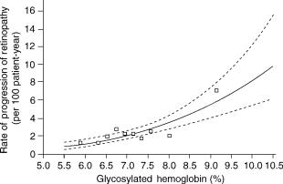
The objective of all forms of therapy for diabetes is the maintenance of blood glucose near normal levels (1). The Diabetes Control and Complications Trial (or DCCT) and counterpart studies such as the United Kingdom Prevention of Diabetes Study (UKPDS) have clearly demonstrated (Fig. 1) that lower mean blood glucose levels resulting from aggressive treatment can lead to a reduced incidence and progression of retinopathy, nephropathy, and other complications of the disease (2,3). These prospective studies showed definitively that there exists a cause-and-effect relationship between poor blood glucose control and the complications of diabetes. As convenient means for frequent glucose assay were not available at the time, glucose control was assessed in these trials by glycosylated hemoglobin levels (HbA1c), which indicate blood glucose concentrations averaged over the previous 3 month period. Although HbA1c levels are useful for assessment of longitudinal blood glucose control, the values indicate only averaged blood glucose, rather than blood glucose dynamics (i.e., how blood
Figure 1. The results of the DCCT (2). Results show that improved glucose control, measured by a reduction in the fraction of glycosylated hemoglobin, leads to reduced long-term complications of diabetes. (Copyright # 1993, Massachusetts Medical Society.)
394 GLUCOSE SENSORS
glucose changes with time), and cannot be used for immediate adjustment of therapy (4). There is general agreement that frequent determination of glucose by a sensing method that is convenient and widely acceptable to people with diabetes would allow a finer degree of control. Normalization of blood glucose dynamics may be of equal or greater importance than normalization of average blood glucose. The results of the DCCT and related studies point to the need for practical new approaches to achieve control.
The primary need for a new type of glucose sensor is to facilitate improved treatment of type 1 diabetes. In this case, the insulin producing ability of the pancreas has been partially or fully destroyed due to a misdirected autoimmune process, making insulin replacement essential. The sensor would help avoid the long-term complications associated with hyperglycemia (i.e., above-normal blood glucose) by providing information to specify more timely and appropriate insulin administration. It is now becoming widely appreciated that a new sensor could also be beneficial for people with the more common type 2 diabetes, where a progressive resistance of peripheral tissues to insulin develops, leading to glucose imbalances that can eventually produce long-term clinical consequences similar to type 1 diabetes. Type 2 diabetes is related to obesity, lifestyle, and inherited traits. In recent years, the incidence of type 2 diabetes has increased at extraordinary rates in many populations, to the point of becoming a worldwide epidemic (5). It is estimated that within 10 years, the prevalence of diabetes may approach 210 million cases worldwide (6). This places increased urgency on developing new approaches to managing or preventing the disease where possible, and a meliorating its consequences.
In addition, an automatic or continuous sensor may also have an important role in preventing hypoglycemia (i.e., below-normal blood glucose). Hypoglycemia is caused primarily by a mismatch between the insulin dosage used and the amount of insulin actually needed to return the blood glucose level to normal. Many people with diabetes can reduce the mean blood glucose by adjustment of diet, insulin, and exercise, but when aggressively attempted, this has led to a documented increase in the incidence of hypoglycemia (7). Below-normal glucose values can rapidly lead to cognitive lapses, loss of consiousness, and lifethreatening metabolic crises. In children, there is concern that severe hypoglycemic events may lead to neurologic sequelea (8). A significant percentage of deaths of people under 40 with type 1 diabetes is due to the ‘‘dead-in-bed’’ syndrome (9), which may be linked to nocturnal hypoglycemia. Some experts claim that ‘‘. . . the threat of severe hypoglycemia remains the single most important barrier to maintaining normal mean blood glucose’’ (10). A continuous glucose sensor that does not depend on user initiative could be part of an automatic alarm system to warn of hypoglycemia and provide more confidence to the user to lower mean blood glucose, in addition to preventing hypoglycemia by providing improved insulin dosages. Hypoglycemia detection may be the most important application of a continuous glucose sensor. Ultimately, a glucose sensor may also be useful in the prediabetic state to indicate
behavior modification for reduction of metabolic stress on the pancreas.
Beyond applications in diabetes, it has recently been shown that stricter glycemic control during surgery and intensive care can reduce mortality in non-diabetic patients and significantly shorten the hospital stay (11). The exact mechanism of this effect has not been elucidated, but the benefit is closely tied to the extent of glucose control and not simply insulin dosage (12). This is another important application for new glucose sensors.
Alternatives to sensor-based therapies for diabetes are more distant. Several biological approaches to diabetes treatment have been proposed, including pancreatic transplantation, islet transplantation, genetic therapies, stem cell-based therapies, new pharmaceutical strategies, islet preservation, and others. Whole or partial organ and islet transplantation requires discovery of methods for assuring immuno-tolerance that do not rely on anti-rejection drugs and approaches for overcoming the shortage of transplantable pancreatic tissue. Potential therapies based on stem cells, if feasible, require basic research on growth, regulation, and implementation of the cells, and share the immuno-intolerance problem. Genetic therapies are limited by incomplete understanding of the complex genetic basis of diabetes, as well as progress in developing sitespecific gene delivery, activation, and inactivation. Although transplantation, stem cell, and genetic approaches are based wholly on biological materials, it is not certain that the glucose and insulin dynamics resulting from their use will necessarily be near-normal or readily adjustable. Immunotherapeutic approaches for in situ preservation of islets are also being studied but, if eventually feasible, are far off and may require lifetime immune system modulation. The possibility of prevention of type 1 diabetes relies on development of timely methods for early detection of the disease and discovery of an acceptable approach to avoid or interrupt the islet destruction process. Furthermore, prevention will have little value for people who already have diabetes. These alternatives require substantial basic research and discovery, and while often highly publicized, are not likely to be available until far into the future, if eventually feasible.
Although new glucose sensors have the advantage of being closer to clinical introduction, there are certain other advantages as well. First, no anti-rejection medication will be needed. Second, the sensor will provide real-time information about blood glucose dynamics that is not available from other technologies. Third, in addition to real-time monitoring, continuous sensor information may be useful to predict blood glucose ahead of the present (13), a capability not feasible with the other approaches. Real-time monitoring and predictive capabilities may lead to entirely new applications of present therapies. Fourth, the sensor could operate in parallel with various other therapies, should they become available. The glucose sensor will likely have broad application, regardless of whether or when other technologies are introduced.
The sensor is also key to the implementation of the mechanical artificial beta cell. In the ideal configuration, this device would have an automatic glucose sensor, a refillable insulin pump, and a controller containing an
algorithm to direct automatic pumping of insulin based on information provided by the sensor. There has been progress on development of several of the components of this system, including: (1) external insulin pumps, which operate in a substantially preprogrammed mode with minor adjustments by the user based on fingerstick glucose information; (2) long-term implantable insulin pumps that operate in a similar way; (3) models of glucose and insulin distribution in the body that may eventually be useful in conjunction with control systems; and (4) controllers to direct insulin pumping based on sensor information. In contrast to other approaches to insulin delivery, the mechanical artificial beta cell has the advantage that the insulin response can be reprogrammed to meet the changing needs of the user. Development of an acceptable glucose sensor has thus far been the most difficult obstacle to implementation of the mechanical artificial beta cell.
THE IDEAL GLUCOSE SENSOR
The likelihood that glucose monitoring will reach its full potential as a tool for the therapy of diabetes depends on the technical capabilities of candidate sensors and the general acceptance of sensors by people with diabetes. Technical requirements of the sensor system include: specificity for glucose in the presence of interfering biochemicals or physiological phenomena that may affect the signal; sensitivity to glucose and adequate concentration resolution over the relevant range; accuracy as compared to a ‘‘gold standard’’ blood glucose assay; a sufficiently short response lag to follow the full dynamic range of blood glucose variations; reliability to detect mild hypoglycemia without false positives or negatives; and sufficient stability
GLUCOSE SENSORS |
395 |
that recalibration is rarely needed. The specific criteria for sensor performance remain a matter of consensus and may become better defined as sensors are introduced. The general acceptance of new sensors by people with diabetes will be based on such factors as safety, convenience, reliability, automatic or initiative-independent operation, infrequent need for recalibration, and independence from fingersticking.
For the glucose sensor to be a widely accepted innovation, the user must have full confidence in its accuracy and reliability, yet remain uninvolved in its operation and maintenance. Sensor systems under development have yet to reach this ideal, but some promising aspirants are described below. Short of the ideal, several intermediate sensing technologies with limited capabilities may find some degree of clinical application and, if used effectively, may lead to substantial improvements in blood glucose control. Nevertheless, the most complete capabilities will lead to the broadest adoption by users.
GLUCOSE SENSORS AND SENSING METHODOLOGIES
Several hundred physical principles for monitoring glucose have been proposed since the 1960s. Many are capable of glucose measurement in simple solutions, but have encountered limitations when used with blood, employed as implants, or tested in clinically relevant applications. Certain others have progressed toward clinical application. A brief summary of the history of events related to glucose sensor development is shown in Figure 2.
Present Home Glucose Monitoring
A major innovation leading to improved blood glucose management was the widespread use of home glucose
Figure 2. A time-line of some important developments relating to glucose sensors (2,14–24).
396 GLUCOSE SENSORS
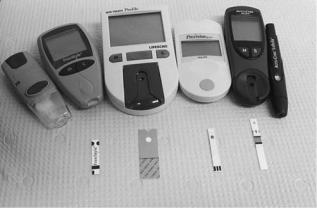
Figure 3. A small collection of home glucose monitoring equipment developed over the past decade. At either end (above) are devices used to puncture the skin for sample collection. Examples of commercial glucose meter (above, center) are also shown. Strips (below) contain immobilized glucose oxidase and are discarded after a single measurement.
monitoring in the 1980s (22). Present commercial versions of this technology are available with respective methods for glucose assay, data presentation and storage, sample volume requirements, and various convenience features (Fig. 3). These devices employ single-use strips based on enzyme methods discussed below. The widespread application of home glucose monitoring has permitted laypeople with diabetes to assume a newfound role in the management of their disease. The present standard-of-care recommends glucose measurement three or more times a day for insulin-dependent individuals (25), but a small number of individuals samples 10 or more times daily. It is generally suspected that the average sampling rate is inadequate and a recent publication noted that only 56% of diabetic individuals sampled their blood glucose once or more daily (26). The general resistance to more frequent sampling may be related to several factors, including: the pain associated with finger puncture, the requirement for user initiative, the general inconvenience of the assay, and unwillingness to carry out nocturnal testing (27).
When sampling is not sufficiently frequent, undetected blood glucose excursions can occur between samples. It has been shown that blood glucose measurements must be obtained every 10 min to detect all blood glucose excursions in the most severe diabetic subjects (28), although slower blood glucose excursions in the majority of people with diabetes may not require sampling at this frequency. The fact that the sample frequency required to detect all glycemic excursions is not clinically feasible with present technology indicates that the dynamic control of blood glucose is currently not practiced in diabetes management.
To compensate for infrequent monitoring, users typically adopt various strategies to estimate blood glucose concentration using subjective ad hoc models. These strategies rely on the most recent reported values of glucose, in conjunction with the timing and content of recent or upcoming meals, insulin therapy, and exercise. The effectiveness of these strategies is limited and the constant attention required to make such estimates represent a
substantial intrusion in lifestyle. Although glucose monitoring by fingersticking is likely to become more acceptable as the sample volume and the pain associated with sample collection are reduced, the problem of infrequent sampling and the requirement for user initiative will continue to be the major obstacles to the improvement of glucose control based on this technology.
Noninvasive Optical Sensing Concepts
Noninvasive optical methods are based on directing a beam of light onto the skin or through superficial tissues, and recording the reflected, transmitted, polarized, or absorbed components of the light (29). A key requirement for success of these methods is a specific spectral region that is sufficiently sensitive to glucose, but insensitive to other similar optically active interfering molecules and tissue structures. Several optical methods allow straightforward glucose measurement in simple aqueous solutions, but are ineffective at detecting glucose in tissue fluid, plasma, or blood. If an optical approach can be validated, a noninvasive sensor might be possible. For this reason, an intensive research effort and substantial industrial investment over the past two decades have gone into investigation of these concepts.
Infrared (IR) absorption spectroscopy is based on excitation of molecular motions that are characteristic of the molecular structure. The near-infrared (NIR) region of the spectrum (750–2500 nm) is relatively insensitive to water content so that the beam penetration depth in tissues can be substantial (30). Trials to identify a clinical correlation between NIR signals and blood glucose have employed various computational methods for analyzing the absorption spectrum. Basic studies have focused on identifying the absorbing species and tissue structures responsible for optical signals. However, after much effort the operating conditions that provide selectivity for glucose have yet to be established, leading one investigator to conclude that ‘‘. . .
signals can be attributed to chance’’ (31).
Raman spectroscopy relies on detecting scattered emissions associated with vibrational molecular energy of the chemical species (as opposed to transmitted, rotational, or translational energy). Early studies compared the measurement in water of three different analytes (urea, glucose, lactic acid) and found that glucose levels could be determined with limited accuracy (32). Raman spectroscopy has been applied in the aqueous humor of the eye (33), which is thought to reflect delayed blood glucose levels over certain ranges (34). As with other optical methods, adequate specificity for glucose in the presence of other molecules remains to be demonstrated.
Measurement of the concentration using polarimetry is based on ability of asymmetric molecules such as glucose to rotate the plane of polarized light (35). This method is limited by the presence of other interfering asymmetric molecules, as well as the thickness and light scattering by tissues in the region of interest (30). Considerable development of polarimetry has centered on measurements in the anterior chamber of the eye (36), but there is yet to be a demonstration of sufficient selectivity under biological conditions.
Attempts at validation of optical sensor concepts have involved two general approaches. One approach endeavors to establish selectivity for glucose by identification of the components of tissues besides glucose that contribute to the optical signal, and determine if the effects of these interfering substances can be eliminated or the observed signals can be reconstructed based on all contributions. This has not yet been successful, in spite of intensive efforts. The impediment is the large number of optically active components in tissues, many of which produce much stronger effects than glucose. A second approach to validation involves identifying an empirical relationship between the observed optical signal in vivo and simultaneously recorded blood glucose concentration. Noninvasive optical approaches have been the premise of several human clinical trials, all of which have been unsuccessful. The prospects for a non-invasive optical glucose sensor are distant.
Implantable Optical Sensor Concepts
Implanted optical sensors offer the prospect of a less congested optical path, at the expense of requiring a more complicated device and confronting the foreign body response. One promising optical concept is based on chemical interaction between glucose and an optically active chemical species that is immobilized in an implanted, glucose-permeable chamber. The interaction creates a change in the optical signal which, under ideal conditions, may indicate glucose concentration. An example is the ‘‘affinity sensor’’ (37), in which glucose competes with a fluorescent substrate for binding with a macromolecule, Con A, resulting in a change in the optical signature. A similar detection strategy has been proposed as part of an implantable intraocular lens (38). There are difficulties with biocompatibility of the implant, design of the chamber, specificity of the optical detector, as well as toxicity and photobleaching of the indicator molecules (38). These systems have yet to be extensively tested.
Tissue Fluid Extraction Techniques
The interstitial fluid that irrigates tissues contains glucose derived from the blood in local capillaries. Several strategies have been devised to extract this fluid for glucose assay.
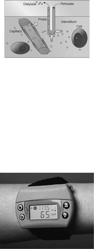
Microdialysis is based on a probe consisting of a fine hairpin loop of glucose-permeable dialysis tubing in a probe that is inserted into subcutaneous tissues (39). A fluid perfusate is continuously circulated through this tubing by a pump contained in an external apparatus (40), collected, and assayed for glucose concentration using an enzyme electrode sensor (Fig. 4). This methodology relies on the exchange of glucose between the microvascular circulation and the local interstitial compartment, transfer into the dialysis tube, and appropriate adjustment of the pumping pressure and perfusion rate (41). The advantage of the system is that a foreign body response of the tissue and local mass transfer resistance are slow to develop due to the sustained tissue irrigation, but drawbacks include the requirement for percutaneous access, the need for frequent relocation of the probe to minimize the chance
GLUCOSE SENSORS |
397 |
Figure 4. Diagram of a microdialysis probe (39). The semipermeable membrane at the probe tip allows exchange of soluble molecules between the probe and surrounding tissue. Samples are continuously collected and analyzed. (Used with permission of BMJ Publishing Group.)
of infection, and management of the external apparatus by the user. This device may find clinical applications for short-term monitoring.
Reverse iontophoresis employs passage of electrica current between two electrodes placed on the surface of the body to extract tissue fluid directly through the intact skin (42). Glucose in the fluid has been measured by an enzyme electrode-type sensor as part of a wristwatch-like apparatus (43) (Fig. 5). With a 2 h equilibration process after placing the device and a fingerstick calibration, the sensor can take measurements as often as every 10 min for 12 h, at which time sensor components must be replaced and the sensor recalibrated (44). This sensor was approved by the Food and Drug Administration (FDA) for indicating glucose trends, but users are instructed to revert to more reliable conventional assays for insulin dosing decisions. Minor skin irritation has been reported as a side effect (45). Although this sensor was briefly available commercially, it
Figure 5. The Glucowatch Biographer (43). An integrated system for sample extraction by reverse iontophoresis and glucose sensing. (Used with permission from John Wiley & Sons, Inc.)
398 GLUCOSE SENSORS
was not successful as a product due to its limited capabilities and inconvenience.
Implantable Enzyme Electrode Sensors
The most promising approaches have been various configurations of the enzyme electrode sensor based on immobilized glucose oxidase coupled to electrochemical detectors. The enzyme catalyzes the reaction:
glucose þ O2 þ H2O ! gluconic acid þ H2O2 ð1Þ
Monitoring of glucose can be based on detection of hydrogen peroxide production, oxygen depletion, or electron transfer via a conductive polymer link, as described below. Enzyme electrode sensors must contact the sample fluid to be assayed, and therefore require either sensor implantation or sample extraction (as in the case of reverse iontophoresis, microdialysis sensors and fingerstick devices). By employing the enzyme, sensors can have a significant advantage over non-enzymatic sensors of being specific for glucose rather than just selective. However, the benefits of enzyme specificity may not be fully realized unless the sensor is properly designed. To achieve the best performance, enzyme electrode sensors must include design features to address enzyme inactivation, biological oxygen variability, mass transfer dependance, generation of peroxide, electrochemical interference, and other effects.
From the perspective of biocompatibility, sensors can be implanted either in direct contact with blood or with tissues. Biocompatibility in contact with blood depends on the surface properties of the sensor as well flow characteristics at the implant site. Implantation in an arterial site, where the pressure and fluid shear rates are high, poses the threat of blood clotting and embolization, and is rarely justified. Central venous implantation is considerably safer, and there are several examples of successfull longterm implants in this site.
Implantation of the sensor in a tissue site is safer, but involves other challenges. The sensing objective is to infer blood glucose concentration from the tissue sensor signal, and factors that affect glucose mass transfer from nearby capillaries to the implanted sensor must be taken into account. These factors include: the pattern and extent of perfusion of the local microvasculature; regional perfusion of the implant site, the heterogeneous distribution of substrates within tissues, and the availability of oxygen. There are also substantial differences in performance between shortand long-term implant applications. In the short term, a dominant wound healing response prevails, whereas in the long term, encapsulation may occur. Definitive studies are needed to establish the real-time accuracy of implanted sensors and determine when recalibration is necessary. Studies should be designed to ascertain whether signal decay is due to enzyme inactivation, electrochemical interference, or tissue encapsulation. More information is needed about the effect of these processes on the sensor signals.
There are >104 technical publications and several thousand patents related to glucose measurement by glucose oxidase-based enzyme electrodes, although only a fraction
of these address implant applications. Rather than an attempt to be comprehensive, comments here are limited to examples of the most advanced approaches intended for implant applications.
Enzyme Electrode Sensors Based on Peroxide Detection.
Detection of hydrogen peroxide, the enzyme reaction product, is achieved by electrochemical oxidation of peroxide at a metal anode resulting in a signal current that passes between the anode and a counterelectrode (46). A membrane containing immobilized glucose oxidase is attached to the anode and, in the presence of glucose and oxygen under certain conditions, the current can reflect glucose concentration.
The peroxide-based sensor design is used in several home glucose monitoring devices and has been highly successful for glucose assay on an individual sample basis. However, it is not easily adapted as an implant, especially for long-term applications. The peroxide-based sensor is subject to electrochemical interference by oxidation of small molecules due to its requirement of a porous membrane and an aqueous pathway to the electrode surface for transport of the peroxide molecule. This factor partially accounts for a documented decay in sensitivity to glucose during sensor use. In addition, this sensor design can incorporate only a limited excess of immobilized glucose oxidase to counter enzyme inactivation, as high enzyme loading reduces peroxide transport to the electrode (47). Coimmobilization of catalase to avoid peroxide-mediated enzyme inactivation is not an option because it would prevent peroxide from reacting with the anode. There are also no means to account for the effects of physiologic variation in oxygen concentration and local tissue perfusion on the sensor response.
There have, nevertheless, been proposals to address some of these challenges. Composite membranes with reduced pore size have markedly reduced electrochemical interference from a variety of species over the shortterm (48). A ‘‘rechargeable’’ enzyme system has been devised for periodically replenishing enzyme activity (49), in which a slurry of carbon particles with immobilized glucose oxidase is pumped between membrane layers of a peroxide electrode from a refillable reservoir. A gascontaining chamber has been proposed (50) to address the ‘‘oxygen deficit’’ (51), or stoichiometric limitation of the enzyme reaction by the relativley low tissue oxygen concnetration. Certain other challenges of the peroxide sensor principle remain to be addressed. As a result of the inherent features of this sensor principle, the peroxidebased sensor may be best suited to short-term implant applications and where frequent sensor recalibration is acceptable.
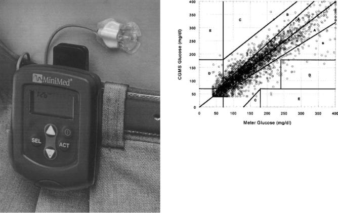
Small, needle-like short-term peroxide-based sensors connected by wire to a belt-mounted monitor have been developed for percutaneous implantation (52) (Fig. 6). The sensor was ultimately intended for insertion by the user for operation up to 3 days at a given tissue site before relocation. Sensors based on peroxide detection have been tested extensively in animals and humans (52–55) and, in some cases have functioned remarkably well, although frequent recalibration was required. In
GLUCOSE SENSORS |
399 |
Figure 6. Components of the MiniMed CGMS system (62). This sensor, based on the peroxide-based sensing principle, is FDA approved for short-term monitoring. (Copyright # 1999, Elsevier.)
some cases, difficulties of two types have been identified (56–61). First, the sensors themselves have not been specific for glucose, sufficiently sensitive, or stable. In some cases where the sensor response in simple buffer solutions was acceptable, ambiguous signals have sometimes resulted when used as an implant. Examples of such responses are: sensor signals that decay over the short term while blood concentrations remain constant, signals that apparently follow blood concentrations during some periods, but not at other times; and identical sensors implanted in different tissue sites in a given subject that sometimes produce opposite signals (55). The peroxidebased subcutaneous sensor was the first commercially available near continuous sensor. In small controlled stu-
dies, use of the sensor was shown to lower HbA1c levels (62). Latter versions of this sensor have been approved by the
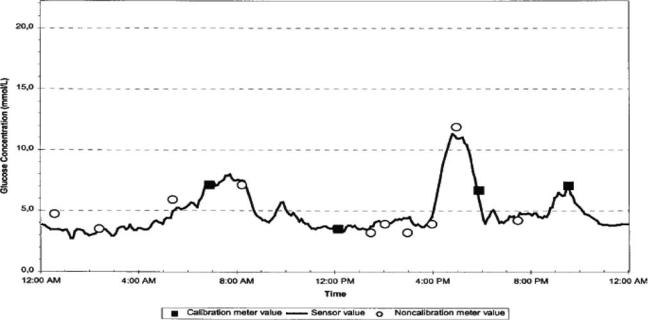
FDA to be used as monitors to alarm for hyperand hypoglycemia in real time. Although there are reservations about its accuracy, the needle sensor has been used in clinical research settings (Figs. 7, 8). A recent study found substantial error in values produced by a prominent commercial needle sensor and concluded that this sensor ‘‘. . .
cannot be recommended in the workup of hypoglycemia in nondiabetic youth’’ (66) and, by extension, to other diabetic subjects. Reasons for these signal deviations are not fully understood.
Figure 7. An approach to sensor validation. Comparison of 2477 glucose values determined by a CGMS sensor system and a standard meter (63). Data pairs were collected during home use from 135 patients. The plot, known as the Clarke Error Grid, has zones with differing clinical implications. This type of plot is widely used to describe glucose sensor performance, but has limited ability to discriminate ineffective sensors (64). (Copyright # 1999, Elsevier.)
Although the needle sensor may be acceptable only to a relatively small group of the most motivated individuals, it represents an advance in glucose sensor technology. Perhaps the most important contribution of the short-term needle sensor has been the revelation to users and clinicians that blood glucose excursions generally occur much more frequently and in a greater number of people than previously thought. This heightened awareness of blood glucose dynamics may lead to a greater appreciation of the need for dynamic control in improved metabolic management.
Long-term peroxide-based sensors have been implanted in the peritoneal cavity of dogs and in humans in conjunction with battery-operated telemetry units (67,68). Although the sensors remained implanted in humans for up to 160 days, the sensitivity to glucose decayed during the study and frequent recalibration was required.
Peroxide-based sensors with telemetry systems have also been implanted in the subcutaneous tissues of human type 1 diabetic subjects to determine if the mere presence of a nonreporting sensor can improve metabolic control (69). Glucose was monitored in parallel by fingerstick throughout the study as a basis for insulin dosage. Study subjects were able to reduce the time spent in hyperglycemia, increase the time spent in normoglycemia and modest hypoglycemia, and markedly reduce the time spent in severe hypoglycemia, but reductions in HbA1c values were not observed. The study was not specifically designed to validate sensor function and a more straightforward and informative study design is needed.
Short-Term Enzyme Electrodes Based on Conductive Polymers. Another principle for glucose monitoring is based on
400 GLUCOSE SENSORS
Figure 8. An example of the CGMS sensor response (65). Squares are reference values utilized in sensor calibration. Circles are additional reference blood glucose values. Reference values were obtained from a standard glucose meter. Values are in mmol L 1. (Used with permission.)
immobilization of glucose oxidase to electron-conducting polymers that can act as ‘‘chemical wires’’, providing a means for direct electron transport between glucose oxidase and the electrode (70). This priniciple eliminates the need for oxygen as a coreactant and, although a porous membrane that can allow passage of ionic current and interferants is still required, the electrode can be operated at lower anodic potentials to reduce electrochemical interference (71). A short-term needle-like version of this sensor for 3 day operation is under development.
Long-Term Enzyme Electrode Sensors Based on Oxygen Detection. Glucose can also be monitored by detecting differential oxygen consumption from the glucose oxidase reaction. In this case, the process is based either on glucose oxidase alone (reaction 1), or a two-enzyme reaction including catalase in excess, which produces the following overall process:
Glucose þ 0:5 O2 ! gluconic acid |
ð2Þ |
The enzymes are immobilized within a gel membrane in contact with the electrochemical oxygen sensor. Excess oxygen not consumed by the enzymatic process is detected by an oxygen sensor and, after comparison with a similar background oxygen sensor without enzymes, produces a differential signal current that is related to glucose concentration.
This approach has several unique features (23). Electrochemical interference and electrode poisoning from endogenous biochemicals are prevented by a porefree silicone rubber membrane between the electrode and the enzyme layer. This material is permeable to oxygen but completely impermeable to polar molecules
that cause electrochemical interference. Appropriate design of the sensor results in sufficient supply of oxygen to the enzyme region to avoid a stoichiometric oxygen deficit (51), a problem that has not been addressed in the peroxide-based sensor system. The differential oxygen measurement system can also readily account for variations in oxygen concentration and local perfusion, which may be particularly important for accurate function of the implant in tissues. Vast excesses of immobilized glucose oxidase can be incorporated to extend the effective enzyme lifetime of this sensor, a feature not feasible with peroxideand conductive polymer-based sensors. Coimmobilization of catalase can further prolong the lifetime of glucose oxidase by preventing peroxide-mediated enzyme inactivation, the main cause of reduced enzyme lifetime (72). This sensor design also avoids current passage through the body and hydrogen peroxide release into the tissues.
A long-term oxygen-based sensor has been developed as a central venous implant (23) (Fig. 9). The sensor functioned with implanted telemetry (73) in dogs for >100 days and did not require recalibration during this period (Fig. 10). The central venous site permitted direct exposure of the sensor to blood, which allowed simple verification of the sensor function without mass transfer complications. This was particularly beneficial for assessing sensor stability. In clinical trials, this system has been reported (74) to function continuously for >500 days in humans with <25% change in sensitivity to glucose over that period. This achievement represents a world record for long-term, stable, implanted glucose sensor operation, although there may still exist hurdles to commercialization.

GLUCOSE SENSORS |
401 |
Figure 9. Animal prototype long-term central venous glucose sensor with implanted telemetry (73). Glucose and oxygen sensors are at the end of the catheters. Telemetry antenna emerges from the top, left. The telemetry body is 2 2.5 in.
These results have lead to several unanticipated conclusions. Although native glucose oxidase is intrinsically unstable, with appropriate sensor design the apparent catalytic lifetime of the immobilized enzyme can be substantially extended (75). The potentiostatic oxygen sensor is remarkably stable (76) and the oxygen deficit, once thought to be an insurmountable barrier, can be easily overcome (51). The central venous implant site, which is uniquely characterized by slow, steady flow of blood, allows for sufficient long-term biocompatibility with blood that the sensor stability can be documented (23). Nevertheless, the
Figure 10. Response of an implanted intravenous sensor to glucose challenges on day 108 after implantation in a dog (23). The solid line is the sensor signal and triangles are venous blood glucose assays. Blood glucose excursions with initial rates of 0.2- 8 mM min 1 were produced by infusions of sterile glucose solutions through an intravenous catheter in a foreleg vein. (Note: 90 mg dL 1 glucose ¼ 5.0 mM.) (Copyright # 1990, American Diabetes Association.)
Figure 11. Close-up view of tissue glucose and oxygen sensor array (77). Sensor array with small (125 mm diameter) independent platinum working electrodes, large (875 mm diameter) common platinum counterelectrodes, and a curved common Ag/AgCl reference electrode. The membrane is not present. (Copyright 2003, American Physiological Society.)
potential for thromoembolic events, anticipated to be rare but potentially significant over many years of sensor use, suggests reservations that may limit clinical acceptance and provides motivation for development of a potentially safer long-term sensor implant in tissues.
Long-term oxygen-based sensors have also been implanted in tissues. The successful central venous sensor cannot be simply adopted for use in the safer tissue site, but certain design features of that sensor which promote long-term function, such as immobilized enzyme design, the stable potentiostatic oxygen sensor, and membrane design to eliminate the oxygen deficit, can be incorporated (Fig. 11).
A systematic approach is required to validate sensor function, based on quantitative experimentation, mass transfer analysis, and accounting for properties of tissues that modulate glucose signals. Several new tools and methods have been developed. A tissue window chamber has been developed that allows direct optical visualization of implanted sensors in rodents, with surrounding tissue and microvasculature, while recording sensor signals (77) (Fig. 12). This facilitates determination of the effects of microvascular architecture and perfusion on the sensor signal. A method has been devised for sensor characterization in the absence of mass transfer boundary layers (78) that can be carried out before implantation and after explantation to infer stability of the implanted sensor. This allows quantitative assessment of mass transfer resistance within the tissue and the effects of long-term tissue changes. A sensor array having multiple glucose and oxygen sensors has also been developed that shows the range of variation of sensor responses within a given tissue (77). This provides a basis for averaging

402 GLUCOSE SENSORS
Figure 12. An implanted glucose sensor and nearby microvasculature(79). Optical image taken in a hamster windowchamber. Sensor diameter is 125 mm.
sensor signals for quantitative correlation to blood glucose concentration.
REMAINING CHALLENGES FOR SENSOR DEVELOPMENT
Although there has been recent progress in sensor development and implementation, certain challenges remain. In many cases, there is need for improvement in data presentation and sensor validation. Standard glucose measurements for validation of sensor signals are often either not reported or are not obtained frequently enough to validate sensor responses. Requirements for sensor recalibration are not always given. Published results are often selected to show what may be ideally possible for a particular sensor rather than what is typical, sometimes conveying the impression that sensors are more accurate than may be the case.
There is a need to understand the effects of physiologic phenomena such as local perfusion, tissue variability, temperature and movement, that modulate sensor responses to glucose and affect measurement accuracy. A detailed understanding of these effects and their dynamics is needed for a full description of the glucose sensing mechanism. Robust sensor designs and modes of operation are required that assure reliable determination of glucose during exercise, sleeping and other daily life conditions.
A complete explanation for the response of every sensor should be sought, whether it is producing ‘‘good’’ or ‘‘bad’’ results, as more can often be learned for sensor improvement from sensors that produce equivocal results than from those that produce highly predictable signals (56). Present definitions as to what constitutes an acceptable sensor are based on narrow technical criteria proposed by sponsors of individual sensors that apply under specific conditions and lead to limited-
Figure 13. Blood glucose prediction based on recently sampled values (13). A 10 min prediction in a non-diabetic, average rms error ¼ 0.2 mM. (Copyright # 1999, American Diabetes Association.)
use approvals by the FDA. There is a need to establish rational criteria for sensor validation and performance specific for the intended use (80). As sensors must be useful for hypoglycemia detection, sensor function must be validated in the hypoglycemic state. Correlation
with HbA1c levels may not be useful for sensor validation, as the detailed information from sensors is
likely to supplant HbA1c as a more comprehensive index of control.
BLOOD GLUCOSE PREDICTION
The ability to monitor blood glucose in real-time has major advantages over present methods based on sample collection that provide only sparse, historical information. There exists, however an additional possibility of using sensor information to predict future blood glucose values. It been demonstrated that blood glucose dynamics are not random and that blood glucose values can be predicted using autoregressive moving average (ARMA) methods, at least for the near future, from frequently sampled previous values (13) (Fig. 13). Prediction based only on recent blood glucose history is particularly advantageous because there is no need to involve models of glucose and insulin distribution, with their inherent requirements for detailed accounting of glucose loads and vascular insulin availability. This capability may be especially beneficial to children. Glucose prediction can potentially amplify the considerable benefits of continuous glucose sensing, and may represent an even further substantial advance in blood glucose management.
CLOSING THE LOOP
Glucose control is an example of a classical control system (Fig. 14). To fully implement this system, there is a need to establish a programmable controller based on continuous glucose sensing, having control laws or algorithms to counter hyperand hypoglycemic excursions, identify performance targets for optimal insulin administration, and employ insulin pumps. The objective is restore optimal blood glucose control while avoiding over-insulinization by adjusting the program, a goal that may not be possible to achieve with alternative cellor tissue-based insulin replacement strategies.
|
|
|
|
|
|
Disturbance |
|
y (sp) |
error |
|
|
|
|
|
y (out) |
|
|
|
|
|
|||
|
Control Law |
|
Control Element |
|
Plant |
Sensor
Figure 14. A simple control system for blood glucose. y (out) is the blood glucose concentration, y (sp) is the desired blood glucose, the natural sensor is in the pancreatic beta cell, the plant is the body over which glucose is distributed, and the disturbance is absorption of glucose from the gut via digestion. The control element can be an insulin pump. The control law is an algorithm that directs the pump in response to the difference between measured and target blood glucose.
Programmable external pumps that deliver insulin to the subcutaneous tissue are now widely used and implanted insulin pumps may soon become similarly available. At present, these devices operate mainly in a preprogrammed or open-loop mode, with occasional adjustment of the delivery rate based on fingerstick glucose information. However, experimental studies in humans have been reported utilizing closed-loop systems based on implanted central venous sensors and intra-abdominal insulin pumps in which automatic control strategies were
GLUCOSE SENSORS |
403 |
employed over periods of several hundred days (81) (Fig. 15). Initial inpatient trials using subcutaneous peroxide sensors to close the loop with an external insulin pump are also underway. There is a need to expand development of such systems for broad acceptance. Extensive reviews of pump development can be found elsewhere (82–84).
These results demonstrate that an implantable artificial beta cell is potentially feasible, but more effort is required to incorporate a generally acceptable glucose sensor, validate the system extensively, and demonstrate its robust response.
CONCLUSIONS
The need for new glucose sensors in diabetes is now greater than ever. Although development of an acceptable, continuous and automatic glucose sensor has proven to be a substantial challenge, progress over the past several decades has defined sensor performance requirements and has focused development efforts on a limited group of promising candidates. The advent of new glucose sensing technologies could facilitate fundamentally new approaches to the therapy of diabetes.
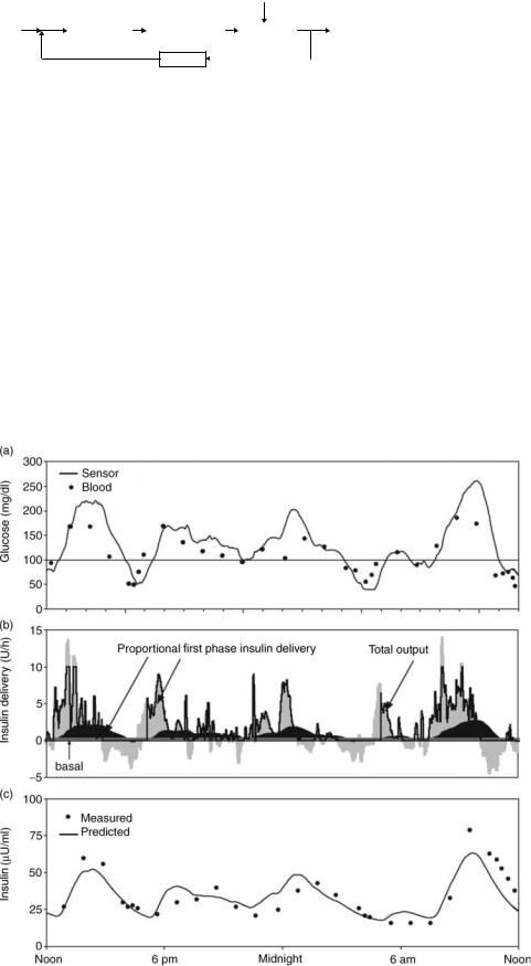
Figure 15. Blood glucose control in humans by an implanted artificial beta cell. A chronic, central venous blood glucose sensor and implanted insulin pump (Medtronic/MiniMed) implanted in a human subject. (a) Plasma (solid circles) and sensor (line) glucose following initiation of closed-loop control (noon). Solid line at 100 mg dL 1 indicates setpoint.
(b) Proportional (medium shading), basal (light shading), and derivative (dark shading) insulin delivery during the closed-loop (solid line indicates total, which is not allowed to go below zero). (c) Plasma (circles) and predicted insulin (solid line) concentrations. (Study performed by Medical Research Group. Copyright 2004, Elsevier.)
404 GLUCOSE SENSORS
ACKNOWLEDGMENTS
This work was supported by grants from the National Institutes of Health and the Technology for Metabolic Monitoring Initiative of the Department of Defense. D.G. holds equity interest in GlySens, Inc., a company dedicated to the development of a new glucose sensor, and is a scientific advisor. This arrangement that has been approved by the University of California, San Diego in accordance with its conflict of interest policies.
BIBLIOGRAPHY
Cited References
1.Cahill Jr GF, Etzwiler LD, Freinkel N. Editorial: ‘‘Control’’ and diabetes. N Engl J Med 1976;294(18): 1004–1005.
2.The effect of intensive treatment of diabetes on the development and progression of long–term complications in insulin-dependent diabetes mellitus. The Diabetes Control and Complications Trial Research Group. N Engl J Med 1993;329(14): 977– 986.
3.Stratton IM, et al. Association of glycaemia with macrovascular and microvascular complications of type 2 diabetes (UKPDS 35): prospective observational study. Bmj 2000; 321(7258):405–412.
4.Nathan DM, et al. The clinical information value of the glycosylated hemoglobin assay. N Engl J Med 1984;310(6): 341–346.
5.Skyler JS, Oddo C. Diabetes trends in the USA. Diabetes Metab Res Rev 2002;18(3 Suppl):S21–S26.
6.Zimmet P, Alberti KG, Shaw J. Global and societal implications of the diabetes epidemic. Nature (London) 2001;414 (6865):782–787.
7.Egger M, et al. Risk of adverse effects of intensified treatment in insulin-dependent diabetes mellitus: a meta-analysis. Diabet Med 1997;14(11):919–928.
8.Rovet JF, Ehrlich RM. The effect of hypoglycemic seizures on cognitive function in children with diabetes: a 7-year prospective study. J Pediatr 1999;134(4):503–506.
9.Sovik O, Thordarson H. Dead-in-bed syndrome in young diabetic patients. Diabetes Care 1999;22(2 Suppl):B40– B42.
10.Santiago JV. Lessons from the Diabetes Control and Complications Trial. Diabetes 1993;42(11):1549–1554.
11.van den Berghe G, et al. Intensive insulin therapy in the critically ill patients. N Engl J Med 2001;345(19):1359– 1367.
12.Van den Berghe G, et al. Outcome benefit of intensive insulin therapy in the critically ill: Insulin dose versus glycemic control. Crit Care Med 2003;31(2):359–366.
13.Bremer T, Gough DA. Is blood glucose predictable from previous values? A solicitation for data. Diabetes 1999;48 (3):445–451.
14.Clark Jr LC, Lyons C. Electrode systems for continuous monitoring in cardiovascular surgery. Ann NY Acad Sci 1962; 102:29–45.
15.Chang KW, et al. Validation and bioengineering aspects of an implantable glucose sensor. Trans Am Soc Artif Intern Org 1973;19:352–360.
16.Slama G, Bessman SP. [Results of in vitro and in vivo use of a prototype implantable glucose sensor]. J Annu Diabetol Hotel Dieu 1976; 297–302.
17.Albisser AM, et al. An artificial endocrine pancreas. Diabetes 1974;23(5):389–396.
18.Thomas LJ, Bessman SP. Prototype for an implantable insulin delivery pump. Proc West Pharmacol Soc 1975;18:393– 398.
19.Clemens AH, Chang PH, Myers RW. The development of Biostator, a Glucose Controlled Insulin Infusion System (GCIIS). Horm Metab Res 1977; (7 Suppl):23–33.
20.Skyler JS, et al. Home blood glucose monitoring as an aid in diabetes management. Diabetes Care 1978;1(3):150– 157.
21.Pickup JC, et al. Clinical application of pre-programmed insulin infusion: continuous subcutaneous insulin therapy with a portable infusion system. Horm Metab Res 1979; (8) (Suppl):202–204.
22.McCall AL, Mullin CJ. Home monitoring of diabetes mellitus–a quiet revolution. Clin Lab Med 1986;6(2):215– 239.
23.Armour JC, et al. Application of chronic intravascular blood glucose sensor in dogs. Diabetes 1990;39(12):1519–1526.
24.Sage Jr BH. FDA panel approves Cygnus’s noninvasive GlucoWatch. Diabetes Technol Ther 2000;2(1): 115–116.
25.Clinical Practice Recommendations 2005. Diabetes Care 2005;28(1 Suppl):S1–S79.
26.Lojo J, et al. Prevantive Care Practices Among Persons with Diabetes, United States, 1995 and 2001. Morbidity Mortality Weekly Rep, Center Disease Control Prevention 2002;51(43): 965–967.
27.Bennion N, Christensen NK, McGarraugh G. Alternate site glucose testing: a crossover design. Diabetes Technol Ther 2002;4(1):25–33; discussion 45–47.
28.Gough DA, Kreutz-Delgado K, Bremer TM. Frequency characterization of blood glucose dynamics. Ann Biomed Eng 2003;31(1):91–97.
29.McNichols RJ, Cote GL. Optical glucose sensing in biological fluids: an overview. J Biomed Opt 2000;5(1):5–16.
30.Cote GL. Noninvasive and minimally-invasive optical monitoring technologies. J Nutr 2001;131(5):1596S–604S.
31.Arnold MA, Burmeister JJ, Small GW. Phantom glucose calibration models from simulated noninvasive human near-infrared spectra. Anal Chem 1998;70(9):1773–1781.
32.Goetz Jr MJ, et al. Application of a multivariate technique to Raman spectra for quantification of body chemicals. IEEE Trans Biomed Eng 1995;42(7):728–731.
33.Steffes PG. Laser-based measurement of glucose in the ocular aqueous humor: an efficacious portal for determination of serum glucose levels. Diabetes Technol Ther 1999;1(2):129– 133.
34.Cameron BD, Baba JS, Cote GL. Measurement of the glucose transport time delay between the blood and aqueous humor of the eye for the eventual development of a noninvasive glucose sensor. Diabetes Technol Ther 2001;3(2):201– 207.
35.Blass DA, Adams E. Polarimetry as a general method for enzyme assays. Anal Biochem 1976;71(2):405–414.
36.Baba JS, et al. Effect of temperature, pH, and corneal birefringence on polarimetric glucose monitoring in the eye. J Biomed Opt 2002;7(3):321–328.
37.Schultz JS, Mansouri S, Goldstein IJ. Affinity sensor: a new technique for developing implantable sensors for glucose and other metabolites. Diabetes Care 1982;5(3): 245–253.
38.March WF, Ochsner K, Horna J. Intraocular lens glucose sensor. Diabetes Technol Ther 2000;2(1):27–30.
39.Muller M. Science, medicine, and the future: Microdialysis. Bmj 2002;324(7337):588–591.
40.Meyerhoff C, et al. On line continuous monitoring of subcutaneous tissue glucose in men by combining portable glucosensor with microdialysis. Diabetologia 1992;35(11): 1087–1092.
41.Hoss U, et al. A novel method for continuous online glucose monitoring in humans: the comparative microdialysis technique. Diabetes Technol Ther 2001;3(2):237–243.
42.Tamada JA, Bohannon NJ, Potts RO. Measurement of glucose in diabetic subjects using noninvasive transdermal extraction. Nat Med 1995;1(11):1198–1201.
43.Potts RO, Tamada JA, Tierney MJ. Glucose monitoring by reverse iontophoresis. Diabetes Metab Res Rev 2002;18 (1 Suppl):S49–S53.
44.Lenzen H, et al. A non-invasive frequent home blood glucose monitor. Practical Diabetes Int 2002;19(4):101– 103.
45.Tierney MJ, et al. The GlucoWatch biographer: a frequent automatic and noninvasive glucose monitor. Ann Med 2000; 32(9):632–641.
46.Bindra DS, et al. Design and In vitro studies of a needle-type glucose sensor for subcutaneous monitoring. Anal Chem 1991;63(17):1692–1696.
47.Jablecki M, Gough DA. Simulations of the frequency response of implantable glucose sensors. Anal Chem 2000;72(8):1853–1859.
48.Ward WK, et al. A new amperometric glucose microsensor: in vitro and short-term In vivo evaluation. Biosens Bioelectron 2002;17(3):181–189.
49.Xie SL, Wilkins E. Rechargeable glucose electrodes for long-term implantation. J Biomed Eng 1991;13(5):375– 378.
50.Clark Jr LC. Membrane Polarographic Electrode System and Method with Electrochemical Compensation. US pat 3,539,455. 1970.
51.Gough DA, Lucisano JY, Tse PH. Two-dimensional enzyme electrode sensor for glucose. Anal Chem 1985;57(12):2351– 2357.
52.Mastrototaro JJ. The MiniMed continuous glucose monitoring system. Diabetes Technol Ther 2000;2(1 Suppl): S13–S18.
53.Rebrin K, et al. Subcutaneous glucose predicts plasma glucose independent of insulin: implications for continuous monitoring. Am J Physiol 1999;277(3 Pt. 1): E561– E571.
54.Gross TM, Mastrototaro JJ. Efficacy and reliability of the continuous glucose monitoring system. Diabetes Technol Ther 2000;2(1 Suppl):S19–S26.
55.Metzger M, et al. Reproducibility of glucose measurements using the glucose sensor. Diabetes Care 2002;25(7):1185– 1191.
56.Gough DA, Armour JC. Development of the implantable glucose sensor. What are the prospects and why is it taking so long? Diabetes 1995;44(9):1005–1009.
57.Shichiri M, et al. Telemetry glucose monitoring device with needle-type glucose sensor: a useful tool for blood glucose monitoring in diabetic individuals. Diabetes Care 1986;9 (3):298–301.
58.Abel P, Muller A, Fischer U. Experience with an implantable glucose sensor as a prerequisite of an artificial beta cell. Biomed Biochim Acta 1984;43(5):577–584.
59.Moatti-Sirat D, et al. Towards continuous glucose monitoring: In vivo evaluation of a miniaturized glucose sensor implanted for several days in rat subcutaneous tissue. Diabetologia 1992;35(3):224–230.
60.Johnson KW, et al. In vivo evaluation of an electroenzymatic glucose sensor implanted in subcutaneous tissue. Biosens Bioelectron 1992;7(10):709–714.
GLUCOSE SENSORS |
405 |
61.Kerner W, et al. The function of a hydrogen peroxide-detect- ing electroenzymatic glucose electrode is markedly impaired in human sub-cutaneous tissue and plasma. Biosens Bioelectron 1993;8(9–10):473–482.
62.Bode BW, et al. Continuous glucose monitoring used to adjust diabetes therapy improves glycosylated hemoglobin: a pilot study. Diabetes Res Clin Pract 1999;46(3):183–190.
63.Gross TM, et al. Performance evaluation of the MiniMed continuous glucose monitoring system during patient home use. Diabetes Technol Ther 2000;2(1): p. 49–56.
64.Gough DA, Botvinick EL. Reservations on the use of error grid analysis for the validation of blood glucose assays. Diabetes Care 1997;20(6): p. 1034–1036.
65.Kerssen A, de Valk HW, Visser GH. The Continuous Glucose Monitoring System during pregnancy of women with type 1 diabetes mellitus: accuracy assessment. Diabetes Technol Ther 2004;6(5): p. 645–51.
66.Mauras N, et al. Lack of accuracy of continuous glucose sensors in healthy, nondiabetic children: results of the Diabetes Research in Children Network (DirecNet) accuracy study. J Pediatr 2004;144(6):770–775.
67.Gilligan BJ, et al. Evaluation of a subcutaneous glucose sensor out to 3 months in a dog model. Diabetes Care 1994;17(8): 882–887.
68.Updike SJ, et al. A subcutaneous glucose sensor with improved longevity, dynamic range, and stability of calibration. Diabetes Care 2000;23(2):208–214.
69.Garg SK, Schwartz S, Edelman SV. Improved glucose excursions using an implantable real-time continuous glucose sensor in adults with type 1 diabetes. Diabetes Care 2004; 27(3):734–738.
70.Csoregi E, Schmidtke DW, Heller A. Design and optimization of a selective subcutaneously implantable glucose electrode based on ‘‘wired’’ glucose oxidase. Anal Chem 1995;67(7): 1240–1244.
71.Heller A. Implanted electrochemical glucose sensors for the management of diabetes. Annu Rev Biomed Eng 1999;1:153– 175.
72.Tse PHS, Leypoldt JK, Gough DA. ‘‘Determination of the Intrinsic Kinetic Constants of Immobilized Glucose Oxidase and Catalase’’. Biotechnol Bioeng 1987;29:696–704.
73.McKean BD, Gough DA. A telemetry-instrumentation system for chronically implanted glucose and oxygen sensors. IEEE Trans Biomed Eng 1988;35(7):526–532.
74.Medtronic Minimed talk at the Diabetes Technology and Therapeutics Conference, S.F., CA. 2003.
75.Gough DA, Bremer T. Immobilized glucose oxidase in implantable glucose sensor technology. Diabetes Technol Ther 2000; 2(3):377–380.
76.Lucisano JY, Armour JC, Gough DA. In vitro stability of an oxygen sensor. Anal Chem 1987;59(5):736–739.
77.Makale MT, et al. Tissue window chamber system for validation of implanted oxygen sensors. Am J Physiol Heart Circ Physiol 2003;284(6):H2288–H2294.
78.Makale MT, Jablecki MC, Gough DA. Mass transfer and gasphase calibration of implanted oxygen sensors. Anal Chem 2004;76(6):1773–1777.
79.Makale MT, Chen PC, Gough DA. Variants of the tissue/ sensor array chamber. Am J Physiol Heart Circ Physiol 2005;286, in press.
80.Bremer TM, Edelman SV, Gough DA. Benchmark data from the literature for evaluation of new glucose sensing technologies. Diabetes Technol Ther 2001;3(3):409–418.
81.Steil GM, Panteleon AE, Rebrin K. Closed-loop insulin delivery-the path to physiological glucose control. Adv Drug Deliv Rev 2004;56(2):125–144.
406GLUCOSE SENSORS
82.Saudek CD. Implantable Pumps. 3rd ed. International Textbook of Diabetes Mellitus; 2004.
83.Selam JL. External and implantable insulin pumps: current place in the treatment of diabetes. Exp Clin Endocrinol Diabetes 2001;109(2 Suppl):S333–S340.
84.Vague P, et al. The implantable insulin pump in the treatment of diabetes. Hopes and reality?. Bull Acad Natl Med 1996; 180(4):831–41. discussion 841–843.
See also FIBER OPTICS IN MEDICINE; OPTICAL SENSORS; OXYGEN SENSORS;
PANCREAS, ARTIFICIAL.