Добавил:
Опубликованный материал нарушает ваши авторские права? Сообщите нам.
Вуз: Предмет: Файл:
Wiley - Encyclopedia of Medical Devices and Instrumentation - Vol. 3.pdf
Скачиваний:
296
Добавлен:
10.08.2013
Размер:
16.56 Mб
Скачать

47.Sleisenger MH, Fordtran JS, editors. Gastrointestinal Disease. Philadelphia: Saunders; 1983. p 1599–1616.

48.Sleisenger MH, Fordtran JS, editors. Gastrointestinal Disease. Philadelphia: Saunders; 1983. p 1617–1626.

49.Silvis SE, editor. Therapeutic Gastrointestinal Endoscopy. New York: Igaku-Shoin; 1985. p 241–268.

50.Huizinga E. On esophagoscopy and sword swallowing. Ann Otol Rhinol Laryngol 1969;78:32–34.

51.Shaheen N, Ransohoff DF. Gastroesophageal reflux, Barrett esophagus, and esophageal cancer: scientific review. JAMA 2002;287:1972–1981.

52.Spechler SJ. Clinical practice. Barrett’s Esophagus. N Engl J Med 2002;346:836–842.

53.Sampliner RE. Updated guidelines for the diagnosis, surveillance, and therapy of Barrett’s esophagus. Am J Gastroenterol 2002;97:1888–1895.

54.Wolfsen HC, Hemminger LL, DeVault KR. Recurrent Barrett’s esophagus and adenocarcinoma after esophagectomy. BMC Gastroenterology 2004;4:18.

55.Jean M, Dua K. Barrett’s Esophagus: Best of Digestive Disease Week 2003. Curr Gastroenterol Rep 2004;6:202– 205.

56.Hunt RH, Waye JD, editors. Colonoscopy. London: Chapman & Hall; 1981. p 11–18.

57.Ohligisser M, Sorokin Y, Hiefetz M. Gynecologic laparoscopy, a review article. Obstet Gynecol Surv 1985;40:385–396.

58.Robinson HB, Smith GW. Application for laparoscopy in general surgery. Surg Gynecol Obstet 1976;143:829–834.

59.Devaux BC, Joly L, Page P, Nataf F, Turak B, Beuvon F, Trystram D, Roux F. Laser-assisted endoscopic third ventriculostomy for obstructive hydrocephalus: Technique and results in a series of 40 consecutive cases. Lasers Surg Med 2004;34:368–378.

60.Cram GP, Copeland ML. Nucl Instrum Methods Phys Rev B 1998;144:256.

61.Edwards G et al. Free-electron-laser-based biophysical and biomedical instrumentation. Rev Sci Instrum 2003;74:3207– 3245.

62.Paparella MM, Shumrick DA, editors. Otolaryngology Philadelphia: W.B. Saunders; 1980. p 2410–2430.

63.Ballenger JJ, editor. Diseases of the Nose, Throat, Ear, Head, and Neck. Philadelphia: Lea & Febiger; 1985. p 1293–1330.

64.Steiner W. Techniques of diagnostic and operative endoscopy of the head and neck. Endoscopy 1979;1:51–59.

65.Vaughan CW. Use of the carbon dioxide laser in the endoscopic management of organic laryngeal disease. Otolaryngol Clin North Am 1983;16:849–864.

66.Pfalz R, Hibst R, Bald N. Suitability of different lasers for operations ranging from the tympanic membrane to the base of the stapes. Adv in Oto-Rhino-Laryngol 1995;49:87–94.

67.Schade G, Leuwer R, Kraas M, Rassow B, Hess M. Laryngeal morphometry with a new laser ‘clip on’ device. Lasers Surg Med 2004;34:363–367.

68.Future Trends in Medical Device Technology: Results of an Expert Survey. Available at http://www.fda.gov/cdrh/ost/ trends/TOC.html.

Further Reading

Ponsky JL. Atlas of Surgical Endoscopy. St. Louis (MO): MosbyYear Book; 1992.

Barkin J, O’Phelan C, editors. Advanced Therapeutic Endoscopy. New York: Raven Press; 1990.

Ovassapian A. Fiberoptic Airway Endoscopy in Anesthesia and Critical Care. New York: Raven Press; 1990.

Niemz M. Laser-Tissue Interactions: Fundamentals and Applications. 2nd ed. Berlin Heidelberg: Springer-Verlag; 2002.

ENGINEERED TISSUE

189

Infrared Fiber Systems. Available at http://www.infraredfibersy stems.com.

Polymicro Technologies, LLC. Available at http://www.polymicro. com.

Omniguide Communications, Inc. Available at http://www.omniguide.com.

LightLab Imaging, Inc. Available at http://www.lightlabimaging. com.

Given Imaging. Available at www.givenimaging.com.

Intuitive Surgical, Inc. Available at www.intuitivesurgical.com.

See also ESOPHAGEAL MANOMETRY; FIBER OPTICS IN MEDICINE; MINIMALLY INVASIVE SURGERY.

ENGINEERED TISSUE

GREGORY E. RUTKOWSKI

University of Minnesota-Duluth

Duluth, Minnesota

INTRODUCTION

History of Tissue Engineering

In 1988, researchers gathered at the Granlibakken Resort in Lake Tahoe, CA under the sponsor ship of the National Science Foundation (NSF) to develop the fundamental principles of tissue engineering as an emerging technology. Based on an earlier proposal by Dr. Y.C. Fung to develop an Engineering Research Center focused on the engineering of living tissues, NSF held several meetings that led to the decision to designate tissue engineering as a new emerging field. A formal definition was finally agreed upon at the Granlibakken workshop. Based on this meeting, tissue engineering is defined as ‘‘the application of the principles and methods of engineering and the life sciences toward the fundamental understanding of structure–function relationships in normal and pathological mammalian tissues and the development of biological substitutes to restore, maintain, and improve function’’ (1). This was further refined in 1992 by Eugene Bell who developed a list of more specific goals:

1. Providing cellular prostheses or replacement parts for the human body.

2. Providing formed acellular replacement parts capable of inducing regeneration.

3. Providing tissue or organ-like model systems populated with cells for basic research and for many applied uses such as the study of disease states using aberrant cells.

4. Providing vehicles for delivering engineered cells to the organism.

5. Surfacing nonbiological devices (2).

These discussions eventually culminated in the pioneering review article by Langer and Vacanti in 1993 (3). The general strategies to create engineered tissue would include the isolation of cells or cell substitutes, the use of tissue-inducing substances, and development of threedimensional (3D) matrices on which to grow tissue.

190 ENGINEERED TISSUE

Much of tissue engineering owes its beginnings to reconstructive surgery and internal medicine. In the sixteenth century, Gaspare Tagliacozzi developed a method for nasal reconstruction using flaps of skin taken from the arm and grafted onto the injury site (4). With the scientific development of the germ theory of disease and the sterile techniques that were introduced, modern surgery became established as a means to treat patients with internal injuries. In the late nineteenth century, surgeons used veins and arteries as conduits to enhance the nerve regeneration (5). World War I saw improvements in reconstructive surgery as doctors were able to hone their skills due to the number of soldiers injured in battle. Reconstructive surgery had it limitations in terms of the availability of biological material. By the 1940s, much progress had been made in understanding the function of the immune system is accepting tissue from a donor. This eventually led to the first successful organ transplant (kidney) in 1954. The next 50 years, would see tremendous advances in organ transplants as well as in immunosuppressive drug therapy.

An alternative to organ transplant has been the development of artificial devices to mimic biological function. The mid-nineteenth century also saw the rise in the use of prosthetic limbs that would initiate the use of artificial devices to replace biological functions. Artificial limbs were used as far back as the Dark Ages to assist knights heading off to battle. The intervening centuries saw improvements over such devices through the use of stronger, lighter materials, and a better understanding of biomechanics

(6). Besides limbs, artificial devices have also been invented to replace the function of certain internal organs. The dialysis machine was created in the 1940s to assist patients with acute renal failure. In 2001, an implantable artificial heart was first used in a human. While many advances have been made in artificial devices, some of the drawbacks include the breakdown of the artificial materials, a lack of interaction with the human body, and the inability to self-renew.

The modern era of tissue engineers seeks to overcome the limitations of reconstructive surgery, organ transplantations, and prosthetic devices by creating functional, completely biocompatible tissues and organs. Since the late 1980s, the field of tissue engineering has grown exponentially and continues to draw scientists from diverse fields, from the biological and medical sciences to engineering and materials science.

THEORY

Tissue engineering adds to the modern health care system by providing the tools to assist in the repair of tissue and organs damaged by injury and disease. An exact replica of the tissue could potentially be grown in the lab and later inserted into the patient. Alternatively, precursors may be placed in the body with the expectation that it will develop into fully formed functional tissue. Also, devices may be implanted into the body to encourage the regeneration of already existing tissue in the body.

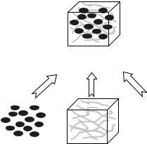
Engineered tissue is created by combining relevant cells and chemical factors within a 3D matrix that serves as a

. .. .. ......... .

.. . . . .. . .. .

Engineered

Tissue

Cells

Scaffold

. .. .. ......... .

.. . . . .. . .. .

Chemical

Factors

Figure 1. Basic components of engineered tissue.

scaffold (Fig. 1). Sterile cell culture techniques allows for the expansion of cells in vitro to obtain sufficient quantities for use in engineered tissue. Cells can originate from the patient, another donor, or even animal tissue. As our understanding of biochemical cues during development expands, tissue formation can be better controlled through the delivery of pertinent growth factors or through the interaction of complex multiple cell cultures. Scaffold materials may be biological (collagen, extra cellular matrix) or synthetic. Synthetic materials can be designed to mimic the extra cellular matrix and may be infused with chemical factors to support tissue regeneration. Since the discovery and availability of synthetic polymers in the 1940s, scientific advances have made these materials biodegradable and biocompatible.

Cellular Processes

During the formation of tissue, either during development or in the lab, cells undertake many different processes. In order to maintain the integrity of the tissue, the cells tissue must adhere to others as well as to the surrounding matrix material. Certain cells must migrate through the 3D space in order to properly position themselves within the tissue. Once in position, they must also continue to multiply to provide adequate tissue growth. Vascularization and innervation of the tissue is required to complete integration of the engineered tissue with its surroundings. Tissue derived from donor material also has immune concerns.

Development. Engineered tissue is best created by mimicking the processes that occur during development

(7). Following fertilization, the zygote divides until the blastocyst is formed. The early embryonic tissue is comprised of two cell phenotypes: epithelial cells and migratory mesanchymal cells. The transformation between these cells is regulated by growth factors, the extra cellular matrix, and intracellular signaling. As the embryo develops, these cells will become diversified as they commit to various tissue forms. These cells will eventually differentiate into the various cell types found in the body.

Morphogenesis describes the cascade of events that leads to the spatial pattern formation of the developing

embryo. Regeneration of tissue mimics this process. Specific chemical factors, called morphogens, provide information on the pattern via diffusion. The combination of various morphogens due to diffusion leads to complex pattern formation. One well-studied example is the development of bone. Several bone morphogenic factors have been isolated and observed to affect the bone and cartilage morphogenesis and formation. Also, BMPs have been implicated in the development of tissue as diverse as skin, eye, heart, and kidney. These morphogens have the potential to assist in the engineering of bone tissue.

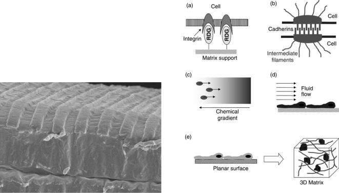
Adhesion. Tissue is held together by the adhesion processes between different cells and the cells and extracellular matrix. The cell–cell interactions consist of tight, anchoring, and communication junctions. Tight junctions are composed of transmembrane proteins that form connections between cells. More contact points will decrease the permeability between the cells so that the tissue can become essentially impermeable. This is typically found with epithelial cells, as in the intestinal, reproductive, and respiratory tract, where the barrier that is formed is essential to function.

Anchoring junctions are loosely held connections that take advantage of the cytoskeleton. With adherens junctions, actin filaments of the cytoskeleton are connected and linked to integrins on the cell exterior. These integrins form focal contacts that interact with cadherins found on the surface of other cells. The focal contacts can also interact with extracellular domains. With the involvement of the cytoskeleton, these junctions can also affect cell function by providing a mechanism to signal changes in cell growth, survival, morphology, migration, and differentiation.

Desmosomes are similar to adherens junctions, but they involve intermediate filament protein, such as vimentin, desmin, and keratin. These junctions connect with other cells via cadherins. A similar junction called the hemidesmosome behaves in the same manner, but connects with basal lamina proteins via integrin.

Communication junctions are those that provide direct communication between cells. These are large proteins that form pore structure that connects the two cells. These large pores (connexons) allow the transport of molecules between cells. These junctions are commonly found between neurons for rapid signal conduction.

In order to connect with other cells or the extracellular matrix, the junction proteins must interact with some receptor molecule. Integrins will bind with the amino acid sequence arginine-glycine-aspartic acid (RGD). This sequence is found in several proteins, such as collagen and fibronectin and these peptides can be incorporated onto other surfaces in order to improve cell adhesion. Cadherin– cadherin binding is mediated via Ca2þ. Without the presence of Ca2þ, the connection is subject to proteolysis. The Ig-like receptors contain motifs found in immunoglobulins. These receptors can interact with neural cell adhesion molecule (N-CAM) and are present during development. Finally, selectins are specific receptor types that are expressed during the inflammatory response. The lectin domain found in oligosaccharides on neutrophils allows

ENGINEERED TISSUE

191

these cells to interact with endothelial cells along the surface of blood vessels.

Migration. During embryogenesis, diffusible factors and ECM composition are important factors in the pattern formation of tissue development. The migration of cells is necessary for the formation of tissue not just for development, but also during regeneration. Cell migration is also observed in other body functions. Angiogenesis, or blood vessel formation, involves the migration of endothelial cells into new tissues. For immune responses, B and T cells patrol the body ready to attack invaders. Tumor invasion and, more importantly, metastasis relies on the migration of cancer cells into other parts of the body. Controlling migration can help to control the spread of cancer.

Signals from various factors can lead to the release of cells from their contact with other cells or with the extracellular matrix. Once released, different mechanisms can affect the migration pattern of the cell depending on the cell type. Cells may move in random directions. The movement can be modeled by Brownian motion. In this case, motion is due to collisions with other particles. This motion is characterized by the time between collisions, the mean free path (average distance before hitting another particle) and the average speed. The characteristics are dependent on the density of the particles.

Of more relevance is the issue of directed migration. Direct migration depends on some sort of gradient. In response to some kind of stimulus, cells may move toward or away from the source of the stimulus. The stimulus may affect speed, direction, or both. Chemotaxis is the general term describing the response of cells to a chemical gradient. The strength of the effect is dependent on the absolute concentration as well as the steepness of the gradient.

Growth. Mitosis is a tightly controlled process to regulate cell growth and depends on signals from the cell’s environment. During mitosis, the deoxyribonucleic acid (DNA) is replicated and copies are separated as the cells divide into two exact copies. This process repeats itself depending on the extracellular signaling and the properties of the cell itself. Certain cells, such as pancreatic beta cells, are locked in the cell cycle and mitosis is arrested. In order to overcome this barrier, stem cells can be used to generate new differentiated cells. Also, cells may be converted into a precursor cell form that can undergo proliferation before switching back to the differentiated form.

Cell growth relies on the availability of essential nutrients. As the need for nutrients increases during cells proliferation, the availability becomes reduced barring some mechanism for distributing the nutrients to the growing tissue. Distribution of nutrients occurs naturally in the human body in the form of the network of blood vessels. The lack of such a vasculature for engineered tissue limits the effective size that tissue can grow.

Vascularization. For most engineered tissue to become integrated into the human body, it must become vascularized by the body’s own blood vessel network. The body provides a natural mechanism for neovascularization by the process of wound healing. When tissue has been

192 ENGINEERED TISSUE

damaged, a cascade of events is initiated to block the bleeding wound and encourage healing. Platelets, fibrin, and fibronectin first form a mesh plug. Mast cells release chemotactic agents to recruit other cells as part of the inflammatory response. Keratinocytes migrate to the site of the injury and begin to proliferate. Vascular endothelial growth factor (VEGF) and fibroblast growth factor (FGF) are released to encourage blood vessel formation. Once the new tissue has been vascularized, remodeling of the tissue occurs to complete the healing process.

For engineered tissue, two options are used to ensure vascularization and promote tissue growth and integration. The engineered tissue can be designed to contain a vascular network in vitro that would then be connected to the body’s own network when it is implanted. This presents a significant engineering challenge in trying to create several different types of engineered tissue in one construct simultaneously. An alternative method is to engineer the tissue to recruit blood vessels from the body’s existing framework. This has been accomplished though the controlled release of VEGF from the biodegradable support matrix of the engineered tissue (8).

Innervation. To become fully integrated into the body, certain tissues and organs must also reconnect with the body’s own nervous system. Several organs of the body form connections with the sympathetic nervous system. These connections are important to regulation of the organ. Skeletal muscle tissue makes sensory and motor connections via the peripheral nervous system. In any case, the cells of these engineered tissues need to make synaptic connection with the axons of the relevant neurons. Because the new tissue is being engineered to replace that which has been lost to injury or disease, the neural framework may not be available for integration. If it is present, the neurons may be encouraged to regenerate toward the tissue and form new connections. Another complex, but theoretically possible, option may be to engineer the tissue with neural connections that can be later integrated into the existing nervous system. Also, artificial devices may be integrated into the system to provide the appropriate control of the tissue function.

Immune Concerns. As with organ transplants, engineered tissue also has concerns of rejection by the immune system. The major histocompatibility complex I (MHC I) present on the cells of the engineered tissue are recognized by the T cells of the immune system as a foreign body. This would eventually lead to cell lysis. The primary means of preventing rejection, though, is the use of immune suppressant drug therapy. While this may be acceptable, for a patient receiving a life saving organ transplant, it is not for those receiving engineered tissue. Certain tissues, such as cartilage, may not interact with the immune system. Metabolic tissues that only interact chemically can be physically separated from the environment. Some engineered tissues may contain cells that are only temporary until the body’s own cells can take over. In these cases, immune suppressant drug may be a viable option. For most other tissues, methods are being developed for side stepping the immune system.

Because the cells of the immune system are formed within the bone marrow, one method is to transplant the bone marrow from the cell and tissue donor along with the organ. The donated bone marrow can form a chimera with the patient’s existing bone marrow to allow the adaptation of the immune system to the new tissue (9).

At the molecular level, the antigen of the foreign cell can be blocked or completely eliminated. In order to block the antigen, a fragment of antibody to the antigen can be added to the tissue to mask the foreign cells from the patient’s own immune system (10). This effect is temporary as the fragments will eventually separate from the antigen. Another drawback is that is may not counter all the mechanisms of immune response. The antigen can also be removed by placing an inactive form of the antigen gene into the cells (11). A gene can also be added to the cells to produce a protein that will inhibit rejection (12). Finally, oligonucleotides can be added to hybridize with either ribonucleic acid (RNA) or DNA in order to inhibit the transcription or translation of the antigen molecule (13).

Microencapsulation is a means of physically separating the cells from the environment. For this method, cells are surrounded by a porous synthetic material. As long as the membrane coating remains intact, the cells are isolated from the immune system. The pore size can be adjusted to allow chemical interaction with the environment while preventing cellular interaction. The encapsulation should allow nutrients to permeate through the membrane and reach the cells. This was first used in clinical studies for the encapsulation of xenogenic pancreatic islets (14).

Cell Types. While the response of the immune system is of great importance to the success of the engineered implant, the source of cells and their application will determine the best method for immunomodulation. Cells may come from the patients themselves (autologous), from a human donor (allogeneic), or from another species (xenogeneic). Each type has its own advantages and disadvantages.

Autologous cells are derived from the patient, expanded in culture, and then placed back into the patient. These cells are completely biocompatible with the patient and this eliminates the need for any immune modulation. Genzyme, for example, has developed a successful protocol for the repairing articular cartilage. One drawback to using autologous cells is that it requires a period of time to expand the cells. This would not be acceptable for patients needing an immediate tissue replacement. As a result, the engineered tissue does not have off-the-shelf availability. Depending on the tissue and the amount of damage, the amount of cells that may be harvested from the patient may be insufficient to form tissue.

Allogeneic cells can help to overcome some of the drawbacks to autologous cells because the cells come from donor sources that may be pooled together. This can provide off-the-shelf availability, but at the expense of immune rejection. The cells can be engineered to become immune acceptable or immunosuppressive drug therapy may be used. These cells are also well suited for tissues that do not interact with the patient’s vasculature system or may only be used until the native tissue regenerate.

Advanced Tissue Science developed a skin replacement tissue (Dermagraft) with cells derived from circumcision surgeries.

Xenogeneic cells are derived from nonhuman species. An animal source can provide an unlimited amount of cells, but they provoke an acute immune response within minutes of implantation into the body. One useful application of such cells is the encapsulation of pancreatic islets (14,15). The membrane can be adjusted to allow the islets to regulate blood sugar and insulin levels while protecting the cells from the immune system. Another drawback to xenogeneic cells is the threat of transmission of animal viruses and well as endogenous retroviruses that may interact with the patient.

Cells that have been isolated may be modified to alter their characteristics before being incorporated into engineered tissue. Primary cells that are subcultured eventually become cell lines. These cell lines may be finite or continuous. The cells may also be normal or transformed compared to the primary cell from which it is derived. Transformation is associated with genetic instabilities that may lead to a change in the phenotype. These changes may impart different growth characteristics for the cell, such as immortalization, anchorage independence, and overall growth rate. The genetic material may be altered to effect gene expression. Changes in protein expression may effect the secretion of chemical factors or even the formation of extracellular matrix. In the worst case, cells may become cancerous and prove to be detrimental to the patient. Examples of stable, well-characterized cell lines used in tissue engineering include HEK-293 for nerve growth factor secretion (16), and 3T3-L1 for adipose tissue formation (17).

Primary cells as well as cell lines can be artificially transformed by introducing new genes by the process of transfection. In this process, a new gene is carried by some vector, such as a liposome or virus, to the host cells. The gene is transferred into the cells and delivered into the nucleus where it can be transcribed. This method allows the creation of cells with desirable characteristics for engineered tissue. Such cells may secrete growth factor that will enhance tissue formation. Examples of engineered tissue utilizing transfected cells include insertion of hBMP-4 gene in bone marrow stromal cells for bone tissue engineering (18) and aquaporin gene transfection in the LLC-PK1 cell line for a bioartificial renal device (19), secretion of neuronal growth factors from fibroblasts to enhance axonal regeneration (20).

Regardless of the source, primary cells have limitations depending on their age and genetic makeup. Adult cells may only have a limited number of cell divisions before they reach senescence or even become cancerous. Transformed cell lines may allow for continuous growth, but without proper control can become a problem for the patient. These cells are typically used to enhance regeneration of existing tissue, but do not become integrated into the body. Because of these issues, a universal cell that can develop into multiple tissue types has an infinite capacity to proliferate without loss of function, and if immune acceptable would be ideal for engineered tissue. Stem cells come closest to meeting these criteria.

ENGINEERED TISSUE

193

Fertilized

Egg

Totipotent

Blastocyst


Amniotic

Inner Cell


Embryonic


Sac


Mass


Stem Cells


All Cell


Types


Fetus


Embryonic


Germ Cells


Pluripotent


Hematopoietic

Blood


Adult

Stem Cells

Cells

Spermatocyte


Neural

Neurons


Stem Cells

Stem Cell


Astrocytes


Oligodendrocytes


Mesenchymal


Sperm


Stem Cells

Bone


Cartilage

Unipotent


Muscle


Multipotent

Tendon


Fat


Figure 2. Stem cell potency. Relationship of stem cells to various stages of development. Cells and tissue derived from stem cells are shown in italics.

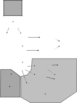
Stem Cells. The potency of stem cells is defined by their ability to differentiate into one or more cell genotypes (see Fig. 2). Unipotent stem cells give rise to only one cell type (ex. Spermatogonia). Multipotent stem cells are more functional and can differentiate into multiple cell types. Embryonic stem cells are considered pluripotent in that they can form all the cell types found in an embryo and adult. The fertilized egg develops into an embryo as well as the amniotic sac and is considered totipotent. Embryonic stem cells hold the most promise for engineering tissue, but more work must be done on the basic principles of stem and progenitor cell biology as well as on the control of differentiation.

Multipotent stem cells obtained from embryonic or adult sources are considered the optimal choice for tissue engineering applications because of their ability to form multiple tissue type. Hematopoietic stem cells (HSCs) are the most well characterized of stem cells. While isolated from bone marrow, HSCs can differentiate to form skeletal muscle, cardiac muscle, hepatocytes, endothelial cells, and epithelial cells. Multipotent stem cells can be found in several other tissues, such as brain, heart, pancreas, retina, liver, and lung, and skin.

To take full advantage of the functionality of stem cells, more work needs to be done to elucidate the mechanisms for differentiation. Once established, stem cells can be encouraged to differentiate into the required cell type.

194 ENGINEERED TISSUE

The mechanism may rely on an environmental cue as to whether they are physical contact, chemical, or chemotactic in nature. The ability of stem cell to proliferate, integrate, and differentiate also depends on the methods of identifying, isolating, and expanding. Protocols for stem cell culture need to be developed and optimized to ensure the cells achieve their full potential.

Since some stem cells are harder to obtain than others, getting stem cells to transdifferentiate from one form to another would allow for further flexibility. Evidence suggests that bone marrow stromal cells may convert to neural stem cells (21) and neural stem cells to hematopoietic stem cells (22). Recent evidence, though, points to the fusion of stem cells with other cells instead of true transdifferentiation (23).

Another source for stem cells is from embryos discarded from in vitro fertilization clinics. Human embryonic stem cells have an even greater capacity to form tissues than the multipotent stem cells. These cells have been isolated from humans and protocols for long-term cultures have successfully been developed (24). The use of embryonic stem cells has raised many ethical concerns because of the destruction of the fetus from which they are derived. This has led to legislation tightly regulating their use. To avoid these concerns, several proposals have been suggested to obtain embryonic stem cells without compromising the potential for life (25).

Stem cells have been used successfully for tissue engineering applications. Stem cells have been seeded on scaffolds to form cartilage, small intestine, and bone (26). Neural stem cells have also been used for repair of spinal cord injuries (27). Embryonic stem cells have also been seeded onto scaffolds where the native vasculature integrated into the engineered tissue (28).

Instead of preseeding cells on scaffolds, stems cells may also be injected directly to the site of injury to promote tissue regeneration. Clinical studies in nonhuman primates have shown that neural stem cells can enhance repair of spinal cord injuries (29). Stem cells have also been used to regenerate damaged cardiac muscle tissue (30). Embryonic stem cells may also be used to regenerate pancreatic beta cells in order to alleviate diabetes (31). Mesanchymal stem cells also show some promise for the repair of articular cartilage for those suffering from osteoarthritis (32).

Scaffolds

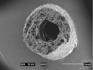
Primary cells will form monolayer cultures when dissociated from tissue. In order to encourage the cells to form engineered tissue, they must be placed in a 3D matrix that acts as a support scaffold. Ideally, these scaffolds should be compatible with the cells as well as being biodegrade. The scaffold should have a high porosity to ensure the diffusion of nutrients and other chemical factors as well as provide room for cell migration and proliferation. The material should have a higher surface area to ensure adequate room for cell attachment. The matrix should maintain its structural integrity until tissue integration has been completed. For certain applications, the final construct may need to be formed into a specific 3D shape (see Fig. 3).

Figure 3. Example of a scaffold for engineered tissue. This scaffold is designed for nerve regeneration. The cylindrical conduit was manufactured by using the solvent casting method (see below) with particle leaching. Poly(vinyl alcohol) rods were coated with a suspension of salt crystals in a poly(lactic acid) (PLA)–chloroform solution. Dissolution of the rods permitted the formation of the large central pore through which nerve tissue could regenerate. A porosity of 85% was obtained by controlling the volume fraction of salt crystals.

For tissue engineering, scaffolds may come in different forms. An acellular matrix can be used to recruit cells from the host tissue (33). With this form, the immune response is not a concern though inflammation may occur. For most applications, cells must be seeded onto the scaffold to ensure successful tissue formation. Cells can be seeded onto collagen gels that mimic the naturally occurring extracellular matrix (34,35). Cells can also be encouraged to self-assemble in culture. These systems are ideal for tissue that forms simple shapes, such as sheets and cylinders (36,37). Biodegradable polymers provide a flexible means of creating scaffolds with properties desirable for tissue formation.

Natural Polymers. Macromolecules found with the extracellular matrix provide chemical and structural support for living tissue. Because of their function in tissue formation, these natural polymers provide an excellent scaffold for engineering new tissue. These natural polymers have some limitations in their application. The mechanical properties and degradation rates cannot be controlled as well as synthetic polymers (38). Also, material derived from donors may elicit an immune response (39). Many of these limitations can be overcome by chemical modification of the materials as well as creating composite materials of natural polymers or natural–synthetic polymers.

Collagen is a large family of proteins that make up much of the extracellular matrix. At least 19 different types have been isolated. Fibrillar collagen is comprised of several collagen types that combine to form extended structures that assist is maintaining tissue integrity. The relative amounts of collagen are characteristic of the tissue type. Collagen has been used for cartilage (40), nerve regeneration (41), gallbladder engineering (42), corneal tissue (43),

and skin (44). It can be cross-linked to enhance mechanical properties (45). Magnetically aligned collagen fibers can also be used to enhance nerve regeneration (46).

Matrigel is comprised of proteins found in the basal lamina. The basal lamina, or basement membrane, is a specialized area of the extracellular matrix found at epithelial–stromal boundaries. It is composed of collagen, laminin, nidogen, and perlecan, (a heparin sulfate proteoglycan). It has been used for spinal cord repair (47) vascular network formation (48), and cardiac tissue (49).

Alginates are synthesized naturally by brown seaweeds as well as some bacteria. They are comprised of units of mannuronic and guluronic acid. The proportion and repeat unit sequence is dependent on the organism of origin. Alginates will form a gel in the presence of calcium ions. Alginates have been used for cartilage (50), cardiac tissue (51), and liver engineering (52).

Fibrin is formed from the polymerization of fibrinogen that occurs during the wound healing process. The process is controlled by the presence of thrombin. Fibrin has been used for cardiovascular repair (53), bone (54), cartilage (55), and skin (56). Like collagen, the fibrin chains can be cross-linked to alter the characteristics of the matrix (57).

Chitosan is derived from chitin, a polysaccharide that comprises the exoskeleton of crustaceans. The amide group on chitin is replaced by an amine group. Since chitosan is not protein based, it does not elicit an immune response. Chitosan has been used for the engineering of cartilage (58) and bone (59). It has also been combined with other materials to create novel composite matrices (60,61).

Besides isolating single components as scaffolding material, intact decellularized tissue can also be used. In this method, tissue is obtained from a donor and the cells are destroyed chemically using compounds, such as sodium dodecyl sulfate. This methods is currently used to engineer blood vessels (62) and heart valves (63). Decellularized tissue may also be applied to the engineering of other types of tissues, such as utilizing porcine skin for urinary tract reconstruction (64).

Synthetic Polymers. While natural polymers are an excellent choice for scaffolds because of their biocompatibility, synthetic polymers offer more flexibility. Composites with natural polymers can also be made to take advantage of their properties as well. Biodegradable polymers also have the additional benefit of decomposing into nontoxic metabolites that can be removed from the body as waste. The molecules can be modified to obtain mechanical properties and appropriate degradation rates that are well suited to the specific application.

The most common polymers in use are PLA and poly- (glycolic acid) (PGA). These are the first two polymers to obtain approval by the Food and Drug Administration (FDA) for use in medical implants. During degradation, the polymer is hydrolyzed to form lactic and glycolic acids. Since PLA is more hydrophobic than PGA, it has a slower degradation rate. As such, the degradation rate of the polymers can be easily adjusted by changing the ratio of lactic acid to glycolic acid within copolymers of PLA and PGA, poly(lactic-co-glycolic acid) (PLGA). The degradation

ENGINEERED TISSUE

195

rate is also affected by the overall molecular weight and extent of crystallinity of the polymer. During degradation, the polymer swells with water. The nonspecific interactions between the water and polymer lead to hydrolysis at random locations along the polymer chain. This leads to bulk degradation of the matrix that can be detrimental to the release profile of drugs. The hydrophobicity of PLA also makes the matrix less amenable for cell adhesion. Adsorption of proteins, such as laminin (65), to the matrix surface encourages cell adhesion. Amines can also be incorporated into the polymer chain to control protein and cell attachment (66), because many types of tissue have been engineered using PLA, PGA, as well as combinations of their stereoisomers and copolymers (see Ref. 67 for a review).

The limitations due to bulk degradation can be overcome using matrices based on polyanhydrides. These polymers degrade only at the surface. When used for drug delivery, the release profiles can be affected just by altering the shape of the matrix. For tissue engineering, these polymers provide a mechanism for renewing the surface to allow new areas for cells to grow. As the surface degrades, cells will slough off providing a fresh surface for cell growth. One example of such a polymer, recently approved by the FDA for use in the treatment of glioblastoma multiformae, is derived from bis(p-carboxyphenoxy propane) and sebacic acid.

While PGA, PLA, and their copolymers provide much flexibility for developing scaffolds that have particular degradation properties, they are fairly brittle. This makes them unsuitable for use in certain engineered tissues, such as tendon and ligament, where mechanical stresses may be present. Polycaprolactone (PCL) is also a polyester, but its longer monomer unit provides greater elasticity. The polymer and degradation product is nontoxic, which has led to its FDA approval for use as a long-term contraceptive implant (68). Other elastomeric polymers are poly-4-hydro- xybutyrate and polyhydroxyalkanoate, which have been used in various engineered tissues (69).

Certain polyesters have been developed to degrade into biocompatible products that make them suitable for drug delivery and tissue engineering applications. Poly(propylene fumarate) (PPF), for example, will degrade into fumaric acid, a natural component of the Kreb’s cycle, and 1,2- propandiol, a common drug diluent. Mechanical properties can be improved through the use of chemical cross-linkers or certain ceramic composites (70). A tyrosine-based polycarbonate is another polyester that has been used in bone engineering (71). This osteoconductive polymer has excellent structural properties and has been shown to promote bone growth (72).

Like polyanhydrides, poly(ortho esters) can be designed for surface degradation. Their use in drug delivery (73) also make them suitable candidates for tissue engineering. Some poly(ortho esters) may degrade into acidic byproducts that can autocatalyze the degradation process, which may be useful for systems where fast degradation of the scaffold is desired.

In contrast to most hydrocarbon-based polymers, poly- (phosphazenes) consist of a phosphorous and nitrogen chain. These polymers undergo hydrolysis to form

196 ENGINEERED TISSUE

phophate and ammonium salts. Ethyl glycinate substituted poly(phosphazenes) have shown promise for osteoblast attachment (74).

Polyurethanes have long been used for medical implants because of their good biocompatibility and mechanical properties. They have been used in long-term implants, such as cardiac pacemakers and vascular grafts. Though not degradable, these polymers can be cross-linked with degradable compounds to create scaffolds for engineered tissue (75). The main disadvantage is the toxicity of the degradation byproducts especially for cross-linkers based on diisocyanates.

Poly(amino acids) have good potential as a scaffold material because they are biocompatible and release amino acids as their degradation product. Poly-L-lysine is a common example that is adsorbed to surfaces in order to improve cell adhesion. Enzymatic degradation makes the breakdown of the polymer difficult to control. These polymers also have high reactivity and moisture sensitivity. Along with being expensive to produce, these materials have limited use for engineered tissue. An alternative is to create a pseudo-poly(amino acid), where the amino group in the polymer backbone is replaced with another nonamide linkage. This can improve the stability and mechanical properties of the polymer (76).

Hydrogels. Hydrogels are a subcategory of biomaterials defined by their ability to retain water within their polymeric matrix. Because of the high water content, hydrogels have mechanical properties similar to that of soft tissue. This may limit the application of hydrogels to certain tissues, but the environment closely simulates the environment of native tissue. Hydrogels are created by the crosslinking of water soluble polymers while in an aqueous environment. The cross-linking can be initiated chemically or via exposure to light of a particular wavelength. Natural polymers, such as fibrin and collagen, can be used to create hydrogels. For synthetic hydrogels, polyethylene glycol is a popular choice because of its biocompatibility, hydrophilicity and customizable transport properties (77).

Cells can also be entrapped within the matrix during the gelling process that permits a more uniform distribution. Cell entrapped in hydrogels include chondrocytes (78), fibroblasts (79), and smooth muscle (80). Photoinitiated cross-linking can be used to create cell–matrix composites in situ (81). In such systems, cells still maintain their viability (82). This process can be used to create a cell– polymer matrix that fits exactly into the shape of the tissue defect.

Scaffold Fabrication. Once a biomaterial has been chosen that has the properties crucial to the particular tissue to be engineered, it must be fabricated into an appropriate matrix in which the cells can survive and form the tissue. Several factors, such as porosity and pore structure, surface area, structural strength, and shape, are relevant to the design of a suitable scaffold. In general, a high porosity is important to the formation of tissue because it provides space for the cells to grow, as well as allows nutrients to diffuse into the matrix and promote cell survival (3). For certain situations, though, the porosity

should be optimized to ensure that growth factors important to tissue regeneration are retained within the matrix (83).

The strength of the scaffold is important to ensure that the scaffold will protect the regenerating tissue until it becomes integrated into the body. Also, some engineered tissue, such as bone and cartilage, may require a strong scaffold in order to retain integrity while functioning under physiological loading conditions. The structural strength is dependent on the mechanical properties of the polymer as well as the processing of the scaffold. A high surface area will ensure adequate contact for the cell adhesion. Depending on the polymer, the surface may have to be modified to improve adhesion. Depending on the application, the scaffold may need to be processed to form a particular 3D shape. For example, cylinders can be used as vascular grafts and for nerve regeneration.

Several processing techniques are available for the fabrication of scaffolds (Table 1). These techniques allow for the control of porosity and pore structure as well as the contact area for cell adhesion. In fiber bonding, a polymer is dissolved in a suitable solvent and fibers from another polymer are suspended within the solution (84). The solvent is then removed by vacuum drying. Heat is slowly applied to the composite material to cause the fibers to bond to one another. The other polymer is again dissolved leaving behind the bonded fiber matrix. To ensure success in this process, solvents must be chosen that do not dissolve the fibers and the fibers must have a melt temperature lower than that of the matrix polymer.

A similar method, called particle leaching, involves the incorporation of particles suspended within the polymer solution (85). As with fiber bonding, the particles are locked within the polymer matrix after vacuum drying is done to remove the solvent. In this method, though, the particles

Table 1. Summary of Various Scaffold Fabrication

Methods

Fabrication Method

Characteristics


Solvent casting

No thermal degradation


Residual solvent may harm cells

Fiber bonding

High porosity


Thermal degradation


Limited solvent–polymer combination

Particle leaching

Porosity easy to control


Entrapment of particle with matrix


Brittle foams

Gas foaming

No organic solvents


Noncontinuous pores

Freeze drying

Small pore sizes


Low temperature

Phase separation

For entrapment of small


bioactive molecules


Low temperature

Extrusion

Long fibers


Thermal degradation

Membrane lamination

Three dimensional shapes

3D printing

Slow processing


Control of shapes

In situ polymerization

Limited polymer–cell combinations


Injectable, shape to fit defect


are removed via dissolution leaving behind a porous matrix. Typically, salt or sugar crystals, which are soluble in water, are suspended within a relatively hydrophobic polymer. The porosity can be controlled by altering the amount of particles suspended in the matrix. If the amount of particles is too low, they may become trapped within the polymer and remain undissolved. The foams that are formed tend to be brittle and also may require prewetting with ethanol to promote fluid infiltration. Instead of suspending the particles in a solution, they can be placed into a polymer melt that is subsequently cooled and the particles later dissolved away. A major drawback to this process is the thermal degradation of the polymer that can greatly affect its mechanical properties.

With gas foaming, carbon dioxide is added to the solid polymer at a very high pressure so that it infiltrates the matrix (86). When the pressure is rapidly dropped, the gas will expand within the polymer creating a porous foam. This method eliminates the need for an organic solvent whose presence may be detrimental to cell survival and tissue formation. One disadvantage is that the pores may not connect. This can be overcome by using this method in combination with particulate leaching.

Freeze drying can also be used to create porous polymer matrices (87). A polymer dissolved in an organic solvent can be mixed with water to form an emulsion. The emulsion is then quenched in liquid nitrogen and the water is removed by freeze drying. This method can create a matrix with 90% porosity and very high surface area, but the pore size may be too small.

Bioactive molecules may be added to the polymer matrix so that their release can enhance tissue formation. In many cases, though, the bioactive molecule may be incompatible with the organic solvent or thermal conditions used in the processing of the polymer matrix. Phase separation may be used to overcome this problem (88). In this method, the bioactive molecule is dispersed within the polymer solution and the solution is cooled until two liquid phases are formed. The two immiscible liquids are quickly frozen and the solvent is removed by sublimation. Removal of the solvent-rich phase leads to the highly porous structure while the bioactive molecule remains trapped within the solid polymer phase. The cold temperatures used in this process ensure that the bioactive molecule is not adversely affected. This method works well with small molecules, but can be difficult with large proteins.

For certain structural applications, such as bone, the mechanical strength of the polymer matrix is important to the success of the engineered tissue. In these cases, the matrix should have a high compressive strength to help the tissue maintain its integrity until the formation of the engineered tissue. To improve the mechanical properties of the polymer, hydroxyapatite fibers may be included to reinforce the polymer matrix (89).

Specific 3D shapes may be required for certain engineered tissue. Simple shapes like rods and cylinders can be formed via thermal extrusion (90). Particulates can be added to create porous structures as with the particulate leaching methods described earlier (91). The extrusion process may also lead to thermal degradation of the polymer matrix. Membranes formed by other techniques may

ENGINEERED TISSUE

197

be cut into particular patterns and stacked to form specific 3D shapes (92). The membranes can be bonded through the use of a suitable solvent. The 3D printing is a method akin to rapid prototyping that can also be used to form specific shapes (93). In this method, a polymer powder is spread into an even layer, and solvent is sprayed in a specific pattern to bind the powder together. Another powder layer is added and the solvent is sprayed on to bind the powder to the previous layer. The process is repeated until the entire structure has been formed. Using this method, features as small as 300 mm can be formed.

When specific shapes are not required, in situ polymerization can be used to deliver cells and matrix molecules to the site of the of tissue defect. This process can be used to create a cell–polymer matrix to precisely fill the site of the defect. Poly(propylene fumarate) has been used as bone cement (94). Hydrogels containing chondrocytes have been used for cartilage engineering (95).

To ensure proper cell adhesion and growth, the surface of the polymer must be suitable for cell attachment. The wettability and surface free energy greatly influence the extent of cell adhesion. A more hydrophilic surface is necessary to ensure cells will attach to the surface. This must be balanced with the cells needed to interact with surrounding cells in order to form tissue. Surface eroding polymers, such as polyanhydrides, can also renew their surface providing additional area for cell adhesion, which helps to promote cell growth. The surface of the polymer may need to be modified. Amphipathic molecules may be adsorbed to the surface. For example, the polar side chains of poly-L-lysine provide a suitable surface for cell attachment. Extracellular matrix molecules, such as laminin, fibronectin, and collagen, can also be adsorbed to the surface. These molecules contain the amino acid sequence (RGD), which binds to integrins present on the cell surface. The RDG along with IKVAV and YIGSR (found on laminin) can also be covalently bonded to the polymer to create sites for cell adhesion.

The surface of the scaffold can also be modified using microfabrication techniques, these methods can be used to alter the surface chemistry for improved cell adhesion or to create micron scale structural features to affect cell function (96). Besides altering the surface chemistry, microfabrication techniques can also be used to create microscale morphological features on the surface. Microscale features can effect the attachment, motility, and proliferation of fibroblasts in culture (97). The texture of the surface can also influence cardiac myocytes at the molecular level, such as protein localization and gene expression (98). Grooved surfaces can also be used to physically align cells and direct nerve regeneration (99).

Photolithography is a technique commonly used in the production of computer chips. With this method, a special type of photopolymer called a photoresist is coated onto the surface of the scaffold material. Separately, metal is coated onto a glass substrate and laser etched to form a mask in the pattern of interest. The mask is placed over the photoresist and ultraviolet (UV) light is shown through to polymerize the photoresist into the pattern of interest. A solvent is used to remove the unpolymerized photoresist and expose the scaffold. The surface can now be modified

198 ENGINEERED TISSUE

by several methods. Compounds, such as collagen and laminin, can be adsorbed to the surface. Small molecules, such as the RGD tripeptide, can be covalently bonded to the surface. The surface can also be exposed to oxygen plasma to make the area more hydrophilic. Once modified, the remaining photoresist can be removed using another solvent. The success of this method depends on the choice of appropriate solvents and photoresist to use in conjunction with the scaffold and surface modifiers. The major drawback of this technique is that it can only be applied to planar surfaces and relies on the use of expensive specialized equipment. Lithography can also be used to create elastomeric stamps. These stamps can be used to place molecules onto the substrate in a defined pattern. The stamp can be used repeatedly, which helps to reduce the cost compared to traditional photolithography.

Reactive ion etching builds on the premise of photolithography to create microstructural features on the surface (see Fig. 4). Using glass or quartz for the substrate, metal can be deposited onto the patterned photoresist. When the photoresist is removed, the surface is exposed to ion plasma that etches into the substrate leaving deep grooves in the surface. The substrate can now be used as a mold. The polymer matrix can be dissolved and cast onto the mold and dried. When the matrix is lifted from the mold, it will contain the micropatterned structure. As with photolithography the matrix can only be planar. Despite this disadvantage, this method has been used to improve nerve regeneration in bioartificial nerve grafts (100).

Surface of the biodegradable matrix can be etched directly using the process of laser ablation. In this method, a laser beam is used to etch grooves into the substrate. The laser is pulsed rapidly to allow the dispersion of thermal energy and thus reduce the degradation of the polymer matrix.

Another method for depositing molecules in specific patterns is through the use of microfluidics. In this method, a microscale channel system is placed on top of the substrate. Various solutions are passed through the channel and molecules are allowed to interact with the substrate surface. The channel system is removed leaving behind the patterned substrate.

Signals

Once cells have been seeded onto a biodegradable matrix, signals must be provided to ensure the cells will continue to grow and form fully functional tissue (see Fig. 5). These signals must mimic the environment in which tissue naturally regenerates or develops. Signals may come from contact with the extracellular matrix or other cells. Diffusible factors may be delivered to the cells spatially and transiently. Mechanical and electrical stimuli may also promote tissue formation for certain cell types. Cues may also be provided based on the spatial arrangement of the cells.

Extracellular Matrix. When forming tissue, cells will interact with various proteins that make up the ECM. The ECM is comprised of collagens, proteoglycans, hyaluronic acid, fibronectin, vitronectin, and laminin. Integrins that are found on the surface of cells can interact with short amino acid sequences located on various ECM proteins. For example, cells will adhere to the arginine-glycine-aspartic acid-serine sequence (RGDS) found in fibronectin (101). Integrins are transmembrane proteins that also interact with cytoskeletal proteins like actin. In response to changes in the ECM, they modulate signals to the cells that result in a change in cell function. Certain amino acid sequences have also been implicated in the interactions between integrins and the ECM for specific cell types. These include REDV for endothelial cell adhesion (102), IKVAV for neurite outgrowth (103), and LRE for synaptic development (104).

Figure 4. Microfabrication of polymer surface using a combination of reactive ion etching and compression molding. Grooves are 10 mm wide and 3 mm deep.

Figure 5. Examples of signals used to control tissue formation.

(a) Cell–matrix interaction (RGD–integrin binding). (b) Cell–cell interaction (desmosome). (c) Diffusible factors (chemotaxis).

(d) Mechanical forces (shear). (e) Spatial organization.

The primary purpose of the ECM is to anchor the cells so that tissue can form. The connection can ensure that polarized cells are oriented in the correct direction. When cells die, the ECM may retain its integrity until new cells can grow and form tissue. The composition of the ECM varies among tissues. The differences help to define the boundaries between tissue types as well as promote a microenvironment that is best suited for that particular tissue. Soluble regulatory factors may also be stored within the ECM. Disruption of ECM, and thus the tissue, lead to the release of these regulatory factors in order to affect cell function. The ECM can affect functions, such as cell growth, differentiation, and apoptosis. During development, the ECM plays a role in the repatterning of the epithelial-mesanchymal transformation. The physical connect between the cells and the ECM also helps with the modulation of signals due to mechanical stresses.

Diffusible Factors. During development or tissue regeneration, soluble diffusible factor help to control various cell functions, such as proliferation, adhesion, migration, and differentiation. Examples include the various families of growth factors and morphogenic proteins as well as nutrient to ensure cell survival. These factors may be expressed by the cell itself (autocrine) or it may come from a nearby cell (paracrine) or a remote site (endocrine). These factors can elicit changes in the cell cycle, promote differentiation, encourage cell motility and regulate the synthesis of DNA and protein.

During normal development or regeneration, these factors are secreted from various cells and delivered to the target tissue. In order to mimic this effect in engineered tissue, various methods may be used. Prior to implantation, the engineered tissue may be subject to a medium that is supplemented with factor to promote tissue growth. The scaffold for the engineered tissue may have soluble factors incorporated into the matrix. As the substrate degrades, the factors are released in a controlled manner. Molecules may also be immobilized onto the surface of the scaffold in order to alter cell function. Immobilized growth factors can still impart their effect on the cell without being internalized by endocytosis. This provides a mechanism to potentiate the effect of the growth factor.

For controlled release, the scaffold has a dual purpose to provide structural support for the engineered tissue as well as deliver signaling factors at the appropriate time and dose. Other particles that provide controlled release, such as microspheres, may also be incorporated into the engineered tissue. Using a combination of chemical factors with different release profiles can mimic the developmental processes.

Spatial Organization. The spatial arrangement of the cells in culture can have a direct impact on their function. Three-dimensional scaffolds provide support, but also encourage cells to display the appropriate phenotype for tissue formation. Hepatocytes are prone to loss of phenotype when cultured as a monolayer (105). Chondrocytes exhibit fibroblast behavior in monolayers compared to 3D systems. The 3D cultures secrete a great amount of type II collagen that is essential for cartilage formation (106).

ENGINEERED TISSUE

199

The shape of the scaffold is an important consideration for the regeneration of certain tissue types. Nerve tissue engineering uses tubular conduits to help guide the regenerating axons. A luminal diameter that is 2.5 times the diameter of the nerve bundle is optimal for axon extension (107).

Mechanical Modulation. When grown in static cultures, cells tend to settle and form monolayers. This can lead to a loss of phenotype that would be detrimental to tissue formation. To prevent this disaggregation, cells can be cultured in microgravity (108). Microgravity is achieved through the used of specialized bioreactor systems that keeps the cell-scaffold matrix suspended within the media. The net mechanical forces on the tissue are essentially zero.

Mechanical stress may need to be imparted on the engineered tissue in order to ensure the proper development. Shear stresses can encourage the alignment of fibrillar extracellular matrix proteins, such as collagen. This effect will encourage the cell to align with the matrix (109). Alignment of collagen can also be achieved through the use of magnetic forces (46). Pulsatile shear stresses can also effect the orientation of endothelial cells in engineered blood vessels (110). This cyclic stress lead to the orientation of the cells along the circumference of the blood vessel as compared to cells aligned in the axial direction for constant shear loading.

Physical loading of tissue can also impact the mechanical properties of tissues. Signals are modulated via the interaction between the cells and the ECM. Cyclic loading can improve the tensile strength of tissues, such as arteries, heart valves, ligament, muscle, and bone (111). Hydrostatic pressure can also effect the formation of tissue. Cyclic pressure can alter the metabolic function of chondrocytes to produce greater amounts of type II collagen for cartilage formation (112).

Electrical Signals. Besides chemical and mechanical signals, cells will also respond to electrical stimuli, such as electric fields and direct current. Electric fields describe the amount of force exhibited by a charged particle. These forces can have an impact on materials that can be induced to carry a charge, such as cells, or individual ionic molecules. Electric fields can be used to move polarizable materials (dielectrophoresis) or encourage the motion of ions (iontophoresis). Application of direct current (dc) can also be used to mimic the conduction of electrical signals. The effect depends on the tissue type and its conductivity as well as the type of current (alternating or direct), the frequency and magnitude of voltage, and the uniformity of the electric field.

Because the neuromuscular system relies on electrical signaling for its functions, such stimulation can affect tissue formation. Muscle will atrophy when not exposed to an electrical stimulus. Providing an electrical stimulus to muscles until new neural connection can be made will greatly improve the likelihood of success for the nerve regenerative process. Neurons themselves may also respond to electrical stimuli by altering the effect of biochemical cure on growth cone dynamics (113). Myocytes will

200 ENGINEERED TISSUE

develop into functional heart tissue under electrical stimulation (114). Muscle progenitor cells also show improved contractility when exposed to electrical stimulation (115).

Other tissues that do not typically exhibit electrical behavior may still respond to such stimuli. Corneal epithelium has been found to emit dc electrical fields with injured tissues. These cells will migrate toward the cathode in the presence of artificial electrical fields (116). Direct and alternating current (ac) electrical fields can also be used to reduce wound area and wound volume comparatively (117,118).

Cell-to-Cell Interactions. Cells can communicate directly with each other through physical contact via tight junctions, gap junctions, cadherins, and desmosomes or through chemical interactions using soluble factors. Such contact is important for architectural support and for inducing a favorable phenotype. In complex tissue containing multiple cell types, one cell type can act as a physical support for other cells. For example, smooth muscle cells help to support various tubular tissues, such as bladder ducts and blood vessels. Other cells may provide molecular signals to control the metabolic processes in other cells. Schwann cells secrete growth factors that stimulate neurons to regenerate. Without this chemical support, some cells may cease to proliferate and eventually die.

Bioreactor Technology

Once the appropriate cell source, scaffold, and signaling molecules have been selected, the engineered tissue construct may require a specialized bioreactor in which to grow. The bioreactor can provide the vehicle for supplying chemical factors, as well as place mechanical stresses on the tissue if necessary. Liquid media optimized for tissue growth supplies nutrients to the growing tissue and waste products are removed.

A perfusion bioreactor describes the general type of system where cells are retained. The cell-scaffold construct is held in place and media is continuously fed into the bioreactor. The tissue may be physically pinned in place or fixed due to a balance of gravitational and shear forces. Spinner flasks provide mixing through the use of stirrers (119). The engineered tissue is pinned in place while the stirrer keeps the fluid well mixed. The mixing ensured that adequate nutrients are delivered to the cells and wastes are quickly removed to prevent toxic buildup.

Several tissues may require mechanical stimuli in order to achieve full functionality. Novel bioreactor systems have been developed to provide these stresses. Pulsatile shear forces have been used to create engineered arteries with burst strengths comparable to native vessels (110). Placing chondrocytes under cyclic hydrodynamic pressure can enhance the formation of articular cartilage (120). Mechanical cyclic stretching can also improve the strength of engineered ligaments (121).

Tissue Properties

Biomechanics. Engineered tissue should have the same mechanical properties of native tissue in order to achieve full functionality. This issue is especially important with

tissue, such as bone and cartilage. Mechanical properties can also influence the tissue architecture by altering the structure of the extracellular matrix. The cytoskeleton interactions with the extracellular matrix can also be affected by the mechanical properties of the tissue. This can alter cell growth, movement, and metabolic function.

Certain tissues also interact with the nervous system based on mechanical signals. The alveoli and passageways in lungs respond to inhalation (122). Stretch receptors in the urinary bladder can detect when the organ must be emptied. Sensory receptors in the skin can detect touch. Engineered tissue must account for physical interaction with other tissue as well as the outside environment.

The influence of biomechanics can be seen at multiple spatial dimension. At the lowest level, biomechanics is influenced by the forces of individual molecules. The individual proteins of the extracellular matrix determine its mechanical properties. The formation and dissolution of chemical bonds can also alter the strength of the tissue. At the cellular level, the plasma membrane and cytoskeleton influence the physical properties of the cell. Cells can also interact with their environment via mechanotransduction pathways. The adhesion, aggregation, and migration of cells will affect the overall tissue and its mechanical properties. At the tissue level, cells interact with the extracellular matrix to create a material with specific physical properties that are essential to its function. An example of this hierarchy can be seen in tendon tissue (123). Individual collagen molecules are bundled together to form microfibers and fibrils. These fibrils combine with fibroblasts to create fascicles that in turn comprise the tendon tissue.

The mechanical properties of structural tissue can be defined by the relationship between stress and strain. As a certain level of stress is placed on the tissue, it will deform. This deformation relative to the initial length defines the strain. In many cases, stress is proportional to strain. The constant of proportionality is called the Young’s modulus. For some tissue-like bone, the Young’s modulus may vary from location to location (124).

Tissue exhibits characteristics of viscoelastic behavior. As with most biological materials, tissue is slow to deform in response to stress. This slow deformation is known as creep. When stress is quickly placed on tissue and the tissue deforms, the amount of force needed to maintain the deformation decreases over time. This phenomenon is known as stress relaxation. This viscoelastic behavior may be due to the presence of interstitial fluid or the movement of collagen fiber as with cartilage. Subtissue components, like lamellae in bone, may slip relative to one another. Also, some tissue may undergo internal friction due to repeated motions as is seen in tendons and ligaments.

Besides normal forces due to stress, shear forces can also influence the physical properties of tissue. Shear forces are present in tissue where fluid flow is involved. For example, blood flow will influence the properties of endothelial cells in veins and arteries. Compressive forces cause the flow of interstitial fluid in cartilage.

Structural tissue must be engineered to respond to the shear and normal forces it will encounter when placed into the body. In some cases, such as bone, the material placed

into the site of injury must meet the mechanical requirements of the surrounding tissue. If not, the engineered tissue will become damaged thus reducing the chances of recovery.

Biocompatibility. Ideally, the cells and scaffolds that are used in engineered tissue would not provoke an immune or inflammatory response. Unless cells are obtained from the patients themselves, the possibility of rejection remains. The possibility of rejection is dependent on the access that the immune system has with the implanted engineered tissue. For tissue that lacks an established vasculature, for example, cartilage and epidermis, the immune response is limited. Also, certain engineered tissues may utilize cells for a short period of time until the patient’s own cells recover. For example, nerve regeneration may be promoted through the use of donor Schwann cells that may become unnecessary once the nerve has recovered. Barring these special cases, methods must be developed to protect the tissue from the immune system.

As with organ transplants, the primary method of controlling the immune response to engineered tissue is through the use of immunosuppressant therapy. These drugs are required for the life of the patient. To avoid side effects and the long-term implications of having a suppressed immune system, methods must be developed to mask the cells. Bone marrow from the donor can be transplanted with the engineered tissue to create a chimera. The combined bone marrow can reduce the likelihood of rejection.

At the molecular level, surface antigens can be altered to prevent rejection. Fragments of antibodies that do not elicit cytotoxic T cell attack may be used to make the antigen from the patients own immune system. This is only temporary and may not prevent all immune mechanisms. Gene ablation can be used to create cells where the antigen coding gene has been inactivated. Alternatively, a gene may be added that is used to synthesize a protein that inhibits rejection. The DNA or RNA that codes for the antigen can be blocked with oligonucleotides that hybridize to prevent transcription or translation.

For cells whose function is primarily metabolic, the cells can be encapsulated to isolate them from the immune system. Encapsulation is accomplished by surrounding the cells with an artificial membrane. The pores in the membrane must be small enough to prevent interaction with the immune system, but large enough to allow nutrients and therapeutic proteins to pass through. The membrane must also be able to withstand any mechanical stress that may disrupt the membrane and allow interaction with the immune system.

Besides the immune system, the implantation of engineered tissue will, to some extent, elicit an inflammatory response. The response initiates a cascade of chemical reactions that alters the gene expression of circulating blood cells (granulocytes, platelets, monocytes, and lymphocytes), resident inflammatory cells (e.g., macrophages, mast cells) and endothelial cells, In conjunction with the cellular activation, many compounds (e.g., growth factors, cytokines, chemokines) are released. The overall

ENGINEERED TISSUE

201

effect is characterized by increased blood flow to the tissue that increases temperature and causes redness, swelling, and pain. This creates an environment that isolates the inflamed tissue from the body and promotes wound healing.

During the wound healing, damaged tissue is cleared by the circulating blood cells, A fibrin and fibronectin meshwork is created to act as a substrate for migrating and proliferating cells. Granular tissue containing fibroblasts, myofibroblasts, monocytes, lymphocytes, and endothelial cells forms at the site of the injury. The endothelial cells lead to the formation of new blood vessels (angiogenesis). The tissue undergoes remodeling as tissue regenerates to replace what had been lost. At the end of the process, fibroblast, myofiborblast, and endothelial cells will undergo programmed cells to reduce scarring and return the tissue to its original form.

Engineered tissue and biomaterials implanted in the body can be affected by the inflammatory and wound healing processes. During the granulation process, fibroblasts may migrate to the implant and encase it. This may isolate the engineered tissue from the surrounding tissue and prevent the integration of the engineered tissue with the native tissue. The additional layer of cells will reduce the diffusion of nutrients to the implant. Also, vascularization may be reduced further, hampering regeneration. Though, the layer of fibroblasts may be problematic, the complete lack of fibroblasts may also indicate some level of toxicity from the implant. The ideal implant will enhance the wound healing process while ensuring the engineered tissue does not become isolated.

Cryopreservation

To engineer tissue that can be delivered on demand, methods must be developed for long-term storage. Cryopreservation can be used not only for the storage of the final tissue product, but also for the cellular components. When new cells are isolated for use in engineered tissue, they may be preserved for latter expansion to prevent senescence or DNA mutation. The original tissue from which cells are derived may also be stored for later use. The isolated cells can be preserved while a sample is screened for infectious agents as well as the ability to form functional tissue. Cells must be banked in accordance with FDA regulations to ensure their genetic stability. During the production stage, the engineered tissue may be preserved at various steps for later quality control testing. The engineered tissue can be stored at hospitals to ensure availability

The protocol for cryopreservation depends on the cooling rate as well as the addition of compounds to reduce cell damage. If the cooling rate is too low, osmosis will drive water from the cell leading to severe dehydration. A high cooling rate will promote intracellular ice formation. The expansion of the ice forming inside the cell may lead to damage of the plasma membrane. Additives may be used to prevent cell damage caused by the increased ionic concentration in the unfrozen part of the tissue. Additives may be permeating [dimethyl sulfoxide (dmss) and ethylene glycol] or nonpermeating [poly(vinyl pyrrolidone) and starch]. The use of permeating additives would necessitate additional

202 ENGINEERED TISSUE

steps to remove the additive in preparation of the engineered tissue for implantation. One drawback to nonpermeating additives is that the osmotic stress due to high extracellular concentrations may lead to dehydration of the cells. The viability of the cells that have been preserved will depend not just on the method, but also on the cell concentrations and the type of cell.

Regulations

During the 1990s, the FDA recognized the need to develop regulations for products derived from tissue engineering principles. Engineered tissue is renamed by the FDA as tissue engineered medical products (TEMPs), which would include products used to replace damage tissue. Organs for transplantation and blood were excluded from this designation. Examples include artificial skin, bone graft, vascular grafts, nerve grafts and metabolic assist devices.

The primary concerns of TEMPs were disease transmission, controls of product manufacturing to ensure product integrity, clinical safety and efficacy, promotional claims, and monitoring the industry. The characteristics that determine the level risk include the cells source, the viability of cells and tissue, homologous function, manipulation of the implant chemically or genetically, systemic versus local effects, the long-term storage of the device, and the combination of the cells with other cells or drugs.

The first rule (63 FR 26744 Establishment Registration and listing of manufacturers of Human Cellular and Tis- sue-based Products) proposed the registration of the various established companies involved in the manufacturing of TEMPs in order to provide better communication between the FDA and industry. The second rule (Suitability Determination for Donors of Human Cellular and Tis- sue-Based Products, Final Rule 5/24/04 changes to 21 CFR 210, 211, 820) is designed to make changes to the regulations for Good Manufacturing Practices to require industry to test cells and tissue in order to prevent the transmission of disease and the unwitting use of contaminated tissue. The thirds rule (Current Good Tissue Practice for Manufacturers of Human Cellular and Tissue-based Products; Inspection and Enforcement, final rule 5/25/05 changes to 21 CFR 820) defines the methods, facilities, and controls used in the manufacturing of TEMPs.

EXAMPLES OF ENGINEERED TISSUE

Engineered tissue implants are designed from a combination of cells, a biodegradable scaffold, and chemical signal. Once a need has been established for an implant, the system must be designed to be compatible with the patient. The implant relies on an adequate source of cells that can form functional tissue. Alternatively, the implant may be designed to recruit cells from the patient. Since the human body is not a static system, the engineered tissue must be able to interact with the patients existing tissue. Cells must continue to survive. The structure of the scaffold must maintain its integrity until the tissue is fully integrated. The tissue structure should be achieved very quickly and be fully accepted by the host. The implant should foster cell migration into and out of the new tissue.

The implant should seamlessly integrate with surrounding tissue.

Tissue can be categorized into three main types: structural, metabolic, or combination. Structural tissue provides physical strength and a stable structure. The strength is determined by the extracellular matrix supported by the cells. Examples of structural tissue include bone, ligament, and vascular grafts. Metabolic-type tissues are defined as having functions based on the secretion or absorption of chemicals. These can be subdivided into tissues that respond to some stimulus (pancreas) and those that function independent of stimuli (liver). Combined tissue exhibit characteristics of both structural and metabolic tissue (skin).

Metabolic

Pancreas. The pancreas controls the blood sugar level through the regulation of insulin. The loss of this function leads to diabetes mellitus and requires daily injection of insulin. Over the long term, diabetes can damage other tissue such as eyes, kidneys, and nerve. Islets of Langerhans comprise only 1–2% of pancreatic tissue, but are the cells that regulate insulin levels. The islets are comprised of different cell types that lack the capacity to expand. As a result, islets must come from donor material.

The number of organ donors cannot supply sufficient islets for fill the demand of diabetes patients. Xenografts offer a viable alternative. Pig pancreatic tissue is currently used for insulin production and so is considered a good candidate for islet transplantation. To prevent an acute immune response, the islets must be encapsulated. Glucose and insulin along with other small molecules would diffuse across the membrane to allow chemical interaction while preventing antibodies from initiating an immune response.

Pancreatic islets have been encapsulated in various polymers. A biodegradable hydrogel-based coating has been developed for timed degradation of the capsule (125). The polymer can be modified to degrade at the same time that the islets reach the end of their functional life. The cells would then be removed by the immune system. Additional islets can be injected into the patient as necessary. A bioartificial device incorporating microencapsulated pancreatic islets can be connected to the vasculature to allow chemical interaction between the islets and the blood stream (126).

An alternative to the bioartificial pancreas is to inject islets directly into the existing pancreatic tissue. Islets have successfully been transplanted into diabetic patients (127). For this method to be successful, the islets must be completely biocompatible with the patient. Progenitor cells have been isolated from pancreatic tissue that exhibits the capacity to differentiate into cells similar to beta cells (128). Evidence indicates that adult stem cells may also develop into pancreatic cells without showing any indication of transdifferentiation (129). More work, though, needs to be done to create a universally acceptable stem cell derived pancreatic islet.

Liver. The primary cause of liver damage is cirrhosis due to alcohol consumption and hepatitis. This damage will

prevent the proper metabolism of nutrients as well as toxic substance that must be removed from the body. When damage is severe enough, the only treatment available is an organ transplant, but due to the shortage of liver donors, many patients die waiting. Partial liver transplants use a single donor liver that is divided and transplanted into multiple patients. This technique demonstrates the ability of implanted tissue to assist the native liver (130). Individual cultured hepatocytes may also be transplanted to assist in liver function (131). Cells are placed in organs with a high level of vascularization (liver, pancreas, spleen). This enhances the chemical interaction between the hepatocytes and the blood stream. Like islets, the hepatocytes may also be microencapsulated to eliminate the need for immune suppressant therapy.

Until an organ becomes available, a patient can be sustained through the use of a liver assist device. Several different liver assist devices have been developed (132). In these systems, hepatocytes are placed within a bioartificial device and allowed to contact blood for plasma. The cells are retained behind some form of membrane and small molecules are allowed to diffuse through and become metabolized. The technology behind liver assist devices may eventually lead to the development of a whole engineered organ.

Future research is geared toward creating an engineered organ. Techniques will need to be developed to ensure a high level of vascularization through the engineered organ. Another method is to create a totally implantable liver assist device to act as a permanent liver replacement. The system will need to be engineered to be self-contained and small enough to fit into the abdomen of the patient.

Structural Tissue

Bone. The main purpose of bone is to provide structural support for the human body. Bone also helps to protect the internal organs of the body. It provides attachment sites for muscles to allow locomotion.

The primary cells found in bone are osteoclasts, osteoblasts, and osteocytes. Osteoblasts are responsible for the deposition of bone matrix while osteoclasts erode it. The cell dynamics permits the continuous turnover of bone matrix material that helps the tissue quickly respond to damage. Osteocytes can respond to mechanical stimuli and also promote blood–calcium homeostasis. The bone matrix is comprised of 65–70% hydroxyapatite. The remainder is composed of organic molecules, such as collagen 1, fibronectin, and various glycoproteins, sialoproteins and, proteoglycans (133).

Autologous bone grafts are the primary method for bone repair. This method has been very successful, but is restricted by the amount of tissue that can be obtained from the patient. Allogenic tissue may be used, but its rate of graft incorporation is much lower. Also, rejection of the tissue may be problematic and may introduce pathogens. Metals and ceramics offer an alternative to the grafts, but cannot become fully integrated into the existing tissue. This may lead to fatigue failure at the metal–bone interface or breakup of the ceramic material.

ENGINEERED TISSUE

203

For engineered bone tissue, the graft combines scaffold with bone cells and compounds that promote osteoinduction. The most common scaffold is based on natural or synthetic hydroxyapatite (134). These materials have major drawbacks in terms of their brittleness and rapid dissolution rate. Many synthetic and natural polymers have been considered for use as a scaffold (133). Osteoblasts are the primary cell component of engineered tissue because of their ability to synthesize bone matrix. These cells may be isolated from the patient or donor source. Another option is to isolate mesanchymal stem cells from the bone marrow. The cells have been show to differentiate into the various bone cells when exposed to dexamethasone (135). The primary growth factors that have been found to affect bone tissue formation are bone morphogenetic proteins (BMPs), transforming growth factor beta (TGF-), fibroblast growth factors (FGFs), insulin growth factor I and II (IGF I/II), and platelet derived growth factor (PDGF). The VEGF may also be incorporated into the matrix to promote vascularization of the bone tissue.

The bioreactors used for bone tissue engineering have mostly been confined to spinner flask and rotating wall vessels. The rotating wall vessels help to promote cell interactions, but the microgravity imposed by the bioreactor may actually lead to bone loss (136).

Currently, most engineered bone tissue utilizes a combination of mesanchymal stem cells with a highly porous biodegradable matrix (133). Alternatively, multipotent cells isolated from the periosteum seeded on PLGA scaffolds created bone tissue that was well integrated with the native tissue (137). Transfecting bone progenitor cells with BMP-2 can also enhance their ability to create new tissue (138).

Because of the large number of growth factor that can influence bone formation, more work must be done to understand the underlying mechanisms of how these growth factors influence the various bone cell types. New materials with surface modifications are also being evaluated for their ability to control cell differentiation and migration. Rapid prototyping is also emerging as a processing technique to create scaffolds with control spatial arrangement, porosity, and bulk shape (139).

Cartilage. Cartilage is divided into two types: fibrous and articular. Fibrocartilage is used to provide shape, but flexibility to body parts, such as the ear and nose, while articular cartilage is found in joints where bone meets bone. Damage to articular cartilage can lead to painful arthritis and loss of motion. Traditional surgical repair techniques involve removal of damaged cartilage and reshaping the tissue to prevent further damage. Grafts from autologous tissue have not been successful in repair tissue. One reason for the difficulty in repair is the lack of a vasculature. The wound healing process available to other tissue types will not effect cartilage regeneration.

Genzyme has developed a procedure that involves the use of autologous cells that are expanded in culture and reintroduced into the site of injury. This method has had a fairly high success rate and can be used for defects up to 15 cm2. Larger defects require the use of a scaffold to hold the engineered tissue in place. Photomonomers can be

204 ENGINEERED TISSUE

combined with chondrocytes and other growth factors to engineer tissue in vivo (140). Ultraviolet light is then used to initiate polymerization. This method is ideal for irregularly shaped defects and is less invasive. The hydrogel structure of the cross-linked polymer is also ideal for cartilage growth because the chondrocytes have a spherical morphology like that of native cells.

One of the main characteristics of cartilage that gives it strength is the trizonal arrangement of collagen fiber. This characteristic is difficult to mimic in vitro. Polyethylene oxide based photopolymers have successfully been used to encapsulate chondrocytes into discrete layers (141). Chondrocytes also respond to mechanical stresses that will affect its production of extracellular matrix. Cyclic hydrostatic pressure that mimics the forces present in knee joints can increase the synthesis of type II collagen (112). Type II collagen is an important protein in the extracellular matrix of articular cartilage. Fibrocartilage ECM consists primarily of type I cartilage.

Cell culture technology has allowed for the rapid expansion of autologous chondrocytes. Problems associated with immune rejection can be avoided, but two surgeries are required to create the engineered cartilage. Inroads have been to engineer cartilage that is structurally equivalent to native tissue. With proper control of differentiation, stems cells may be injected directly into the injured site to form new cartilage. Advances in immunomodulation technology may render stem cells resistant to the immune system that will eliminate the need for acquiring autologous cells. Also, injectable biomaterials may eventually be created that can be spatially arranged in situ to mimic the trizonal arrangement of collagen.

Blood Vessels. Blood vessels play a major role in delivering nutrient and removing wastes from all parts of the body. Disruptions to the flow of blood can lead to tissue loss downstream from the site of injury. In response to transection of the blood vessel, a cascade of events leads to the clotting of the blood and wound repair. Beside physical damage to the blood vessel, arthrosclerosis can lead to partial or complete blockage of the blood vessel. Within the heart, this can lead to myocardial infarction and even death unless bypass surgery is performed. Artificial grafts are used to bypass the flow of blood around the site of the blockage. This method has only been successful for grafts >6 mm (142). Small diameter grafts are being engineered to fill this gap (143). The major problem with grafting is restenosis, which can lead to failure.

The artery consists of three layers: intimal, medial, and adventitia. The intimal layer is comprised of the endothelial cells that line the inside of the blood vessel. For engineered arteries, these cells are typically obtained from donor material. Advances still need to be made in stem cell research to create new endothelial cells. The smooth muscle cells of the medial layer control the flow of blood via constriction or dilation of the artery. The adventitia layer is made up of fibroblasts and extra cellular matrix. While this layer is commonly left out of engineered arteries, its presence provides additional mechanical strength (37).

For the scaffold, PLA, PGA and their copolymers as well as poly-4-hydroxybutyrate are most commonly used. Engi-

neered arteries have also been created without the use of synthetic materials (37). In these cases, donor arteries have their cells removed. The remaining matrix structure is reseeded with endothelial cells on the inner-lumen and smooth muscle cells on the exterior. Once the cell–scaffold implant has been created, a mechanical stimulus can be used to confer increased strength. Specialized bioreactors that provide cyclic shear flow that mimic the pulsatile flow of blood have been developed for engineering arteries (144).

The ideal engineered artery would have the mechanical strength to withstand the pulsatile shear stresses and blood pressure, would be biocompatible, and would integrate seamlessly with the existing blood vessel. Since autologous vessels may not be practical for patients suffering from arthrosclerosis, an acellular implant would be better suited. Acellular implants have displayed excellent results in animal models (145). These systems have a greater capacity to remodel their structure to better mesh with the native tissue. Cells are recruited from the existing tissue. Better understanding of the remodeling mechanism in humans would help to improve these implants.

Combined

Skin. Skin is considered to be the largest organ of the body. Its primary function is to protect the body from the environment. It helps to regulate fluid content and body temperature. Skin acts as the first line of defense for immune surveillance. Sensory receptor found in the skin help the body examine the environment. When injured, skin has the capacity to self-repair. The common form of injury to skin is a burn. Ulcerations may also form due to venal stasis, diabetes, and pressure sores. Skin continuity may be disrupted due to accidental physical trauma or the removal of skin cancers. Though the skin has the capacity to regenerate, engineered skin may be required to provide physical protection and metabolic regulation until the native tissue covers the wound.

Skin is comprised of two layers: the dermis and epidermis. The epidermis is the outer layer comprised of keratinocytes. This layer protects the dermis layer and helps to regulate heat and water loss. The dermis contains the vasculature, nerve bundles, and lymphatic systems that connect the skin to the rest of the body.

The traditional method to large area skin repair was to obtain skin from intact portions of the body and spread it around to increase the rate of regeneration. This was problematic for patients with severe injuries. Also, the area for harvesting may become damaged as well. An alternative was to use donor cadaver tissue that usually provoked an immune response. To overcome these obstacles, skin may be engineered to be biocompatible with the patient and help to promote the body’s ability to self-repair.

Engineered skin should contain a belayed structure that allows for rapid vascularization and innervation from the body. The dermis layer should promote rapid wound repair. The epidermal layer should have the capacity to protect the body from the environment. The system should become fully integrated into the wound (146).

Initially, engineered skin was designed to act as a wound dressing and not become integrated with the native

tissue. Alloderm, from Life Cell Technologies and approved in 1992, was an acellular dermal matrix derived from cadaver tissue. Integra was approved in 1996 and consisted of a silicone sheet coated with collagen and glycoaminoglycans. The silicone sheet helped to prevent fluid loss, but needed to be removed when the tissue eventually healed. Autologous cultured skin substitutes have been used in combination with Integra for engraftment into burn wounds of pediatric patients. The elastic quality of this engineer skin allowed the new tissue to grow with the patient (147). In 1998, Organogenesis received FDA approval for the first tissue engineer product (Apligraf). The product utilized a collagen gel sheet seeded with fibroblast. Dermagraft (Advanced Tissue Sciences) was the first skin substitute to utilize biodegradable polyglactin. The fiber mesh was coated with fibroblasts that secrete growth promoting factors. More advanced cultured skin substitutes, next generation engineered skin, should have a full thickness bilayered structure that can become integrated into the wound for faster recovery.

For full thickness engineered skin to become integrated into the patient’s own tissue, the dermis layer must promote immediate vascularization. Without a supply of nutrients to the tissue, the epidermal layer would begin to die and slough off (148). The inclusion of fibroblasts and endothelial cells in the dermis layer promoted the formation of a capillary bed that improved neovascularization (149).

Engineered skin can also be used for other functions besides wound repair. Stem cells that are normally present in the epidermis may be transfected and included in engineered skin to produce therapeutic proteins for patient suffering from chronic disorders (150). Engineered skin may contain hair follicle cells to create tissue for hair implants (151). Future engineered skin should include melanocytes to match the skin to the patient’s native tones as well as sweat glands to regulate sweating and sensory receptors that integrate with the existing nervous system.

Nerves. Nerve tissue engineering presents a unique problem not encountered with other tissue. Nerve tissue is comprised of neurons and satellite cells. In the peripheral nervous system, the satellite cells are Schwann cells. These cells secrete growth factors to stimulate nerve regeneration when they lose contact with neurons. In contact with neurons, Schwann cells ensheath or myelinate the axon in order to enhance the conduction of the electrical signal. The central nervous system (brain and spinal column) contains oligodendrocytes that ensheath axons and astrocytes forming the blood–brain barrier. Instead of promoting regeneration, oligodendrocytes are actually inhibitory. Damage to these neurons can lead to the loss of sensory and motor function, paralysis, and even death.

When a nerve becomes severed, the proximal segment of the axon closest to the cell body will extend and enter the degenerated distal portion and continue growing until new synaptic connections are made. If connections are not made in a timely manner, the neuron may lose function and die making the loss of function permanent. The traditional method of nerve repair is to surgically reconnect the two severed ends. When the damage is too extensive to reconnect the tissue, donor tissue from a less critical nerve is

ENGINEERED TISSUE

205

used to bridge the gap. As an alternative, an engineered bioartificial nerve graft can be used to bridge the gap to eliminate the need for the donor material. Several nerve grafts have been developed that utilize different scaffolds materials, various cells, and combinations of growth factor in order to enhance nerve regeneration (152). Clinical trials for peripheral nerve repair have shown success using poly(tetrafluoroethylene) to regenerate nerve tissue with gaps up to 4 cm long (153). The FDA has also approved a PGA conduit (Neurotube, Neuroregen LLC, Bel Air, MD) and a collagen-based nerve tube (NeuraGen, Integra Neurosciences, Plainsboro, NJ) for peripheral nerve repair. For the material that is chosen, the physical parameters of the conduit, such as porosity, can be optimized to ensure adequate nutrient reach the regenerating tissue while retaining growth factor with the implant (83).

Various chemical matrices, such as ECM components, can be added to the conduit to further enhance nerve regeneration (152). While the inclusion of these factors may provide incremental improvements in the overall design, they have not surpassed traditional surgical techniques to regenerate nerves (154). In the 1940s, Weiss, a pioneer in artificial nerve graft research, claimed that the ideal substrate for nerve regeneration is degenerated nerve (155). Degenerated nerve contains the matrix molecules and cellular components that have been naturally optimized to promote nerve regeneration.

Non-neuronal cells may be added to the artificial nerve graft in order to create an environment that mimics the natural nerve regeneration process. Schwann cells can be used to enhance the regeneration of both peripheral and central nerve tissue (156). Though they are not found in the central system, Schwann cells can overcome the inhibitory effect of the oligodendrocytes. Alternatively, fibroblasts transfected to produce nerve growth factor and seeded into an artificial nerve graft have been used to regenerate nerve tissue (157). Neural stem cells used in conjunction with a structured polymer scaffold have led to functional recovery from spinal cord injuries in rat models (27).

The next generation of nerve grafts should regenerate nerve tissue over much longer distances than currently achieved. Grafts containing a matrix comparable to degenerated nerve should make this possible. Microfabrication techniques also show promise for control of the growth of axons at the cellular level (100). Eventually, techniques must be developed to transplant new neural tissue into the existing system. This will become necessary for the innervation of engineered tissue and organs that relay on communication with the nervous system. Additional research is needed to understand the impact of additional neural contact on the overall system. These techniques may make possible a cure for paralysis where nerve tissue has degenerated beyond repair.

FUTURE PROSPECTS

The advances made in tissue engineering since the 1980s are set to transform the standard methods for medical care. The previous section is just a small sampling of the tissues currently under investigation. Just about every tissue in

206 ENGINEERED TISSUE

the body is currently being studied for repair using engineered tissue or cell therapy. Though much has been accomplished, more work still needs to be done.

Stem cells have shown great promise as a universal cell for developing engineered tissue implants. Research adult stem cells, such as mesanchymal and hematopoietic cells, hints at their capacity to act as a pluripotent cell type. Additional work may reveal more cell types that these can differentiate into. Strict regulations may hinder investigation of embryonic stem cells, but additional research in cultivation methods can alleviate ethical concerns. More work still needs to be done to control the differentiation of these cells to prevent the formation of teratomas.

The variety of tissue types being engineered makes development of a single universal scaffold difficult. The mechanical characteristics of individual tissues require scaffolds with specialized properties. Although a few polymers have been accepted by the FDA, more will need to be evaluated to ensure enough options are available for the engineered tissue. New biomaterials should also be investigated to ensure a wide variety of options. For a given class of biomaterials, though, the polymer should be customizable to ensure the appropriate mechanical and degradation properties. Also, the material should be capable of manipulation to enhance adhesion and control tissue formation.

As stem cells increase in importance, more signaling factors to control differentiation will be needed. Such control should not be limited to diffusible factors, but should also include surface bound molecules that can mimic the cell–cell contact interactions that occur during development. Such molecules may be bound to the surface using microfabrication techniques to encourage differentiation of the stem cells into spatially arranged multiple cell types. With advances in gene transfer and other immunomodulation techniques, stem cells may be rendered fully biocompatible with the patient regardless of the donor source.

Several specific examples of engineered tissues were presented earlier. Each tissue type has its own issues that must be overcome for the engineered tissue to be successful. In general, though, many tissues must interact with the existing vascular and nervous systems. As engineered tissue turns to engineered organs, protocols must be developed to ensure adequate nutrient will reach the cells. Vascular endothelial growth factor provides a means of recruiting blood vessels to the new tissue, but would not be useful in growing whole organs in vitro. Bioreactors will need to be designed to accommodate multiple tissue types, including endothelial cells for neovasculature formation. As the mechanisms for stem cell differentiation become better understood, the possibility of growing entire organs may become a reality.

Another general concern for engineered tissues is the connection with the existing nervous system. Muscle and skin interact with the peripheral nervous system. Several internal organs interact with the brain via the autonomic nervous system. For such organs and tissue to be fully integrated and functional, methods must be developed to attract existing neurons. In some cases, nerve tissue may not be present so the engineered tissue may need to include a neural component that can be integrated with the nervous system.

Engineered tissue will ultimately be used to repair practically any tissue in the body. With the almost infinite combinations of biomaterials, growth factors and cells, the only limit to creating new tissue is one’s imagination.

BIBLIOGRAPHY

Cited References

1.Skalak R, Fox CF. Tissue Engineering: Proceedings of a Workshop, Held at Granlibakken, Lake Tahoe, California; 1988 Feb. 26–29. New York: Liss; 1988.

2.Bell E. Tissue Engineering: Current Perspectives. Boston: Birkha¨user; 1993.

3.Langer R, Vacanti JP. Tissue engineering. Science 1993;260 (5110):920–926.

4.Zimbler MS. Gaspare tagliacozzi (1545–1599):Renaissance surgeon. Arch Facial Plast Surg 2001;3(4):283–284.

5.Huber GC. A study of the operative treatment for loss of nerve substance inperipheral nerve. J Morph 1895;11:629–740.

6.NUPOC. Prosthetics History. 9/19 2004;2004(12/29).

7.Birchmeier C, Birchmeier W. Molecular aspects of mesench- ymal-epithelial interactions. Annu Rev Cell Biol 1993;9:511– 540.

8.Ajioka I, Akaike T, Watanabe Y. Expression of vascular endothelial growth factor promotes colonization, vascularization, and growth of transplanted hepatic tissues in the mouse. Hepatology 1999;29(2):396–402.

9.Charlton B, Auchincloss Jr H, Fathman CG. Mechanisms of transplantation tolerance. Annu Rev Immunol 1994;12:707– 734.

10.Faustman D, Coe C. Prevention of xenograft rejection by masking donor HLA class I antigens. Science 1991;252(5013): 1700–1702.

11.Markmann JF, et al. Indefinite survival of MHC class I-deficient murine pancreatic islet allografts. Transplantation 1992;54(6):1085–1089.

12.Platt JL. A perspective on xenograft rejection and accommodation. Immunol Rev 1994;141:127–149.

13.Ramanathan M, et al. Characterization of the oligodeoxy- nucleotide-mediated inhibition of interferon-gamma-induced major histocompatibility complex class I and intercellular adhesion molecule-1. J Biol Chem 1994;269(40):24564–24574.

14.Soon-Shiong P, et al. Insulin independence in a type 1 diabetic patient after encapsulated islet transplantation. Lancet 1994;343(8903):950–951.

15.Soon-Shiong P, et al. Long-term reversal of diabetes by the injection of immunoprotected islets. Proc Natl Acad Sci USA 1993;90(12):5843–5847.

16.McConnell MP, et al. In vivo induction and delivery of nerve growth factor, using HEK-293 cells. Tissue Eng 2004;10(9– 10):1492–1501.

17.Fischbach C, et al. Generation of mature fat pads in vitro and in vivo utilizing 3-D long-term culture of 3T3-L1 preadipocytes. Exp Cell Res 2004;300(1):54–64.

18.Jiang XQ, et al. The ectopic study of tissue-engineered bone with hBMP-4 gene modified bone marrow stromal cells in rabbits. Chin Med J (Engl) 2005;118(4):281–288.

19.Fujita Y, et al. Transcellular water transport and stability of expression in aquaporin 1-transfected LLC-PK1 cells in the development of a portable bioartificial renal tubule device. Tissue Eng 2004;10(5–6):711–722.

20.Jin Y, Fischer I, Tessler A, Houle JD. Transplants of fibroblasts genetically modified to express BDNF promote axonal regeneration from supraspinal neurons following chronic spinal cord injury. Exp Neurol 2002;177(1):265–275.

21.Mezey E, et al. Turning blood into brain: Cells bearing neuronal antigens generated in vivo from bone marrow. Science 2000;290(5497):1779–1782.

22.Bjornson CR, et al. Turning brain into blood: A hematopoietic fate adopted by adult neural stem cells in vivo. Science 1999; 283(5401):534–537.

23.Ying QL, Nichols J, Evans EP, Smith AG. Changing potency by spontaneous fusion. Nature London 2002;416(6880): 545–548.

24.Thomson JA, et al. Embryonic stem cell lines derived from human blastocysts. Science 1998;282(5391):1145–1147.

25.Holden C, Vogel G. Cell biology. A technical fix for an ethical bind? Science 2004;306(5705):2174–2176.

26.Levenberg S, Langer R. Advances in tissue engineering. Curr Top Dev Biol 2004;61:113–134.

27.Teng YD, et al. Functional recovery following traumatic spinal cord injury mediated by a unique polymer scaffold seeded with neural stem cells. Proc Natl Acad Sci USA 2002;99(5):3024–3029.

28.Levenberg S, et al. Endothelial cells derived from human embryonic stem cells. Proc Natl Acad Sci USA 2002;99(7): 4391–4396.

29.Iwanami A, et al. Transplantation of human neural stem cells for spinal cord injury in primates. J Neurosci Res 2005;80(2):182–190.

30.Smits AM, et al. The role of stem cells in cardiac regeneration. J Cell Mol Med 2005;9(1):25–36.

31.Trucco M. Regeneration of the pancreatic beta cell. J Clin Invest 2005;115(1):5–12.

32.Luyten FP. Mesenchymal stem cells in osteoarthritis. Curr Opin Rheumatol 2004;16(5):599–603.

33.Ellis DL, Yannas IV. Recent advances in tissue synthesis in vivo by use of collagen-glycosaminoglycan copolymers. Biomaterials 1996;17(3):291–299.

34.Bell E, Ivarsson B, Merrill C. Production of a tissue-like structure by contraction of collagen lattices by human fibroblasts of different proliferative potential in vitro. Proc Natl Acad Sci USA 1979;76(3):1274–1278.

35.Weinberg CB, Bell E. A blood vessel model constructed from collagen and cultured vascular cells. Science 1986;231(4736): 397–400.

36.Auger FA, et al. Skin equivalent produced with human collagen. In Vitro Cell Dev Biol Anim 1995;31(6):432–439.

37.L’Heureux N, et al. A completely biological tissue-engineered human blood vessel. FASEB J 1998;12(1):47–56.

38.Lee CH, Singla A, Lee Y. Biomedical applications of collagen. Int J Pharm 2001;221(1–2):1–22.

39.Schmidt CE, Baier JM. Acellular vascular tissues: Natural biomaterials for tissue repair and tissue engineering. Biomaterials 2000;21(22):2215–2231.

40.Wakitani S, et al. Repair of large full-thickness articular cartilage defects with allograft articular chondrocytes embedded in a collagen gel. Tissue Eng 1998;4(4):429–444.

41.Liu S, et al. Axonal regrowth through collagen tubes bridging the spinal cord to nerve roots. J Neurosci Res 1997;49(4):425– 432.

42.Atala A. Tissue engineering for bladder substitution. World J Urol 2000;18(5):364–370.

43.Orwin EJ, Hubel A. In vitro culture characteristics of corneal epithelial, endothelial, and keratocyte cells in a native collagen matrix. Tissue Eng 2000;6(4):307–319.

44.Pomahac B, et al. Tissue engineering of skin. Crit Rev Oral Biol Med 1998;9(3):333–344.

45.Elbjeirami WM, Yonter EO, Starcher BC, West JL. Enhancing mechanical properties of tissue-engineered constructs via lysyl oxidase crosslinking activity. J Biomed Mater Res A 2003;66(3):513–521.

ENGINEERED TISSUE

207

46.Ceballos D, et al. Magnetically aligned collagen gel filling a collagen nerve guide improves peripheral nerve regeneration. Exp Neurol 1999;158(2):290–300.

47.Xu XM, Zhang SX, Li H, Aebischer P, Bunge MB. Regrowth of axons into the distal spinal cord through a schwann-cell- seeded mini-channel implanted into hemisected adult rat spinal cord. Eur J Neurosci 1999;11(5):1723–1740.

48.Sieminski AL, Padera RF, Blunk T, Gooch KJ. Systemic delivery of human growth hormone using genetically modified tissue-engineered microvascular networks: Prolonged delivery and endothelial survival with inclusion of nonendothelial cells. Tissue Eng 2002;8(6):1057–1069.

49.Zimmermann WH, Melnychenko I, Eschenhagen T. Engineered heart tissue for regeneration of diseased hearts. Biomaterials 2004;25(9):1639–1647.

50.Stevens MM, Qanadilo HF, Langer R, Prasad Shastri V. A rapid-curing alginate gel system: Utility in periosteumderived cartilage tissue engineering. Biomaterials 2004;25 (5):887–894.

51.Dar A, Shachar M, Leor J, Cohen S. Optimization of cardiac cell seeding and distribution in 3D porous alginate scaffolds. Biotechnol Bioeng 2002;80(3):305–312.

52.Dvir-Ginzberg M, Gamlieli-Bonshtein I, Agbaria R, Cohen S. Liver tissue engineering within alginate scaffolds: Effects of cell-seeding density on hepatocyte viability, morphology, and function. Tissue Eng 2003;9(4):757–766.

53.Mol A, et al. Fibrin as a cell carrier in cardiovascular tissue engineering applications. Biomaterials 2005;26(16):3113– 3121.

54.Karp JM, Sarraf F, Shoichet MS, Davies JE. Fibrin-filled scaffolds for bone-tissue engineering: An in vivo study. J Biomed Mater Res 2004;71A(1):162–171.

55.Fussenegger M, et al. Stabilized autologous fibrin-chondro- cyte constructs for cartilage repair in vivo. Ann Plast Surg 2003;51(5):493–498.

56.Bannasch H, et al. Skin tissue engineering. Clin Plast Surg 2003;30(4):573–579.

57.Pittier R, Sauthier F, Hubbell JA, Hall H. Neurite extension and in vitro myelination within three-dimensional modified fibrin matrices. J Neurobiol 2004.

58.Suh JK, Matthew HW. Application of chitosan-based polysaccharide biomaterials in cartilage tissue engineering: A review. Biomaterials 2000;21(24):2589–2598.

59.Seol YJ, et al. Chitosan sponges as tissue engineering scaffolds for bone formation. Biotechnol Lett 2004;26(13):1037– 1041.

60.Li K, et al. Chitosan/gelatin composite microcarrier for hepatocyte culture. Biotechnol Lett 2004;26(11):879–883.

61.Li Z, et al. Chitosan-alginate hybrid scaffolds for bone tissue engineering. Biomaterials 2005;26(18):3919–3928.

62.Schaner PJ, et al. Decellularized vein as a potential scaffold for vascular tissue engineering. J Vasc Surg 2004;40(1):146– 153.

63.Grabow N, et al. Mechanical and structural properties of a novel hybrid heart valve scaffold for tissue engineering. Artif Organs 2004;28(11):971–979.

64.Kimuli M, Eardley I, Southgate J. In vitro assessment of decellularized porcine dermis as a matrix for urinary tract reconstruction. BJU Int 2004;94(6):859–866.

65.Miller C, et al. Oriented schwann cell growth on micropatterned biodegradable polymer substrates. Biomaterials 2001; 22(11):1263–1269.

66.Cook AD, Hrkach JS, Gao NN, Johnson IM, Pajvani UB, Cannizzaro SM, Langer R. Characterization and development of RGD-peptide-modified poly(lactic acid-co-lysine) as an interactive, resorbable biomaterial. J Biomed Mater Res 1997;35(4):513–523.

208ENGINEERED TISSUE

67.Webb AR, Yang J, Ameer GA. Biodegradable polyester elastomers in tissue engineering. Expert Opin Biol Ther 2004;4(6):801–812.

68.Pitt CG. Biodegradable polymers as drug delivery systems. In: Chasin M, Langer RS, editors. Poly-e-Caprolactone and its Copolym, Ers. New York: Marcel Dekker; 1990.

69.Martin DP, Williams SF. Medical applications of poly-4- hydroxybutyrate: A strong felxible absorbable biomaterial. Biochem Eng J 2003;16:97–105.

70.Temenoff JS, Mikos AG. Injectable biodegradable materials for orthopedic tissue engineering. Biomaterials 2000;21(23): 2405–2412.

71.Tangpasuthadol V, Pendharkar SM, Kohn J. Hydrolytic degradation of tyrosine-derived polycarbonates, a class of new biomaterials. part I: Study of model compounds. Biomaterials 2000;21(23):2371–2378.

72.Muggli DS, Burkoth AK, Anseth KS. Crosslinked polyanhydrides for use in orthopedic applications: Degradation behavior and mechanics. J Biomed Mater Res 1999;46(2):271–278.

73.Daniels AU, et al. Evaluation of absorbable poly(ortho esters) for use in surgical implants. J Appl Biomater 1994;5(1): 51–64.

74.Laurencin CT, et al. Use of polyphosphazenes for skeletal tissue regeneration. J Biomed Mater Res 1993;27(7): 963–973.

75.Zdrahala RJ, Zdrahala IJ. Biomedical applications of polyurethanes: A review of past promises, present realities, and a vibrant future. J Biomater Appl 1999;14(1):67–90.

76.James K, Kohn J. In: Park K, editor. Pseudo-Poly (Amino Acids):Examples for Synthetic Materials Derived from Natural Metabolites. Washington (DC): American Chemical Society; 1997.

77.Peppas NA, Bures P, Leobandung W, Ichikawa H. Hydrogels in pharmaceutical formulations. Eur J Pharm Biopharm 2000;50(1):27–46.

78.Elisseeff J, et al. Photoencapsulation of chondrocytes in poly(ethylene oxide)-based semi-interpenetrating networks. J Biomed Mater Res 2000;51(2):164–171.

79.Gobin AS, West JL. Cell migration through defined, synthetic ECM analogs. FASEB J 2002;16(7):751–753.

80.Mann BK, et al. Smooth muscle cell growth in photopolymerized hydrogels with cell adhesive and proteolytically degradable domains: Synthetic ECM analogs for tissue engineering. Biomaterials 2001;22(22):3045–3051.

81.Anseth KS, et al. In situ forming degradable networks and their application in tissue engineering and drug delivery. J Control Release 2002;78(1–3):199–209.

82.Elisseeff J, et al. Transdermal photopolymerization for minimally invasive implantation. Proc Natl Acad Sci USA 1999;96(6):3104–3107.

83.Rutkowski GE, Heath CA. Development of a bioartificial nerve graft. II. nerve regeneration in vitro. Biotechnol Prog 2002; 18(2):373–379.

84.Mikos AG, et al. Preparation of poly(glycolic acid) bonded fiber structures for cell attachment and transplantation. J Biomed Mater Res 1993;27(2):183–189.

85.Mikos AG, Lyman MD, Freed LE, Langer R. Wetting of poly- (L-lactic acid) and poly(DL-lactic-co-glycolic acid) foams for tissue culture. Biomaterials 1994;15(1):55–58.

86.Mooney DJ, et al. Novel approach to fabricate porous sponges of poly(D,L-lactic-co-glycolic acid) without the use of organic solvents. Biomaterials 1996;17(14):1417–1422.

87.Whang K, Thomas H, Healy KE. A novel method to fabricate bioabsorbable scaffolds. Polymer 1995;36:837–841.

88.Lo H, Ponticiello MS, Leong KW. Fabrication of controlled release biodegradable foams by phase separation. Tissue Eng 1995;1:15–27.

89.Thomson RC, Yaszemski MJ, Powers JM, Mikos AG. Hydroxyapatite fiber reinforced poly(alpha-hydroxy ester) foams for bone regeneration. Biomaterials 1998;19(21):1935–1943.

90.Widmer MS, et al. Manufacture of porous biodegradable polymer conduits by an extrusion process for guided tissue regeneration. Biomaterials 1998;19(21):1945–1955.

91.Thomson RC, Yaszemski MJ, Powers JM, Mikos AG. Fabrication of biodegradable polymer scaffolds to engineer trabecular bone. J Biomater Sci Polym Ed 1995;7(1):23–38.

92.Mikos AG, Sarakinos G, Leite SM, Vacanti JP, Langer R. Laminated three-dimensional biodegradable foams for use in tissue engineering. Biomaterials 1993;14(5):323–330.

93.Park A, Wu B, Griffith LG. Integration of surface modification and 3D fabrication techniques to prepare patterned poly(L-lactide) substrates allowing regionally selective cell adhesion. J Biomater Sci Polym Ed 1998;9(2):89– 110.

94.Peter SJ, Kim P, Yasko AW, Yaszemski MJ, Mikos AG. Crosslinking characteristics of an injectable poly(propylene fumarate)/beta-tricalcium phosphate paste and mechanical properties of the crosslinked composite for use as a biodegradable bone cement. J Biomed Mater Res 1999;44(3):314– 321.

95.Bryant SJ, Durand KL, Anseth KS. Manipulations in hydrogel chemistry control photoencapsulated chondrocyte behavior and their extracellular matrix production. J Biomed Mater Res A 2003;67(4):1430–1436.

96.Andersson H, van den Berg A. Microfabrication and microfluidics for tissue engineering: State of the art and future opportunities. Lab Chip 2004;4(2):98–103.

97.Berry CC, Campbell G, Spadiccino A, Robertson M, Curtis AS. The influence of microscale topography on fibroblast attachment and motility. Biomaterials 2004;25(26): 5781–5788.

98.Motlagh D, Senyo SE, Desai TA, Russell B. Microtextured substrata alter gene expression, protein localization and the shape of cardiac myocytes. Biomaterials 2003;24(14): 2463–2476.

99.Miller C, Jeftinija S, Mallapragada S. Micropatterned schwann cell-seeded biodegradable polymer substrates significantly enhance neurite alignment and outgrowth. Tissue Eng 2001;7(6):705–715.

100.Rutkowski GE, Miller CA, Jeftinija S, Mallapragada SK. Synergistic effects of micropatterned biodegradable conduits and schwann cells on sciatic nerve regeneration. J Neural Eng 2004;1:151–157.

101.Pierschbacher MD, Ruoslahti E. Variants of the cell recognition site of fibronectin that retain attachment-promoting activity. Proc Natl Acad Sci USA 1984;81(19):5985–5988.

102.Hubbell JA, Massia SP, Desai NP, Drumheller PD. Endothelial cell-selective materials for tissue engineering in the vascular graft via a new receptor. Biotechnology (NY) 1991;9(6):568–572.

103.Tashiro K, et al. A synthetic peptide containing the IKVAV sequence from the A chain of laminin mediates cell attachment, migration, and neurite outgrowth. J Biol Chem 1989;264(27):16174–16182.

104.Hunter DD, et al. An LRE (leucine-arginine-glutamate)- dependent mechanism for adhesion of neurons to S-laminin. J Neurosci 1991;11(12):3960–3971.

105.Hsiao CC, et al. Receding cytochrome P450 activity in disassembling hepatocyte spheroids. Tissue Eng 1999;5(3):207–221.

106.Ronziere MC, et al. Ascorbate modulation of bovine chondrocyte growth, matrix protein gene expression and synthesis in three-dimensional collagen sponges. Biomaterials 2003;24(5):851–861.

107.Buti M, et al. Influence of physical parameters of nerve chambers on peripheral nerve regeneration and reinnervation. Exp Neurol 1996;137(1):26–33.

108.Yoffe B, et al. Cultures of human liver cells in simulated microgravity environment. Adv Space Res 1999;24(6):829– 836.

109.Brown RA, et al. Tensional homeostasis in dermal fibroblasts: Mechanical responses to mechanical loading in three-dimensional substrates. J Cell Physiol 1998;175(3): 323–332.

110.Niklason LE, et al. Functional arteries grown in vitro. Science 1999;284(5413):489–493.

111.Fink C, et al. Chronic stretch of engineered heart tissue induces hypertrophy and functional improvement. FASEB J 2000;14(5):669–679.

112.Carver SE, Heath CA. Increasing extracellular matrix production in regenerating cartilage with intermittent physiological pressure. Biotechnol Bioeng 1999;62(2):166–174.

113.Ming G, et al. Electrical activity modulates growth cone guidance by diffusible factors. Neuron 2001;29(2):441– 452.

114.Radisic M, et al. Functional assembly of engineered myocardium by electrical stimulation of cardiac myocytes cultured on scaffolds. Proc Natl Acad Sci USA 2004;101(52): 18129–18134.

115.Cannon TW, et al. Improved sphincter contractility after allogenic muscle-derived progenitor cell injection into the denervated rat urethra. Urology 2003;62(5):958–963.

116.Zhao M, Dick A, Forrester JV, McCaig CD. Electric fielddirected cell motility involves up-regulated expression and asymmetric redistribution of the epidermal growth factor receptors and is enhanced by fibronectin and laminin. Mol Biol Cell 1999;10(4):1259–1276.

117.Bogie KM, Reger SI, Levine SP, Sahgal V. Electrical stimulation for pressure sore prevention and wound healing. Assist Technol 2000;12(1):50–66.

118.Spadaro JA. Mechanical and electrical interactions in bone remodeling. Bioelectromagnetics 1997;18(3):193–202.

119.Sikavitsas VI, Bancroft GN, Mikos AG. Formation of threedimensional cell/polymer constructs for bone tissue engineering in a spinner flask and a rotating wall vessel bioreactor. J Biomed Mater Res 2002;62(1):136–148.

120.Carver SE, Heath CA. Influence of intermittent pressure, fluid flow, and mixing on the regenerative properties of articular chondrocytes. Biotechnol Bioeng 1999;65(3):274– 281.

121.Matsuda N, Yokoyama K, Takeshita S, Watanabe M. Role of epidermal growth factor and its receptor in mechanical stress-induced differentiation of human periodontal ligament cells in vitro. Arch Oral Biol 1998;43(12):987–997.

122.Schumacker PT. Straining to understand mechanotransduction in the lung. Am J Physiol Lung Cell Mol Physiol 2002;282(5):L881–2.

123.Kastelic J, Galeski A, Baer E. The multicomposite structure of tendon. Connect Tissue Res 1978;6(1):11–23.

124.Augat P, et al. Anisotropy of the elastic modulus of trabecular bone specimens from different anatomical locations. Med Eng Phys 1998;20(2):124–131.

125.Lanza RP, et al. Xenotransplantation of cells using biodegradable microcapsules. Transplantation 1999;67(8):1105– 1111.

126.Iwata H, et al. Bioartificial pancreas research in japan. Artif Organs 2004;28(1):45–52.

127.Shapiro AM, et al. Islet transplantation in seven patients with type 1 diabetes mellitus using a glucocorticoid-free immunosuppressive regimen. N Engl J Med 2000;343(4): 230–238.

ENGINEERED TISSUE

209

128.Seaberg RM, et al. Clonal identification of multipotent precursors from adult mouse pancreas that generate neural and pancreatic lineages. Nat Biotechnol 2004;22(9):1115– 1124.

129.Ianus A, Holz GG, Theise ND, Hussain MA. In vivo derivation of glucose-competent pancreatic endocrine cells from bone marrow without evidence of cell fusion. J Clin Invest 2003;111(6):843–850.

130.Chan C, et al. Hepatic tissue engineering for adjunct and temporary liver support: Critical technologies. Liver Transpl 2004;10(11):1331–1342.

131.Selden C, Hodgson H. Cellular therapies for liver replacement. Transpl Immunol 2004;12(3–4):273–288.

132.Kulig KM, Vacanti JP. Hepatic tissue engineering. Transpl Immunol 2004;12(3–4):303–310.

133.Salgado AJ, Coutinho OP, Reis RL. Bone tissue engineering: State of the art and future trends. Macromol Biosci 2004;4(8):743–765.

134.LeGeros RZ. Properties of osteoconductive biomaterials: Calcium phosphates. Clin Orthop 2002;(395)(395):81–98.

135.Pittenger MF, et al. Multilineage potential of adult human mesenchymal stem cells. Science 1999;284(5411):143–147.

136.Droppert PM. The effects of microgravity on the skeletal system–a review. J Br Interplanet Soc 1990;43(1):19–24.

137.Perka C, et al. Segmental bone repair by tissue-engineered periosteal cell transplants with bioresorbable fleece and fibrin scaffolds in rabbits. Biomaterials 2000;21(11):1145– 1153.

138.Lee JY, et al. Effect of bone morphogenetic protein-2-expres- sing muscle-derived cells on healing of critical-sized bone defects in mice. J Bone Joint Surg Am 2001;83-A(7):1032– 1039.

139.He C, Xia L, Luo Y, Wang Y. The application and advancement of rapid prototyping technology in bone tissue engineering. Sheng Wu Yi Xue Gong Cheng Xue Za Zhi 2004;21 (5):871–875.

140.Elisseeff J. Injectable cartilage tissue engineering. Expert Opin Biol Ther 2004;4(12):1849–1859.

141.Kim TK, et al. Experimental model for cartilage tissue engineering to regenerate the zonal organization of articular cartilage. Osteoarth Cart 2003;11(9):653–664.

142.Quinones-Baldrich WJ, et al. Is the preferential use of polytetrafluoroethylene grafts for femoropopliteal bypass justified? J Vasc Surg 1988;8(3):219–228.

143.Schmedlen RH, Elbjeirami WM, Gobin AS, West JL. Tissue engineered small-diameter vascular grafts. Clin Plast Surg 2003;30(4):507–517.

144.Barron V, et al. Bioreactors for cardiovascular cell and tissue growth: A review. Ann Biomed Eng 2003;31(9): 1017–1030.

145.Daly CD, Campbell GR, Walker PJ, Campbell JH. In vivo engineering of blood vessels. Front Biosci 2004;9:1915–1924.

146.Auger FA, et al. Tissue-engineered skin substitutes: From in vitro constructs to in vivo applications. Biotechnol Appl Biochem 2004;39(Pt 3):263–275.

147.Boyce ST, et al. The 1999 clinical research award. cultured skin substitutes combined with integra artificial skin to replace native skin autograft and allograft for the closure of excised full-thickness burns. J Burn Care Rehabil 1999;20(6):453–461.

148.Supp DM, Wilson-Landy K, Boyce ST. Human dermal microvascular endothelial cells form vascular analogs in cultured skin substitutes after grafting to athymic mice. FASEB J 2002;16(8):797–804.

149.Black AF, et al. In vitro reconstruction of a human capillarylike network in a tissue-engineered skin equivalent. FASEB J 1998;12(13):1331–1340.