


18 HYPERBARIC MEDICINE
112.Santamarta D, Diaz Alvarez A, Goncalves JM, Hernandez J. Outcome of endoscopic third ventriculostomy. Results from an unselected series with noncommunicating hydrocephalus. Acta Neurochir (Wien) 2005;147:377–382.
113.Cinalli G, Sainte-Rose C, Chumas P, Zerah M, Brunelle F, Lot G, Pierre-Kahn A, Renier D. Failure of third ventriculostomy in the treatment of aqueductal stenosis in children. J Neurosurg 1999;90:448–454.
114.Hirsch JF, Hirsch E, Sainte Rose C, Renier D, Pierre-Khan A. Stenosis of the aqueduct of Sylvius. Etiology and treatment. J Neurosurg Sci 1986;30:29–39.
See also INTRAUTERINE SURGICAL TECHNIQUES; MICRODIALYSIS SAMPLING;
MONITORING, INTRACRANIAL PRESSURE.
HYPERALIMENTATION. See NUTRITION, PARENTERAL.
HYPERBARIC MEDICINE
BARBARA L. PERSONS
BETH COLLINS
WILLIAM C. LINEAWEAVER
University of Mississippi
Medical Center
Jackson, Mississippi
INTRODUCTION
The goal of hyperbaric oxygen (HBO) therapy is to deliver high concentrations of oxygen under pressure to increase the amount of dissolved oxygen in the blood. The physiologic repercussions of this increased plasma oxygen have widespread effects that translate into a variety of clinical applications. Initially, the use of hyperbaric medicine surrounded acute decompression illness and gas embolism. Later, the increased oxygen under pressure was shown to have use in a variety of clinical situations. The delivered pressure can be two to six times ambient atmospheric pressure (ATM) or atmospheres absolute (ATA) depending on the indication. The current Undersea and Hyperbaric Medical Society approved uses of HBO are shown in Table 1, and many of these indications will be discussed individually.
Table 1. Approved Uses for Hyperbaric
Oxygen Therapy
acute decompression illness gas embolism
carbon monoxide poisioning clostridial gas gangrene necrotizing soft tissue infections
compromised skin grafts and skin flaps crush injury
compartment syndrome acute traumatic ischemias radiation tissue damage refractory osteomyelitis selected problem wounds
acute exceptional blood loss anemia acute thermal burns
intracranial abscess
HBO in the treatment of these conditions is supported by controlled medical trials published in peer-reviewed journals and, as such, is evidence-based. Numerous other experimental uses exist for HBO, such as for stroke and for cardiac ischemia, but these uses have not yet been sufficiently proven to be supported by the Undersea and Hyperbaric Medicine Society or by the American College of Hyperbaric Medicine. The goal of this chapter is to outline the physical principles underlying the use of HBO therapy, to discuss its medical indications for HBO, and to familiarize the reader with the mechanical, safety, and regulatory issues involved in operating a hyperbaric medicine program.
HISTORICAL BACKGROUND
British physician and clergyman Henshaw was the first to use alteration in atmospheric pressure to treat medical conditions when he used his domicilium chamber in 1862. Hyperbaric medicine also surrounded diving and diving medicine. Triger, in 1841, gave the first human description of decompression sickness (1). In 1934, U.S. Naval Submarine Officer Dr. Albert Behnke was the first to use oxygen recompression to treat decompression sickness in naval divers (2). Later, in 1943, Gagnon and Cousteau invented SCUBA (self-contained underwater breathing apparatus). Dr. Boerma, a Dutch thoracic surgeon, removed the blood cells from pigs in 1955 and found they could survive with the oxygen dissolved in plasma by use of HBO. An upsurge in hyperbaric surgery followed in 1956, when Boerma performed cardiovascular surgery in a hyperbaric chamber, which along with hypothermia, allowed for periods of circulatory arrest of 7–8 min. The large chamber developed at Wilhelmina Gasthuis in Amsterdam in 1959, headed by Boerma, allowed a wide variety of research to be carried out on the uses of HBO therapy on many diseases (3). By 1966, it was indicated for the treatment of protection during induced circulatory arrest, homotransplantation, clostridial infection, acute arterial insufficiency, chronic arterial insufficiency, and hypovolemic shock. Shortly thereafter, the advent of cardiopulmonary bypass obviated hyperbaric chambers for cardiac protecton. In 1967, the Undersea Medical Society was founded by the U.S. Navy diving and submarine medical officers. This organization originally focused on undersea and diving medicine but, later, came to include clinical hyperbaric medicine. In 1986, the name was changed to the Undersea and Hyperbaric Medical Society or UHMS with more than 2500 physician and scientist members in 50 countries. More recently, the American College of Hyperbaric Medicine has come to offer board certification to U.S. physicians in the specialty of hyperbaric medicine.
PHYSICS
To understand HBO therapy, one must understand a few basic laws of physics, namely, Boyle’s law, Charles law, Dalton’s law, and Henry’s law. Boyles law explains how gas volume shrinks with increasing pressure. Charles law explains that the volume of a gas decreases with decreasing temperature. Dalton’s law explains that each gas in a

mixture exerts its own partial pressure independently of the others.Henry’s lawexplains that the number ofgasmolecules that will dissolve in a liquid depends on the partial pressure of the gas as well as on the mass of the liquid. These laws themselves will be explained in the first part of this section and the application of the laws to each area of hyperbaric medicine will be explained in the respective section.
Boyle’s Law
Boyles law states if the temperature remains constant, the volume of a gas is inversely proportional to the pressure.
Boyle’s law is stated as PV ¼ K or P ¼ K/V, where P is pressure, V is volume, and K is a constant.
Thus, as in Table 2, the volume of a bubble at 1 Atmosphere or sea level shrinks to one-half of its original volume at 33 feet below sea level (10 m or 2 atmospheres). It shrinks to one-third of its original size at 66 feet below sea level (20 m or 3 atmospheres). Conversely, if a diver were to hold his breath at depth and then ascend, the air in his lungs will expand to three times the volume it occupied at 66 feet below sea level.
Charle’s Law
Charles’ law states that if the pressure remains constant, the volume of a fixed mass of gas is directly proportional to absolute temperature. The volume increases as the temperature increases. For example, a balloon has a volume of 1 L at 20 8C and its volume would expand to 1.1 L at 50 8C.
Charles’ law is stated as V/T ¼ K or V1/T1 ¼ V2/T2, where V is volume of gas 1 or 2 , T is temperature in Kelvin of gas 1 or 2, and K is a constant.
Dalton’s Law
Dalton’s law states that each gas in a mixture exerts its partial pressure independently, which is important in understandinghumanphysiogyand HBO.For example,if a patient is given a high concentration of oxygen and a lower concentration of nitrogen, these gasses will diffuse across membranes and act in the body independently of each other.
Dalton’s law is stated as P(t) ¼ P1þP2þP3, where P(t) is total pressure and P1, P2, and P3 are the individual gas pressures. Gases try to equalize their concentrations across a membrane, which explains how breathing a higher oxygen, lower nitrogen mixture can help nitrogen leave the blood through the lungs as nitrogen gas.
Henry’s Law
Henry’s law states that at a given temperature, the amount of gas dissolved in a solute is directly proportional to the pressure of the gas above the substance.
Table 2. Pressure vs. Volume at Depth
Depth |
Pressure (ATA) |
|
(feet sea water) |
atmospheres |
Gas Volume (%) |
|
|
|
0(sea level) |
1 |
100 |
33 |
2 |
50 |
66 |
3 |
33 |
165 |
6 |
17 |
|
|
|
HYPERBARIC MEDICINE |
19 |
Henry’s law illustrates that when a liquid is exposed to a gas, some of the gas molecules dissolve into it. The number of moles that will dissolve in the liquid depends on the mass of the liquid, the partial pressure of the gas, its solubility in the liquid, the surface area of contact, and the temperature (as that changes with the partial pressure). Thus, more gas, oxygen, or nitrogen will dissolve in tissue fluid at a higher pressure because the partial pressure of each gas increases at a higher pressure.
Henry’s law is stated as p ¼ Kc, where p is the partial pressure of the gas 1, c is its molar concentration, and K is the Henry’s law constant, which is temperature-dependent.
An example of Henry’s law is dissolved carbon dioxide in soda, which bubbles out of solution as the pressure decreases. Another example is exemplified by water when it is heated. Long before it boils, bubbles of air form on the side of the pan, which is an example of the gas coming out of solution as the temperature is raised.
For treatment of decompression illness (DCI) and gas embolism (GE), increased pressure alone as well as the increased oxygen pressure facilitate treatment. Both of these conditions are a result of gas bubbles in the tissues or gas bubbles in the blood causing blockage of vessles or ischemia of tissues. Therefore, shrinking the bubbles with increased pressure allows them to be removed or minimized by the body, which is the principle of Boyle’s law, that the volume of a gas varies inversely with pressure. If one has a bubble occluding an important vessel or lodged in a joint, it will shrink as the pressure increases as in Table 2. The blood can accommodate an increased amount of dissolved gas with increased atmospheric pressure as explained by Henry’s law. Henry’s law states that the amount of gas that will dissolve in a liquid is proportional to the partial pressure of the gas in contact with that liquid as well as to the atmospheric pressure. Atmospheric pressure at sea level is 760 mmHg, and the normal atmosphere consists of 21% oxygen and 79% nitrogen. (see Fig. 1) (4). The pressure of a gas dissolved in plasma relates to its solubility in a liquid as well as to its partial pressure. A gas bubble caught in the tissues or in the systemic circulation either because of an air embolus or because of decompression sickness is significantly decreased under hyperbaric conditions, as illustrated by Table 3. By Boyles law, the pressure alone shrinks the bubble to a fraction of its’ original size. Dalton’s law states that total pressure of a mixture of gases is equal to the sum of the pressures of each gas. Each gas is acting as if it alone were present, which explains how the oxygen under pressure creates a situation whereby the higher dissolved oxygen surrounds the bubble and causes the diffusion of nitrogen out of the bubble, called nitrogen washout. Simply put, each gas attempts to have equal concentration of particles, in this case, nitrogen and oxygen on each side of the gas bubble. Nitrogen, therefore, diffuses out of the bubble and shrinks in size. Hyperbaric oxygen enables treatment of the two conditions where air or nitrogen bubbles become lodged in the tissues.
PHYSIOLOGY
Hyperbaric oxygen therapy oxygen enters the systemic circulation through the alveoli in the lungs, and the
20 |
HYPERBARIC MEDICINE |
|
|
|
|
Table 3. Oxygen Levels During Hyperbaric Oxygen Treatment Breathing Air (4) |
|
||||
|
|
|
|
|
|
ATA Chamber |
Pressure Chamber |
PO2 (mmHg) Chamber |
PAO2 (mmHg) Lung |
O2 ml/dl vol % Plasma |
|
|
|
|
|
|
|
1 |
|
760 |
160 |
100 |
0.31 |
2 |
|
1520 |
319 |
269 |
0.83 |
2.36 |
|
1794 |
377 |
322 |
1.00 |
2.82 |
|
2143 |
450 |
400 |
1.24 |
3 |
|
2280 |
479 |
429 |
1.33 |
4 |
|
3040 |
638 |
588 |
1.82 |
5 |
|
3800 |
798 |
748 |
2.32 |
6 |
|
4560 |
958 |
908 |
2.81 |
|
|
|
|
||
Breathing 100% Oxygen |
|
|
|
||
|
|
|
|
|
|
1 |
|
760 |
760 |
673 |
2.08 |
2 |
|
1520 |
1520 |
1433 |
4.44 |
2.36 |
|
1794 |
1794 |
1707 |
5.29 |
2.82 |
|
2143 |
2143 |
2056 |
5.80 |
3 |
|
2280 |
2280 |
2193 |
6.80 |
4 |
|
3040 |
100% oxygen is not used above 3ATA to minimize the risk of oxygen toxicity. |
||
5 |
|
3800 |
|
|
|
6 |
|
4560 |
|
|
|
|
|
|
|
|
|
diffusion is mediated by the pressure differential between the alveolar oxygen content and the oxygen content of venous blood. Alveolar oxygen content is 100 mmHg and venous oxygen content is 40 mm Hg (5). Normally, 97% of oxygen iscarried in the arterial blood bound to hemoglobin molecules and only 3% of the oxygen is dissolved in plasma, illustrated by the formula for oxygen content in arterial blood. CaO2 ¼ (1.34 Hb SaO2) þ (0.003 PaO2), where CaO2 is oxygen content, Hb is Hemoglobin in grams, and SaO2 is arterial O2 saturation expressed as a fraction not a percentage (0.95, not 95%). 1.34 is the realistic binding capacity of hemoglobin, although 1.39 is the actual binding capacity. 0.003 times the PaO2 is the amount of oxygen soluble in plasma at normal atmospheric pressure. With hyperbaric oxygen, the amount of oxygen dissolved in arterial blood is dramatically increased. Conversely, the amount carried by hemoglobin remains about the same as that achieved by inspiring oxygen at 1 atmosphere absolute (ATA) (4). As measured in ml O2 per deciliter of whole blood, the oxygen content increases significantly under hyperbaric conditions as shown in Table 3. The oxygen content of blood increases from 0.31 at 1 atmosphere to 6.80ml/dl vol% at 3 atmospheres. Note that 100% oxygen is not used at pressures greater than 3 ATA to minimize the risk of oxygen toxicity. The alveolar type 1 cells in the lungs and the neurons in the brain are sensitive to excessive concentrations of oxygen. Again, to prevent oxygen toxicity, which can lead to alveolar damage or seizure, 100% oxygen is not given at pressures greater than 3 ATA. Even in these pressure ranges, air breaks are given to patients and they breathe air as opposed to concentrated oxygen under pressure for 10 minutes during many of the protocols, which in theory, gives the xanthine oxidese system a chance to deal with the current load of free radicals in the lungs and tissues and the Gaba amino buteric acid (GABA) depletion in the brain a chance to normalize. As mentioned, the blood’s ability to carry more dissolved oxygen molecules under higher atmospheric pressure is a function of Henry’s law. It states that the amount of gas that will dissolve in a liquid is propor-
tional to the partial pressure of the gas in contact with that liquid as well as to the atmospheric pressure. Thus, more gas, oxygen, or nitrogen will dissolve in tissue fluid at higher atmospheric pressure. %X is the percentage of gas X dissolvedinthe liquid,P(t)isthe atmosphericpressure,and P(X) is the partial pressure of gas X.
TRANSCUTANEOUS OXYMETRY (TcPO2 OR TCOM)
In order for the patient to benefit from HBO therapy, they must have adequate perfusion of blood to the affected area. This perfusion can be assessed prior to hyperbaric oxygen therapy by checking transcutaneous oxygen tension, TcPO2. Transcutaneous oxygen tension values of less than 40, which increase to more than 100 mmHg while breathing 100% oxygen or to more than 200 during HBO therapy, will likely benefit from hyperbaric oxygen therapy (6). The detailed mechanisms by which the elevated oxygen tension is felt to improve wound healing and the body’s ability to combat bacterial pathogens will be discussed under each indication for HBO therapy. Briefly, Hunt and Pa: (7) showed, in 1976, that increased oxygen tension stimulated collagen synthesis and fiberblast proliferation. Then, studies revealed improved ability of leukocytes to clear infected wounds of bacteria with hyperbaric oxygen (8,9). Then, in 1989, Zamboni et al. (10) showed that HBO therapy in the reduction of tissue flap necrosis was a systemic phenomenon that involved inhibition of neutrophil adherence and prevention of arteriolar vasoconstriction thought to be via a nitric oxide-mediated mechanism. In addition, it increases platelet-derived growth factor Beta (PDGF B), vascular endothelial growth factor (VEGF), Epidermal growth factor (EGF), and other factors.
APPROVED INDICATIONS
The following indications are approved uses of hyperbaric oxygen therapy as defined by the Hyperbaric Oxygen
Therapy Committee of the Undersea and Hyperbaric Medical Society (Table 1). Most of these indications are also covered by Medicare and many are covered by major insurance companies. The indications include air or gas embolism, decompression illness, carbon monoxide poisioning, Clostridial myositis and myonecrosis (gas gangrene), crush injury, compartment syndrome and other acute traumatic ischemias, decompression sickness, problem wounds, exceptional blood loss anemia, intracranial abscess, necrotizing soft tissue infections, refractory osteomyelitis, delayed radiation injury, compromised skin grafts and flaps, and thermal burns. Many of these indications will be specifically discussed in the following sections.
HYPERBARIC CHAMBER BASICS
Hyperbaric oxygen therapy is a feature offered in many hospitals, medical centers, and in specialty situations such as diver rescue stations and oil rigs. Over 500 hyperbaric chambers exist in the United States alone. When a patient is refered for one of the listed emergent or nonemergent indications to undergo hyperbaric oxygen therapy, a complete workup of the patient should be performed if possible prior to hyperbaric oxygen therapy. Emergency situations may necessitate an abbreviated exam. The patient should wear only cotton medical-cen- ter-provided clothing to prevent static electricity, which could cause a spark and a fire. All foreign appliances should be removed, including hearing aids, lighters, and jewelry. Internal appliances such as pacemakers are usually safe under hyperbaric conditions. The patient will then be premedicated with a benzodiazapine such as valium if needed for anxiety. Hyperbaric oxygen therapy can be administered through monoplace or multiplace chambers. Monoplace chambers accommodate a single patient within a pressurized environment of 100% oxygen (Fig. 1). These chambers are often constructed as acrylic cylinders with steel ends and can withstand pressures of up to 3 ATA. Multiplace chambers are usually constructed of steel and can withstand pressures up to 6 ATA (Fig. 2). They can accommodate two or more people and often have the capacity to treat ventilated or critically ill patients. Some are even large enough to accommodate operating teams, and the 100% oxygen is delivered to the patient via face mask, hood, or endotrachial tube. Depending on the indication, patients will require from 1 to 60 treatments. Most commonly, treatments are delivered to the patient for 60–90 min at 2.8–3.0 ATA five days a week for the protocol duration.
CONTRAINDICATIONS
Six absolute contraindications exist to hyperbaric oxygen therapy.
1.Untreated pneumothorax – An untreated pneumothorax can be converted to a tension pneumothorax with administration of HBO.
2.History of spontaneous pneumothorax.
HYPERBARIC MEDICINE |
21 |
Other
1%
Oxygen
21%
Nitrogen
78%
Figure 1. Relative composition of air.
3.Bleomycin – History of the chemotherapy agent Bleomycin, which can cause pneumonitis, especially if the patient is exposed to HBO.
4.Doxyrubicin.
5.Disulfiram – (antibuse) Blocks production of superoxide dismutase, which protects the patient from oxygen toxicity.
6.Cisplatin/Carboplatin – An anticancer agent that interferes with DNA synthesis.
7.Mefanide (Sulfamyelon) – It is a topical ointment for burns and wounds that is a carbonic anhydrase inhibitor and increases the risk of seizure during HBO therapy.
The relative considerations and contraindications are many. In these patients, the hyperbaric physician should consider each patient individually, including their history of thoracic surgery, seizure disorder, obstructive lung disease, congestive heart failure, pulmonary lesions on X-ray or CT scan, seizure disorder, upper respiratory infections
Figure 2. Fire risk.
22 HYPERBARIC MEDICINE
Table 4. Arterial and Venous Air Embolism Etiology
Etiology of Air Embolism
Arterial Air Embolism |
Venous Air Embolism |
Pulmonary Overpressure |
Central Venous Catheters |
Open Lung Biobsy |
Infusion Pumps |
Arterial Catheter |
Neurosurgery |
Angiography |
Laparoscopy |
Surgery |
Liver Transplantation |
Penetrating Chest Trauma |
Neurosurgery |
Pneumothorax |
Pelvic Surgery |
|
Trendelenburg Position |
Cardiopulmonary Bypass |
Necrotizing Enterocolitis |
Dialysis |
Lumbar Spine Spine Surgery |
Autotransfusion |
Air Contrast Salpingogram |
Neonatal Respiratory |
Umbilical Venous Catheters |
Distress Syndrome |
|
Paradoxical, Patent |
|
Foramen Ovale |
|
|
|
(due to risk of barotraumas to the ears), acute viral infections, uncontrolled high fever (as it increases CNS sensitivity to oxygen), reconstructive ear surgery, congenital spherocytosis, history of optic neuritis, recent retinal repair, claustrophobia, acidosis, nicotine, alcohol, and many others.
AIR EMBOLISM
Air embolism is a medical emergency. In the diving community, it is the main cause of death following diving accidents. Early diagnosis followed by definitive treatment are critical in determining the eventual outcome. Treatment is based on compression of the air bubbles by Boyle’s law as well as oxygenation of ischemic tissues and treatment of ischemia reperfusion injury with hyperbaric oxygen. Air embolism can occur in the hospital setting by introduction of air into the systemic circulation by central venous and arterial catheters and other invasive procedures. Interestingly, the first report of a death from air embolism was from France in 1821. The patient was undergoing surgery on his clavicle when the surgeon noted a hissing sound in the area of operation and the patient yelled ‘‘my blood is falling into my heart- I’m dead’’ (11). The patient likely died of a venous air embo-
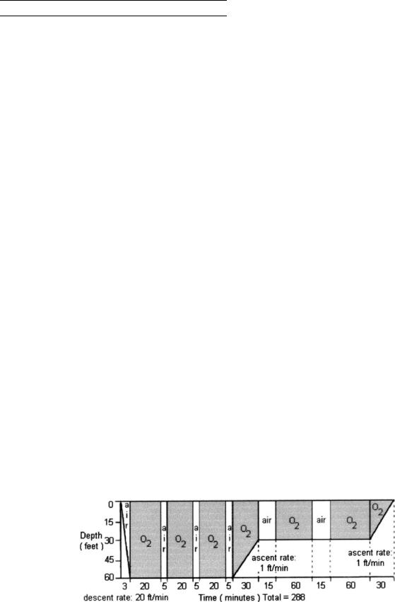
lism obstructing the systemic circulation. Air entry in the systemic circulation occurs following violation of the systemic circulation by any number of mechanisms (Table 4), which can be either by introduction of air into the arterial circulation, as in a lung biopsy, chest trauma, and pulmonary overpressure (diving), or into venous circulation, as in air introduction via central venous catheters, liver transplantation, and neurosurgery. Venous air emboli are more common, whereas arterial emboli are tend to be more serious. Physiologically, the air bubble forms or is introduced into the circulation. The lung usually serves as an excellent filter for air emboli, and can protect the embolism from traveling to the brain. This protective filter may be bypassed by a patent foramen ovale. Approximately 30% of patients have a foramen ovale that is patent by probe. The lung as a filter may be overwhelmed by large quantities of air. The bubble can then lodge in the smaller arteries of the brain causing obstruction. An air embolism is immediately identified as a foreign body, and platelets are activated, which leads to an inflammatory cascade. Hypoxia then develops distal to the obstruction with associated swelling. The embolism is eventually absorbed by the body, but the fibrin deposition at the embolism site may prevent return of blood flow. In order to diagnose an air embolism, it needs to have a high index of suspicion. If air embolism is suspected, the patient should receive a number of immediate measures, including ACLS or ATLS protocols. The patient should be placed on the left side and one should consider draining air from the right atrium with a central venous catheter. Air in the heart can cause a machinery murmur. 100% oxygen should be administered via a face mask or endotrachial tube, and the patient should be hydrated to preserve intravascular volume. Per Dalton’s law, administration of high oxygen will cause nitrogen to diffuse out of the air bubble and shrink in size. Dalton’s law states that total pressure of a mixture of gases is equal to the sum of the pressures of each gas. Each gas acts as if it alone were present. Dalton’s law is stated as P(t) ¼ P1 þ P2 þ P3. The inspired 100% oxygen will also maximize oxygenation of the tissues as much as possible under normal atmospheric pressure. Next, the patient should be emergently treated with hyperbaric oxygen. If a chamber is available that can provide compression to 6 ATA with air or a mixture of 50% nitrogen and 50% air, treatment should immediately be performed following the U.S. Navy protocol (Fig. 3).
Figure 3. U.S. Navy decompression treatment.

DECOMPRESSION ILLNESS
By definition, decompression illness (DCI), also called bends or caisson disease, occurs when gas bubbles exit in the blood or body tissues. At depth, more gas can dissolve in the tissues than in the blood. When the diver ascends too quickly, the gas comes out of solution and forms bubbles in the tissues and in the blood, much like popping a soda can and releasing its pressure causes the carbon dioxide bubbles to come out of the solution. In 1994, Diver’s Alert Network (DAN) recorded 1164 divingrelated injuries and 97 diving-related deaths, many related to DCI (12). The severity of DCI depends on the volume and location of gas bubbles. The range of symptoms is from vague constitutional complaints or limb pain to cardiopulmonary arrest and coma, the pathophysiology of which is explained by Henry’s law. As previously explained, Henry’s law is p ¼ Kc, where p is the partial pressure of the gas 1, c is its molar concentration, and K is the Henry’s law constant, which is temperature-depen- dent. Thus, simply stated, more inert gas can be dissolved in a liquid at higher atmospheric pressure and, conversely, less under lower atmospheric pressure, which occurs on decompression when gas must be removed from tissues, and rapid decompression leads to bubble formation. Using the Henry’s law equation, one can calculate the estimated amount of nitrogen a diver must clear from the bloodstream (about 5 L) in rising from 100 ft to the surface. The amout would be approximately 750 ml nitrogen assuming room temperature, which is a significant volume of nitrogen that must be eliminated from the divers bloodstream. The onset of symptoms is usually rapid and 75% of patients experience symptoms within 1 h of decompression and 90% within 12 h of decompression (13). A small number of patients may present even later, particularly if they have flown in commercial aircraft after diving and not followed the recommendation of the major diving organizations not to fly within 24 hours of one’s last dive. Interestingly, up to 10% of the inert gas that is absorbed in the tissues is released as bubbles after the diver’s decompression (14). Patients experience symptoms depending on the location and concentration of the
Table 5. Signs and Symptoms of Decompression Illness
Symptoms of |
Signs of |
Decompression Illness |
Decompression Illness |
|
|
Unusual fatigue |
Blotchy skin rash |
Skin itching |
Paralysis, muscle weakness |
Pain in joints/ |
Difficulty urinating |
muscles of arms, |
|
legs or torso |
|
Dizziness |
Confusion, personality |
|
changes, bizarre behavior |
Vertigo |
Amnesia, |
Ringing in the ears, (Tinnitus) |
Staggering |
Numbness, tingling |
Coughing up bloody, |
and paralysis |
frothy sputum |
Shortness of breath (Dyspnea) |
Collapse or unconsciousness |
|
Tremors |
|
|
HYPERBARIC MEDICINE |
23 |
bubbles (see Table 5). Bubbles forming in or near joints cause the joint pain of a classical ‘‘bend.’’ These musculoskeletal effects are called type 1 DCS. When these effects occur in the spinal cord or brain, numbness, paralysis, and disorders of higher cerebral function may result. If large numbers of bubbles enter the venous bloodstream, congestive symptoms in the lung and circulatory shock can then occur. These pulmonary and neurologic effects are termed type 2 DCS. Treatment should involve immediate administration of 100% oxygen, which facilitates nitrogen washout by the previously explained principles of Dalton’s law. Rehydration as well as advanced cardiac or trauma life-support protocols should be followed by transfer to a hyperbaric facility emergently. The patient should be treated with hyperbaric oxygen following U.S. Navy guidelines (Fig. 3), even if the inspired oxygen and rehydration alone have improved the patient’s signs and symptoms because tiny bubbles may be left that can cause tissue necrosis. Hyperbaric oxygen shrinks the size of the mostly nitrogen-filled bubbles by the principles of Boyle’s law, and the increased pressure also increases the partial pressure of the gas by Dalton’s law, hastening complete elimination of the bubble (Table 2). If the U.S. Navy (Fig. 3) recompression regimen fails to lead to symptom resolution, the Diver’s Alert Network or a medical expert on DCI should be contacted, and one of a number of recompression tables may be followed.
PROBLEM WOUNDS
The management of problem wounds should always include infection control, debridement, aggressive wound care, and correction of perfusion and oxygenation deficiencies. When an oxygenation deficiency of the wound is found, in the face of nonreconstructable vascular disease, hyperbaric oxygen should be considered as an adjunctive therapy. An increase in tissue oxygen tension by HBO therapy enhances wound healing by increasing neutrophil bactericidal capacity, inhibiting toxin formation in and even killing some anaerobes, encouraging fibroblast activity, and promoting angiogenesis (15). In normal physiology, the oxygen gradient across a wound is essential to stimulate these components of healing. Oxygen consumption is relatively low in wounds, and microvasculature damage and peripheral vasoconstriction increase diffusion distances. Partial pressure via Dalton’s law is the driving force of diffusion. Hyperbaric oxygen creates a steep tissue oxygenation gradient, providing a stronger stimulus than lactate or moderate hypoxia, to initiate and facilitate wound healing (16,17). These stimulated factors are thought to include platelet-derived growth factor B (PDGF-B), Vascular endothelial growth factor (VEGF), Epidermal growth factor, and others. Several clinical studies support the use of hyperbaric oxygen to promote wound healing. Perhaps the studies involving diabetic lower extremity wounds have been most informative. Several studies have shown an increased number of healed wounds, decreased wound size, and decreased rates of amputation among patients receiving hyperbaric
24 HYPERBARIC MEDICINE
oxygen therapy as an adjunctive treatment (18,19). Baroni et al. reported in a controlled study that a significant number of subjects receiving HBO went on to heal their wounds and fewer required amputation when compared with subjects not receiving HBO (20). In another study involving 151 diabetic patients with wounds of the lower extremity, Oriani et al. showed that 130 of these patients completely healed their wounds with adjunctive HBO (21). When compared with conventionally treated wounds, HBO-treated patients had an accelerated rate of healing, reduced rate of amputation, and an increased rate of completely healed wounds on a long-term basis (21). Transcutaneous Oxymetry (TcPO2) is currently the best tool available to evaluate tissue hypoxia and to select patients appropriate for HBO therapy. It can also be used to monitor progress during hyperbaric oxygen therapy. TcPO2 measurements should be taken with the patient breathing room air. A value of greater than 50 mmHg around the wound site indicates that the wound has adequate oxygenation and hyperbaric oxygen is not likely to improve healing. Values below 40 at the wound site should be considered for HBO therapy. Patients with marginal TcPO2 should be further tested while breathing 100% oxygen. TcPO2 values of greater than 100 while breathing 100% oxygen is an indicator that they are likely to respond to HBO therapy. If this challenge TcPO2 is less than 100, they still may benefit if the tested TcPO2 at the wound site is greater than 200 mmHg while they are breathing 100% oxygen at 2.0 ATA in the hyperbaric chamber (22). A TcPO2 value of less than 30 around a wound that does not exhibit this response, which indicates vascular compromise and the patient should be considered for revascularization if possible. Of note, 96% of limbs with TcPO2 values below 30 mmHg had abnormal arteriograms. It is also important to follow TcPO2 values weekly, and diabetic patients may have normal or falsely elevated noninvasive Doppler studies and a low TcPO2, implying satisfactory perfusion and inadequate oxygenation of the wound and, as such, may pose a diagnostic delimma. The diabetic patient with normal noninvasive Doppler and low TcPO2 will respond best to HBO. HBO therapy should be reserved for those diabetic wounds not responding to traditional management of debridement, antibiotics, and general wound care, including vascular reconstruction. The use of HBO therapy is necessary in only 15–20% of these patients. HBO therapy increases wound oxygen tension, enhancing host antibacterial mechanisms and promoting wound healing and is reserved for wounds in which the primary etiologies are tissue hypoxia or infection (13). Treatments are delivered at 2.0–2.4 atmospheres for 90–120 min once or twice daily. When serious infections are present, patients are typically hospitalized and given IV antibiotics and hyperbaric treatments twice daily five days a week. The TcPO2 values should be checked weekly because hyperbaric oxygen facilitates angiogenesis by a nitric oxide and vascular endothelial growth factor Beta (VEGF-B). When the room air TcPO2 is greater than 40 mmHg, the hyperbaric oxygen therapy can safely be discontinued. HBO is an adjuvant treatment; therefore, diabetic control, debridement, and aggressive wound treatment are given first priority.
When the wound bed has adequate granulation tissue, application of grafts can shorten morbidity, hospital stay, and health-care costs. The underlying problem in failure of a wound to heal is usually hypoxia and infection. Hyperbaric oxygen treatments in selected patients can facilitate healing by increasing tissue oxygen tension, thus providing the wound with a more favorable environment for healing. Therefore, hyperbaric oxygen therapy can be an important component to any comprehensive wound care program.
COMPROMISED FLAPS AND GRAFTS
Skin grafts and flaps with adequate blood supply do not require HBO. Hyperbaric oxygen therapy is extremely useful in situations where the skin grafts or flaps suffer from compromised microcirculation or hypoxia.
Flaps
The benefits of HBO on flaps develop from a systemic elevation in oxygen tension (23–25). In addition, HBO therapy prevents neutrophil adherence and subsequent vasoconstriction following ischemia. Too often, a compromised flap is allowed to progress over the days following surgery until visible signs of necrosis obviate the use of HBO, because delayed treatment with HBO cannot revive dead tissue. The resulting disappointment, as well as the associated patient dissatisfaction, can be avoided by rapid diagnosis of the flap problem and early involvement of the hyperbaric physician. The keys to successful treatment of compromised flaps with HBO are accurate diagnosis of the specific flap problem and appropriate and expedient initiation of hyperbaric oxygen treatment. Awareness of the different etiologies of flap compromise is necessary to plan for effective HBO treatment. A random flap with distal necrosis is completely different from a free flap with total venous occlusion. Proper classification of flaps, different etiologies of flap compromise, and understanding of how HBO is thought to effect ischemia reperfusion injury defines which patients will benefit from HBO. Flap classification is based on an assessment of blood supply, tissue composition, and method of movement. Each of these elements must be evaluated, but it is blood supply that is most important. The blood supply to the flap is either axial, based on a named vessel, or random, based on the subdermal plexus. Commonly, flap compromise occurs when the surgeon tries to mobilize tissue outside the defined arterial supply, when there is a pedicle problem exists, or when free flaps are exposed to prolonged ischemia. The tissue composition of a flap may include skin, subcutaneous tissue, fascia, muscle, bone, other tissues, or a combination of these. Flap composition is very important because different tissue types have different tolerances to ischemia. For instance, a myocutaneous flap will be more susceptible to ischemia than a fasciocutaneous flap, because muscle is much more sensitive to ischemic injury than fascia and skin (26). In those circumstances where a prolonged primary ischemia or any secondary ischemia resulting from vessel thrombosis and revision anastomosis exists, the flaps will undergo ischemia reperfusion injury.
When treating compromised flaps, a multimodalityapproach should be initiated. This approach should include the use of vasodilators if arterial vasospasm is suspected, removal of sutures if tension or compression are suspected, dextran and pentoxifylline for rheological purposes, medicinal and chemical leeching for venous congestion, and the early use of hyperbaric oxygen if blood flow can be documented. The use of HBO therapy is appropriate only when the flap problem has been defined, documented perfusion of the flap exists, appropriate surgical salvage measures have been first considered, and HBO therapy can be performed in an expedient manner. Specifically with respect to free flaps, extended primary ischemia time greater than 2 h or any secondary ischemia time may result in partial or total flap necrosis. This injury is usually reversible if recognized early and treated expeditiously. Essentially, it is ischemia reperfusion injury. Numerous research studies support the use of HBO in the salvage of compromised free tissue transfers (27,28). A rat free-flap model showed similar improvement in flap survival (27). A clinical study evaluated free-flap salvage in the face of prolonged primary or any secondary ischemia (28). Salvage was significantly better in the HBO treatment group vs. controls, but only if initiated within 24 h. Free flaps compromised by prolonged primary or secondary ischemia have responded favorably to HBO treatment with complete salvage, in most cases, if HBO is started early. The treatment regimen is 2.0–2.4 ATA, 90 min q 8 h x 24 h, then q 8–12 h x 48 h (29). Treatment duration is based on clinical evaluation.
Grafts
Skin grafts are anatomically different from flaps in that skin grafts lack an inherent blood supply. Skin grafts are composed of avascular tissue that depends entirely on the recipient bed for oxygenation. HBO is useful in preparing the recipient bed and in promoting healthy granulation tissue to support split-thickness skin grafts. One controlled study showed a significant improvement in skin graft survival from 17% to 64% with the addition of HBO treatment. Although literature exists to support the use of HBO for composite grafts, a study by the University of Mississippi Medical Center found no significant effect of HBO on rat-ear composite grafts larger than 1 cm (30,31) Further research is needed to better understand the effects of HBO on composite graft survival. The rational for use of HBO in crush injury, compartment syndrome, frostbite, and other traumatic ischemias is similar to those for compromised flaps as they are all cases of ischemia and ischemia reperfusion injury.
CRUSH INJURY, COMPARTMENT SYNDROME, AND OTHER ACUTE TRAUMATIC ISCHEMIAS
These conditions are trauma-related situations in which the underlying pathophysiology is that of ischemia reperfusion (IR) injury. Ischemia times of greater than 4 h willresult in some degree of permanent necrosis. The physiologic basis of IR injury has become better under-
HYPERBARIC MEDICINE |
25 |
stood in recent years. Most of the animal research centers around the production of oxygen-free radicals. Although the endothelial xanthine oxidase pathway has received much attention in the literature (32), more recent evidence supports the fact that neutrophils are a more important source of oxygen-free radicals via membrane NADPH oxidase and degranulation. Also, neutrophil adhesion is felt to cause ischemia reperfusion IR-asso- ciated vasoconstriction.
A perceived paradox exists related to HBO for IR injury. The less-informed observer often does not understand why HBO improves reperfusion injury and might think HBO instead increases free radical formation. (An oxygen-free radical is an oxygen molecules with an unpaired electron in its outer shell.) During ischemia, ATP is ultimately degraded to hypoxanthine and xanthine, which are anaerobic metabolites. With reperfusion, oxygenated blood is reintroduced into the ischemic tissue, and the hypoxanthine and xanthine plus oxygen creates oxygen-free radicals. Superoxide and hydroxyl oxygen-free radicals are formed, which can cause extensive tissue damage. The authors believe that the major mediator of damage is, in fact, neutrophil adherence to postcapillary venules significant and progressive vasoconstriction occurs in arterioles adjacent to leukocyte-damaged venules. Neutrophil adherence and vasoconstriction lead to a low flow state in the microcirculation and then vessel thrombosis, which is the endpoint of IR injury. The leukocyte-damaged venule is thought to be responsible for the arterial vasoactive response. HBO inhibits neutrophil adherence to the endothelial cells and thereby inhibits the ultimate thrombosis of microvessels, but the complete mechanism is still poorly understood, but is thought to involve the elevation in nitric oxide mediated by an increase in nitric oxide syntase (33). Free radical formation is not felt to be worsened with HBO as fewer adherent neutrophils actually exist to contribute to the neutrophil oxygen-free radicalgenerating system.
Treatment with hyperbaric oxygen in the face of IR injury carried the concern that that providing extra oxygen would increase free radical production and tissue damage. This query has been resolved by studies that have shown that HBO actually antagonizes the ill effects of IR injury in a variety of tissues (33–35). One of the first studies evaluating HBO and IR injury showed that HBO, immediately upon reperfusion, significantly improved skin flap survival following 8 h of global ischemia in a rat axial skin flap model with increased microvascular blood flow during reperfusion. Free-flap survival improves with HBO treatment during reperfusion even following ischemia times of up to 24 h (36). Hyperbaric oxygen administered during and up to 1 h following 4 h global ischemia significantly reduced neutrophil endothelial adherence in venules and also blocked the progressive arteriolar vasoconstriction associated with reperfusion injury (37). HBO inhibited in vitro beta-2-integrin (CD18)-induced neutrophil adherence function, but did not alter other important neutrophil functions such as oxidative burst or stimulus-induced chemotaxis and migration. This latter finding is very important, because HBO, through its action on the CD18 adhesion molecule,
26 HYPERBARIC MEDICINE
blocks the neutrophil adherence associated with IR injury without interfering with other neutrophil functions that would increase the risk of infectious complications. Initially, the focus in acute ischemia caused by trauma should be restoration of blood supply. The authors, therefore, recommend HBO therapy for all patients with muscle ischemia time greater than 4 h and skin ischemia time greater than 8 h. The major effects of IR injury are felt to occur within the first 4–7 h of reperfusion. 2 ATA hyperbaric oxygen increases the tissue oxygen tension 1000%. Treatment protocol is 2.0–2.5 ATA for 60 min, q 8 h x 24 h, then q 8–12 h x 48 h with clinical re-evaluation. If progressive signs of ischemic injury are still present, the treatment is continued at 2.0 ATA, q 12 h for 2–3 more days. Usually, 72 h of treatment is adequate as long as the first treatment is initiated within 4 h of surgery.
RADIATION TISSUE DAMAGE AND
OSTEORADIONECROSIS
1.2 million cases of invasive cancer are diagnosed yearly, half of which will receive radiation therapy and 5% of which will have serious radiation complications, which represents 30,000 cases per year of serious radiation sequellae (38). HBO is also well studied for its use in treating osteoradionecrosis in conjunction with adequate debridement of necrotic bone. Carl et al. also reported success is applying HBO to 32 women with radiation injury following lumpectomy and radiation compared with controls (39). Feldmeier and his colleagues reviewed the literature and found no evidence to support the potentiation of malignant cells or the engancement of cancer growth (40). The treatment protocol is 2.5 ATA for 90 min daily for 20–50 treatments. HBO can also be used as a radiosensitizer and are as much as three times more sensitive to radiation kisses than are hypoxic cells (41).
REFRACTORY OSTEOMYELITIS
Chronic refractory osteomyelitis (CROM) is infection of the medullary and cortical portions of the bone that persists or recurs following treatment with debridement and antibiotics. The principles of treatment are fairly simple. First, the dead bone is debrided and bone cultures should be taken along with administration of appropriate antibiotics. Next, the interface or cicatrix, which separates the compromised bone from adequate blood supply, is removed. Finally, hypoxia in the wound must be corrected, which may be accomplished by HBO. The treatment protocol is 2.0 ATA for 90 min daily for 20–60 treatments. Note that CROM and refractory osteomyelitis require the longest treatment protocols. HBO is believed to oxygenate hypoxic/ischemic tissues, augment host antimicrobial responses, augment osteoclastic activity, and induce osteogenesis in normal and infected bone and antibiotic synergism.
ACUTE THERMAL BURNS
HBO is approved by the USMS but it is not covered by Medicare.Gruberdemonstratedin1970 thattheareaaround and under a third-degree burn was hypoxic and could only be raised by oxygen at increased pressure (42). HBO has been found to prevent extension, reduce edema, increase healing rates, and decrease total cost in several randomized studies (43,44). HBO is also thought to decrease the rate of burn sepsis based on several early studies. The controversy, in part,surroundscurrentguidelinesforearlydebridementand grafting of burns. Once excised, a burn no longer exists and HBO will not be helpful. In case burns are not easily amenable to excision such as flash burns to the face or groin, HBO may be helpful to prevent extension of the burn and to aid healing. Treatment must be started within 24 h. The recommended regimen is 2.0 ATA for 90 min every 8 h on the first day, then every 12 hours for 5 or 6 days.
ACUTE EXCEPTIONAL BLOOD LOSS ANEMIA
Hyperbaric oxygen for treatment of acute blood loss anemia is reserved for those patients whose anemia is not immediately treatable for practical, disease process, or religious reasons, which may include warm antibody hemolytic disease, Jehova’s Witnesses, those with rare blood types, and those who refuse transfusion for other personal reasons. As explained in the physiology section, HBO dramatically increases the amount of solublized oxygen the blood can carry. In fact, Boerema showed, in 1955, that pigs could be exsanguinated to four-tenths of one gram of hemoglobin per deciliter and be maintained in a hyperbaric environment of 3 ATA without hypoxia. The goal in HBO therapy for these conditions is to improve the oxygen depth with the daily or twice daily HBO treatments until the anemia can be improved. In between the treatments, the patients should be maintained a lower FIO2 of inspired oxygen if possible to help reduce oxygen toxicity.
CARBON MONOXIDE POISONING
In 1966, Wada first used HBO to treat survivors of coal mine disasters with carbon monoxide poisoning and burns. The modern-day sources of carbon monoxide include automobile exhaust, home heaters, portable generators, propane engines, charcoal burners and camp stoves, and methylene chloride paint strippers. The initial treatment for carbon monoxide poisoning is 100% oxygen. The administration of 100% oxygen via a nonrebreather mask facilitates the dissociation of CO from hemoglobin to approximately 1.5 h. Hyperbaric oxygen delivered at 2.8–3.0 ATA reduced the halflife of CO-bound hemoglobin further to 23 min. In addition, patients who had one hyperbaric treatment for CO poisoning had 46% neuropsychiatric sequelae at discharge and 50% at one month versus two HBO treatments at 2.8–3.0 ATA having 13% at discharge and 18% at one month. The current recommendation is 3.0 ATA for 90 min with air breaks delivered every 8 h for a total of 3 treatments (called theWeaver protocol). Some authors still feel one treatment may be adequate (45).
CYANIDE POISONING
Hydrocyanide gas or HCN is formed when any number of substances burns, including furniture, asphalt, paper, carpeting (nylon), lighting baths (acrylic), plastic(polystyrene), and insulation (melamine resins). The antidote for cyanide poisoning begins with breathing 100% oxygen, ATLS protocols, and administration of IV sodium thiosulphate and is continued with a slow infusion of sodium nitrate and simultaneous HBO therapy if it is available. The sodium nitrate creates methemoglobin, which can impair the oxygen-carry- ing capacity of hemoglobin. HBO increases the amount of oxygen dissolved in plasma and may offer a direct benefit. The treatment regimin is 3.0 ATA with 30/10 airbreaks.
HYPERBARIC CHAMBER FACILITY DESIGN
AND SAFETY
Over 500 hyperbaric facilities exist in the United States, and the number of hyperbaric chambers is steadily increasing worldwide. Hyperbaric chambers are classified as either monoplace or multiplace. They differ functionally in that the monoplace chamber instills oxygen into the entire chamber environment, whereas in a multiplace chamber, patients breathe 100% oxygen via a breathing mask or oxygen hood and exhaled gases are vented outside the chamber. Monoplace chambers are constructed either as an acrylic cylinder with metal ends or are primarily constructed of metal. Most commonly, the monoplace chambers are formed from an acrylic cylinder from 20 to 40 inches in diameter with tie rods connecting it to end caps. The opening is a rotating lock or a cam action lever closure. Separate oxygen and air sources provide the oxygen sources and air for air breaks during therapy. An oxygen vent must be exhausted outside the building. The through ports on the HBO chamber door allow passage of specially made intravenous monitoring devices and ventilators. The larger diameter monoplace chambers are more comfortable; however, they require more oxygen and can be heavier and more expensive to install. The acrylic chambers can provide a maximum of 3 ATA pressure. Alternatively, some monoplace chambers are constructed mostly of steel with acrylic view ports, which can accommodate pressures of up to 6 ATA and are often used in special situations such as offshore rigs where a compact chamber is needed to treat decompression illness required in U.S. Navy Table 5. Multiplace chambers are much larger and are designed to provide treatment to multiple people or to manage complex conditions. Some can even house operating rooms with special precautions. They are typically made of steel with acrylic view ports and are designed for operation up to 6 ATA or 165 feet of sea water. The gauges are reported in feet of sea water on these multiplace chambers to facilitate the use of dive tables for staff or patients. These multiplace chambers are, therefore, best-suited to treat deep water decompression illness. These chambers can accommodate from 2 to 20 people and have variable configurations including horizontal cylinders, spherical shapes, and rectangular chambers.
HYPERBARIC MEDICINE |
27 |
The primary professional hyperbaric medicine societies in the United States are the Undersea and Hyperbaric Medical Society (UHMS) and the American College of Hyperbaric Medicine. The UHMS has developed a clinical hyperbaric medicine facility accreditation program. This program can be accessed via the UHMS website at http:// www.uhms.org, and it was designed to assure that clinical facilities are:
1.Staffed with well-trained specialists;
2.Using high quality equipment that is properly installed, maintained, and operated to the highest possible safety standards;
3.Providing high quality care;
4.Maintaining proper documentation of informed consent, treatment protocols, physician participation, training, and so on (46).
Safety Elements for Equipment and Facilities
The American Society of Mechanical Engineers (ASME) and the Pressure Vessel for Human Occupancy Committee (PVHO) define the design and fabrication guidelines for hyperbaric chambers. Although not required in all states or worldwide, it is accepted as the international standard (46). Next, the National Fire Protection Association (NFPA) has established a safety standard for hyperbaric facilities. The publication, NFPA 99, Safety Standard for Health Care Facilities, Hyperbaric Facilities, Chapter 20 explains the details of and criteria for equipment associated with a hyperbaric chamber facility. The requirements include fire abatement systems, air quality, and electrical requirements. These requirements apply to any hyperbaric chamber placed within a health-care facility. Each site must have a safety director. It is important to have only cotton clothing and to avoid any sources of sparks or static electricity given the 100% oxygen (Fig. 2). In addition to these guidelines, hyperbaric chambers are pressure vessels and, as such, are subject to boiler and pressure vessel laws. They are also medical devices and, in the United States, are also subject to FDA rules for class II medical devices. A chamber is required to have a clearance from the FDA before the device can be legally marketed or distributed, which is often called a 510 k clearance, denoting the form on which the clearance must be submitted. To check on whether a device has received clearance in the United States, one must contact the manufacturer or the Food and Drug Administration (FDA) most easily via their website, http://www.fda.gov/scripts/cdrh/ cfdocds/cfpmn/dsearch.cfm.
Facilities must develop defined safety protocols and emergency plans that are available through both the Undersea and Hyperbaric Medicine Society (UHMS) and the American College of Hyperbaric Medicine (ACHM).
FRONTIERS AND INVESTIGATIONAL USES
The use of hyperbaric oxygen therapy has, at times, been surrounded with controversy and spurious claims from improving athletic performance to slowing the aging process. It is essential that the hyperbaric medicine
28 HYPERBARIC MEDICINE
physician, staff, and potential patients understand and follow the principles of evidence-based practice, which means prescribing HBO therapy for the conditions proven to benefit from such treatment. The UHMS website, at www.UHMS. org, and AHCM are good resources for additional information as are numerous publications on hyperbaric medicine such as the hyperbaric medicine textbook available through the UHMS website. Investigational uses for hyperbaric oxygen therapy include carbon tetrachloride poisoning, hydrogen sulfide poisioning, sickle cell crisis, spinal cord injury, closed head injury, cerebral palsy, purpura fulminans, intraabdominal and intracranial abscess, mesenteric thrombosis, retinal artery occlusion, cystoid macular edema, bell’s palsy, leprosy, lyme disease, stroke and traumatic brain injury, and brown recluse spider bite. Some of the many investigational uses for HBO therapy may have merit, but these must be rigorously studied using welldesigned trials. As the field of hyperbaric medicine continues to advance, so will our understanding of the complex physiologic effects of delivering oxygen under pressure.
AKNOWLEDGMENT
Special thanks to Bob Bartlet, MD, for his help in preparing this chapter.
BIBLIOGRAPHY
1.Bakker DJ, Cramer FS. Hyperbaric surgery. Perioperative care. p 2.
2.Behnke AR, Shaw LA. Use of hyperbaric oxygen in treatment of compressed air illness. Nav Med Bull 1937;35:1–12.
3.Boerma I. High tension oxygen therapy. Proc Royal Soc Med 1964;57(9):817–818.
4.Bakker DJ, Cramer FS. Hyperbaric Surgery Perioperative Care. p. 67.
5.Jain KK. Physical Physiological, and Biochemical Aspects of Hyperbaric Oxygenation. Textbook of Hyperbaric Medicine. Toronto: Hogrefe & Huber Publishers; 1990. p 11.
6.Matos L, Nunez A. Enhancement of healing in selected problem wounds. In: Kindwall EP, Whelan HT, eds. Hyperbaric Medicine Practice, 2nd ed. Flagstaff AZ; Best; 1999.
7.Hunt TK, Pai MP. The effect of varying ambient oxygen tensions on wound metabolism and collagen synthesis. Surg Gynecol Obstet 1972;135:561–567.
8.Knighton DR, Halliday BJ, Hunt TK. Oxygen as an antibiotic: A comparison of the effects of inspired oxygen concentration and antibiotic administration on in vivo bacterial clearance. Arch Surg 1986;121:191–195.
9.Zamboni WA, Roth AC, Russell RC, Graham B, Suchy H, Kucan JO. Morphologic analysis of the microcirculation during reperfusion of ischemic skeletal muscle and the effect of hyperbaric oxygen. Plast Reconstr Surg 1993;91(6):1110–23.
10.Zamboni WA, et al. The effect of acute hyperbaric oxygen therapy on axial pattern skin flap survival when administered during and after total ischemia. J Reconstr Microsurg 1989;5:343–347.
11.Muth CM. Gas embolism (Review). New Eng J Med 342: 476–482.
12.Divers Alert Network: Report on 1994 Diving Accidents. Durham, NC: Duke University; 1995.
13.Barnard EEP, Hanson JM, et al. Post decompression shock due to extravasation of plasma. BMJ 1966;2:154.
14.Powel MR, Spencer MP, Von Ramm OT. Ultrasonic surveillance of decompression. In: Bennett PB, Elliott DH, eds. The Physiology of Diving and Compressed Air Work, 3rd ed. London Bailliere: Tindall; 1982. pp 404–434.
15.Zamboni WA. Applications of hyperbaric oxygen therapy in plastic surgery. In: Oriani G, Marroni A, eds. Handbook on Hyperbaric Medicine, 1st ed. New York: Springer; 1995. p. 443–484.
16.Hunt TK. The physiology of wound healing. Ann Emerg Med 1988;17:1265–1273.
17.Hunt TK, Hopf HW. Wound healing and wound infection. Surg Clinics N Am 1997;77(3):587–606.
18.Oriani G, Micheal M, Meazza D, et al. Diabetic foot and hyperbaric oxygen therapy: A ten-year experience. J Hyperbar Med 1992;7:213–221.
19.Wattel FE, Mathieu DM, Fossati P, et al. Hyperbaric oxygen in the treatment of diabetic foot lesions: Search for healing predictive factors. J Hyperbar Med 1991;6:263–267.
20.Baroni G, Porro T, Faglia E, Pizzi G, et al. Hyperbaric oxygen in diabetic gangrene treatment. Diabetes Care 1987;10:81–86.
21.Oriani G, Micheal M, Meazza D, et al. Diabetic foot and hyperbaric oxygen therapy: A ten-year experience. J Hyperbar Med 1992;7:213–221.
22.Strauss MB, Bryant BJ, Hart GB. Transcutaneous oxygen measurements under hyperbaric oxygen conditions as a predictor for healing of problem wounds. Foot Ankle Int. 2002 Oct; 23(10):933–7.
23.Zamboni WA, Roth AC, Russel RC, Nemiroff PM, Casas L, Smoot EC. Hyperbaric oxygen improves axial skin flap survival when administered during and after total ischemia. J Reconstr Micro 1989;5:343–347.
24.Hunt TK, Pai MP. The effect of varying ambient oxygen tensions on wound metabolism and collagen synthesis. Surg Gyn Obstet 1972;135:561–567.
25.Niinikoski J, Hunt TK. Oxygen Tension in Human Wounds. J Surg Res 1972;12:77–82.
26.Mathieu D, et al. Pedicle musculocutaneous flap transplantation: prediction of final outcome by transcutaneous oxygen measurements in hyperbaric oxygen. Plast Reconstr Surg 1993;91:329–334.
27.Waterhouse MA, et al. The use of HBO in compromised free tissue transfer and replantation: A clinical review. Undersea Hyperb Med 1993;20(Suppl):54 (Abstract).
28.Perrins DJD, Cantab MB. Influence of hyperbaric oxygen on the survival of split skin grafts. Lancet 1967;1:868–871.
29.Persons BL, Zamboni WA. Hyperbaric oxygen in plastic and reconstructive surgery. In: Bakker DJ, Cramer FS, eds. Hyperbaric Surgery Perioperative Care. Flagstaff, AZ: Best; 2002.
30.Mazelowski MC, Zamboni WA, Haws MF, Smoot EC, Stephenson LL. Effect of hyperbaric oxygen on composite graft survival in a rat ear model. Undersea and Hyperbaric Med Suppl 1995;22:50.
31.McFarlane RM, Wermuth RE. The use of hyperbaric oxygen to prevent necrosis in experimental pedicle flaps and composite skin grafts. Plast Reconstr Surg 1966; 37:422–430.
32.Angel MF, et al. Free radicals: Basic concepts concerning their chemistry, pathophysiology, and relevance to plastic surgery. Plast Reconstr Surg 79:990.
33.Lozano DD, Zamboni WA, Stephenson LL. Effect of hyperbaric oxygen and medicinal leeching on survival of axial skin flaps subjected to total venous occlusion. Undersea Hyperb Med suppl 1997;24:86.
34.Thom SR, Bhopale V, Fisher D, Manevich Y, Huang PL, Buerk DG. Stimulation of nitric oxide synthase in cerebral cortex due to elevated partial pressures of oxygen: an oxidative stress response. J Neurobiol 2002;51(2):85–100.