

- •VOLUME 4
- •CONTRIBUTOR LIST
- •PREFACE
- •LIST OF ARTICLES
- •ABBREVIATIONS AND ACRONYMS
- •CONVERSION FACTORS AND UNIT SYMBOLS
- •HYDROCEPHALUS, TOOLS FOR DIAGNOSIS AND TREATMENT OF
- •HYPERALIMENTATION.
- •HYPERBARIC MEDICINE
- •HYPERBARIC OXYGENATION
- •HYPERTENSION.
- •HYPERTHERMIA, INTERSTITIAL
- •HYPERTHERMIA, SYSTEMIC
- •HYPERTHERMIA, ULTRASONIC
- •HYPOTHERMIA.
- •IABP.
- •IMAGE INTENSIFIERS AND FLUOROSCOPY
- •IMAGING, CELLULAR.
- •IMAGING DEVICES
- •IMMUNOLOGICALLY SENSITIVE FIELD–EFFECT TRANSISTORS
- •IMMUNOTHERAPY
- •IMPEDANCE PLETHYSMOGRAPHY
- •IMPEDANCE SPECTROSCOPY
- •IMPLANT, COCHLEAR.
- •INCUBATORS, INFANTS
- •INFANT INCUBATORS.
- •INFUSION PUMPS.
- •INTEGRATED CIRCUIT TEMPERATURE SENSOR
- •INTERFERONS.
- •INTERSTITIAL HYPERTHERMIA.
- •INTRAAORTIC BALLOON PUMP
- •INTRACRANIAL PRESSURE MONITORING.
- •INTRAOCULAR LENSES.
- •INTRAOPERATIVE RADIOTHERAPY.
- •INTRAUTERINE DEVICES (IUDS).
- •INTRAUTERINE SURGICAL TECHNIQUES
- •ION-EXCHANGE CHROMATOGRAPHY.
- •IONIZING RADIATION, BIOLOGICAL EFFECTS OF
- •ION-PAIR CHROMATOGRAPHY.
- •ION–SENSITIVE FIELD-EFFECT TRANSISTORS
- •ISFET.
- •JOINTS, BIOMECHANICS OF
- •JOINT REPLACEMENT.
- •LAPARASCOPIC SURGERY.
- •LARYNGEAL PROSTHETIC DEVICES
- •LASER SURGERY.
- •LASERS, IN MEDICINE.
- •LENSES, CONTACT.
- •LENSES, INTRAOCULAR
- •LIFE SUPPORT.
- •LIGAMENT AND TENDON, PROPERTIES OF
- •LINEAR VARIABLE DIFFERENTIAL TRANSFORMERS
- •LITERATURE, MEDICAL PHYSICS.
- •LITHOTRIPSY
- •LIVER TRANSPLANTATION
- •LONG BONE FRACTURE.
- •LUNG MECHANICS.
- •LUNG PHYSIOLOGY.
- •LUNG SOUNDS
- •LVDT.
- •MAGNETIC RESONANCE IMAGING
- •MAGNETOCARDIOGRAPHY.
- •MANOMETRY, ANORECTAL.
- •MANOMETRY, ESOPHAGEAL.
- •MAMMOGRAPHY
- •MATERIALS, BIOCOMPATIBILITY OF.
- •MATERIALS, PHANTOM, IN RADIOLOGY.
- •MATERIALS, POLYMERIC.
- •MATERIALS, POROUS.
- •MEDICAL EDUCATION, COMPUTERS IN
- •MEDICAL ENGINEERING SOCIETIES AND ORGANIZATIONS
- •MEDICAL GAS ANALYZERS
- •MEDICAL PHOTOGRAPHY.
- •MEDICAL PHYSICS LITERATURE
- •MEDICAL RECORDS, COMPUTERS IN
- •MICROARRAYS
- •MICROBIAL DETECTION SYSTEMS
- •MICROBIOREACTORS
- •MICRODIALYSIS SAMPLING
- •MICROFLUIDICS
- •MICROPOWER FOR MEDICAL APPLICATIONS
- •MICROSCOPY AND SPECTROSCOPY, NEAR-FIELD
- •MICROSCOPY, CONFOCAL
- •MICROSCOPY, ELECTRON
- •MICROSCOPY, FLUORESCENCE
- •MICROSCOPY, SCANNING FORCE
- •MICROSCOPY, SCANNING TUNNELING
- •MICROSURGERY
- •MINIMALLY INVASIVE SURGICAL TECHNOLOGY
- •MOBILITY AIDS
- •MODELS, KINETIC.
- •MONITORING IN ANESTHESIA
- •MONITORING, AMBULATORY.
- •MONITORING, FETAL.
- •MONITORING, HEMODYNAMIC
- •MONITORING, INTRACRANIAL PRESSURE
- •MONITORING, NEONATAL.
- •MONITORING, UMBILICAL ARTERY AND VEIN
- •MONOCLONAL ANTIBODIES
- •MOSFET.
- •MUSCLE ELECTRICAL ACTIVITY.
- •MUSCLE TESTING, REHABILITATION AND.
- •MUSCULOSKELETAL DISABILITIES.
53.Thornton S. Lens implantation with restored accommodation. Curr Cana Ophthal Prac 1986;4:60–62.
54.Atchison DA. Accommodation and presbyopia. Ophthal Physi Opt 1995;15:255–272.
55.Fisher RF. Presbyopia and the changes with age in the human crystalline lens. J Physiol (London) 1973;228:765–779.
56.Koretz JF, Handelman GH. Modeling age-related accommodative loss in the human eye. Math Mod 1986;7:1003–1014.
57.Schachar RA. Zonular function: A new model with clinical implications. Ann Ophthalmol 1994;26:36–38.
58.Hara T, et al. Accommodative intraocular lens with spring action part 1. Design and placement in an excised animal eye. Ophthal Surg 1990;21:128–133.
59.Gilmartin B. The aetiology of presbyopia: A summary of the role of lenticular and extralenticular structures. Ophthal Physiol Opt 1995;15:431–437.
60.McLeod SD, Portney V, Ting A. A dual optic accommodating foldable intraocular lens. British J Ophthal 2003;87:1083–1085.
61.Cumming JS, Slade SG, Chayet A. AT-45 Study Group. Clinical evaluation of the model AT-45 silicone accommodating intraocular lens. Results of feasibility and the initial phase of a Food and Drug Administration clinical trial. Ophthalmology 2001;108:2005–2009.
62.Kuchle M, et al. Implantation of a new accommodating posterior chamber intraocular lens. J Refract Surg 2002;18:208–216.
See also BIOMATERIALS: POLYMERS; CONTACT LENSES; VISUAL PROSTHESES.
LIFE SUPPORT. See CARDIOPULMONARY RESUSCITATION.
LIGAMENT AND TENDON, PROPERTIES OF
G AZANGWE
RM ASPDEN
INTRODUCTION
Tendons and ligaments are fibrous connective tissues that play a mechanical role in the stability and locomotion of the body by transmitting tension. Unlike muscle, which actively contracts, ligaments and tendons are passive. Tendons transmit mechanical forces from muscle to bone, whereas ligaments join bone to bone. Both tendons and ligaments contain relatively few cells (1), and their extracellular matrices are made up of several components. These components are combined in various proportions, and with different organizations to give mechanical properties appropriate to the function of the particular tendon or ligament. There have been a number of reviews in recent years covering specific ligaments, for example, in the rabbit (2), or tendons, such as the human achilles (3), or aspects of their behavior such as healing and repair (4). In this article, the emphasis is on properties the ligaments have in common, which will provide an insight into how and why they behave as they do. This will be based around their functioning as fiber-reinforced materials whose properties are regulated by the cells they contain that produce and maintain the extracellular matrix.
First, this article considers the components of the tissue, not from a biochemical point of view, but as components
LIGAMENT AND TENDON, PROPERTIES OF |
241 |
that may be combined to produce mechanically stable materials. The constituents are considered in terms of the matrix in which are embedded fibers of collagen and varying amounts of elastin. Following this is a discussion of the ways these components interact in ligaments and tendons to yield composite materials with the required mechanical properties. A consideration of some of the ways in which ligaments and tendons may be damaged, and the mechanisms by which they might recover or be repaired, leads to a final, brief review of their surgical replacement. A small section on work being conducted in order to produce a tissue engineered ligament and tendon is also included.
COMPONENTS
Tendons and ligaments are composed primarily of collagen fibers surrounded by a matrix. Here the matrix refers to all the materials that surround the collagen fibers providing both structural support and a medium for diffusion of nutrients and gases. Note this is in contrast to its use in biological terms in which it generally includes the fibrous components. The matrix contains proteoglycans and adhesive glycoproteins and is highly hydrated (typically 65–80% water) (5,6).
Collagen
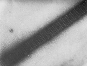
Collagen fibrils are able to reinforce the weak matrix because of their much greater stiffness and strength in tension (7). The collagen molecule is a long, stiff rod made up of three polypeptide chains wound as a triple helix structure (8). Each fibril is like a rope in which linear molecules are packed together with their axes parallel, within a few degrees, to the fibril axis. Molecules are held together by covalent cross-links so that the fibril is very strong in tension. The regular arrangement of molecules along the axial direction in a fibril gives rise to the characteristic periodicity of 67 nm, which may be seen in electron micrographs (Fig. 1). Although to date, 21 genetically different types of collagen have been identified, types
Figure 1. Electron micrograph of collagen fibril from rat tail tendon stained with uranyl formate showing characteristic banding pattern repeated every 67 nm. Micrograph courtesy of Dr D. P. Knight.
242 LIGAMENT AND TENDON, PROPERTIES OF
I and III fibrous collagens dominate in tendons and ligaments.
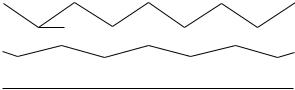
There is a heirarchical structure within tendons (9) that has been reviewed often [e.g., (10)], and that appears to have been based on an earlier description of keratin. In this model, collagen molecules are arranged successively into microfibrils, subfibrils, fibrils, and fascicles that are finally packed into the tendon sheath. Evidence for microand subfibrils is still equivocal, but a principal mechanical structure is the fibril. These generally have a unimodal distribution of diameters in the newborn, typically a few tens of nanometers, but assume a bimodal distribution in mature tendons and ligaments with mode diameters typically 100–300 nm (11,12). The ends of fibrils are rarely seen in electron micrographs, and even when they are seen, it is not clear whether they are artifacts of sectioning. Fibrils appear to grow by end-to-end addition of short fibrils and there is evidence from 12-week old mouse skin that fibril tips may eventually fuse with the central region of other fibrils to create a meshwork (13). It is not known whether this happens in tendon or ligament or whether the fibrils are as long as the tendon or ligament. Fibrils are arranged into fascicles, which are 80–300 mm in diameter and these have a ‘‘crimped’’ morphology that is seen most clearly using polarized light (6,9). This crimp is generally described as a planar zigzag with a sharp change in direction of the collagen fibrils with a periodicity of 200– 300 mm (Fig. 2). On application of a tensile load, initial elongation of the fiber requires a relatively low stress because it simply leads to removal of the crimp (14,15). Once the crimp is removed, the fibers become very stiff. This crimp structure, which is also found in ligaments, explains the characteristic shape of the stress–strain curve for ligaments and tendons (14).
There are many studies of the mechanical properties of tendon and ligament. Most tendons have similar properties, because of their role transmitting forces from muscle to bone and the need to do this most efficiently (16–19). In contrast, every ligament has unique properties that fit it to its function at that particular site in the body. These may range, for example, from the highly extensible ligamentum flavum, helping control spinal posture, to the relatively stiff cruciate ligaments in the knee. Most information on the mechanical properties of collagen has been inferred from experiments on tendon; and though they contain a large proportion of collagen, 70–80% of the dry weight, the fibrils are surrounded by matrix, and therefore
(a)
θ
(b)
(c)
Figure 2. Schematic diagram of the crimp structure in collagen fibers seen from the side. (a) relaxed state [this is grossly exaggerated; the true crimp angle (u) is 158]. As the fiber is stretched, the crimp straightens out (b) until at strains of a0.03, the fiber becomes straight (c), at which point it becomes much stiffer.
the tissue is really a composite material (6). This makes it difficult to separate the behavior of the individual components.
Proteoglycans
The proteoglycans found predominantly in tendon and ligament belong to the small leucine-rich proteoglycan (SLRP) family; decorin, biglycan, lumican, and fibromodulin though there are small amounts of the large proteoglycans aggrecan (20,21) and versican (21,22). The SLRPs all comprise a repeating structure that is rich in leucine residues, between 13 and 16% of all residues (23). They are present in different tissues in differing amounts and appear at different stages of development (24). Their function is poorly understood though gene knockout studies in mice have shown marked osteopenic effects on bone, skin laxity, and irregular collagen fibers in tendon (25). Most of these SLRPs have one or two glycosaminoglycan (GAG) chains of varying lengths attached, generally either dermatan sulfate or chondroitin sulfate, which can interact electrostatically with collagen (26,27). The proteoglycan decorin has been found to be localized in the tissue to a specific region of a collagen fibril (28). It is also reported to play a regulatory role in collagen fibrillogenesis, by affecting fibril radius (13), and increases the strength of uncrosslinked fibers (29,30). In regions where tendons pass over bone and are subjected to compressive loading in addition to tension, a fibrocartilaginous tissue containing large amounts of aggrecan and biglycan develops (31,32). The adhesive glycoproteins include fibronectin and thrombospondin (33–35), both of which contain possible attachment sites for cells. In humans, these were reported to be more common in the tendon sheath than in the fibrous bulk (36) and fibronectin was found to be up-regulated at sites of injury (34).
The matrix is weak in shear; that is, if it is loaded in compression, it tries to slide out sideways unless it is contained. This behavior may be readily seen in jelly (Jello in the United States) and is not surprising given its high water content, since fluids simply flow when sheared. It is not easy to measure the shear strength of matrix. Proteoglycans at similar concentrations have a very low shear strength (37); however, matrix may be stiffer than this because of the interactions between its macromolecular components. An analysis of the behavior of tendon suggests that its matrix would have a shear modulus of 100 kPa (38). Because of this low stiffness in shear, the matrix alone is not well suited to bearing loads. Also, its proportion in ligament and tendon is quite low, 10–20% of the dry weight. The ways in which matrix may transmit stress to the fibers and prevent crack propagation will be discussed later.
Elastic Fibers
Electron microscopy reveals the presence of elastic fibers in ligaments and tendons (39,40). Elastic fibers have two components; elastic fiber microfibrils and elastin. The microfibrils have a diameter of 10–12 nm and consist of glycoproteins. Elastin is composed of mainly hydrophobic nonpolar amino acids with a high content of valine (41).
Elastic fibers are highly extensible, they do not creep and their extension is reversible at high strains. Their mechanical properties are thus very different from collagen. Most of our knowledge of elastic fibers comes from experiments on ligamentum nuchae, a ligament from the cervical spine, which contains 70% elastin by dry weight (42). Elastin closely resembles a rubber in many respects and its mechanical properties are certainly very similar (43). Purified samples of ligamentum nuchae will extend to roughly twice their resting length before breaking.
The extensibility of a tendon or ligament depend in part on the elastin content of the tissue. Ligamentum flavum, from the spine, which may typically be strained to 50% contains roughly 70% elastin, by dry weight (44), whereas tendon, which works at strains <4% contains only 2% elastic fibers by dry weight (1). It is fairly easy to see why highly extensible tissues have a high proportion of elastin, but not quite as easy to explain the presence of elastic fibers in a relatively inextensible tissue such as tendon. A clue is provided by some synthetic fibrous composite materials that contain two different kinds of fiber (45). Here a small proportion of strong, low stiffness fibers added to the composite produces a material that is less susceptible to failure under sudden application of load than one that contains only stiff fibers; that is, it makes the material less brittle. It may be that the small proportion of elastic fibers in tendon provide some protection against the sudden application of load that may occur, for example, if an animal is startled.
Fiber–Matrix Interactions
The combination of strong fibers in a weak matrix leads to materials that are less susceptible to mechanical damage while maintaining a high proportion of the strength of the fibers. In particular, they are less susceptible to sudden failure than a homogeneous material would be; a property called ‘‘toughness’’ (46). This composite nature has been recognized for many years (6) and provides a theoretical framework for understanding the properties of the tissues. It also enables some useful comparisons to be made between relatively simple synthetic composites and biological tissues in which the complexities of composition and structure make modeling very difficult. The aim is to obtain an understanding of how the similarities in the tissues, fibers in a matrix, enable them to function in general terms before considering the differences (in composition, and organization), which give them their specific properties. The function of collagen fibrils and fibers in such a composite is to withstand axial tension, since, like any rope, they have little resistance compression and flexion (7). As the tissue is stretched the matrix will try to flow and this will exert a shear force along the surface of the collagen fibers tending to stretch and orient them (7,47). This length increase, which is normally expressed as a fraction of the original length and is then termed ‘‘strain’’, leads to a restoring force in the fiber that balances the applied force. The behavior is rather like a loaded spring that stretches to enable it to bear load, but returns to its relaxed length on removing the load. Similarly, collagen fibers are able to reinforce a tissue if they are oriented so that an applied
LIGAMENT AND TENDON, PROPERTIES OF |
243 |
load tends to stretch them. The nature of the shear force exerted by the gel is unknown, but two simple models, those of elastic and plastic (or frictional) stress transfer, have been used to investigate stresses in the fibers and the force that has to be generated at the fiber surface to enable them to function in this way (47–49). Fibers that are shorter than the tissue are still able to provide reinforcement (50). Some fibrils observed in tissues (51) and those grown in vitro (52,53) appear to be tapered, rather than having a uniform radius. Analytical and finite element models of idealized single-fiber composites have shown that tapered fibers have two distinct advantages over uniform fibers: the axial stresses within the fiber are more uniformly distributed and they contain a much smaller amount of material, though their effectiveness at reinforcing is just as great. A more uniform stress within the fiber means more of the fiber is carrying a significant stress, thus making better use of the fiber, and avoids the generation of stress concentrations that are potentially damaging and could lead to fiber fracture. In addition, the volume of material in a cone, for example, is only one-third of that in a straight cylinder and, therefore, a tapered fiber incurs a far smaller metabolic cost by the cells to produce it. In straight-cylindrical fibers it has been calculated that interactions at the fiber surface do not have to be great in order to load fully the fiber. In tendon, assuming conservatively only one interaction per 67 nm D-period, it was estimated that fiber–matrix interaction forces of the order of only 10 pN was sufficient to load fully the fiber (47). These forces are similar in magnitude to van der Waals forces or hydrogen bonds. This suggests that permanent bonds or covalent interactions between fiber and matrix are not essential for the mechanical functioning of the tissue though, of course, it does not preclude them. Regulating the interaction between fibers and matrix is clearly important in this model of how the tissues function and decorin, as described above, is a prime candidate for a role in this. Changes in the concentration and orientation of collagen and its interactions with the matrix have been used to explain the dramatic changes in a similar fibrous tissue, the uterine cervix, that occur during parturition (54). The presence of the matrix around the collagen fibrils is also important when it comes to preventing crack propagation. This will be considered in more detail in the context of the tissues themselves.
LIGAMENT
Ligaments are short bands of tough, but flexible, fibrous connective tissue that bind bones together and guide joint motion, or support organs in place. The word ligament is derived from the Latin word ‘‘ligare,’’ which means to bind. Generally, ligaments can be classified into two major subgroups. There are those connecting the elements of the skeletal system (usually crossing joints) and those connecting other soft tissues, such as the suspensory ligaments in the abdomen. This section only considers skeletal ligaments. The main function of the skeletal ligaments, such as the anterior cruciate ligament (ACL) of the knee joint, is to stabilize and control normal kinematics, to prevent
244 LIGAMENT AND TENDON, PROPERTIES OF
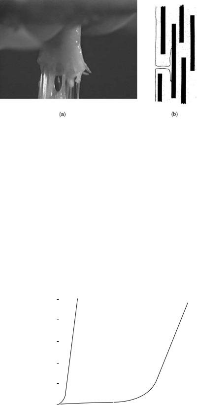
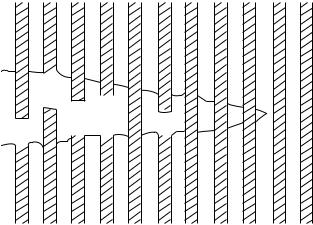
Figure 3. (a) Video image showing a ligament failing under tensile strain. (b) A schematic diagram showing a longitudinal section through a fiber composite illustrating how a weak matrix prevents crack propagation fromonefiber toanother and dissipatesits energy by creating cracks in directions other than across the composite.
abnormal displacements and rotation that may damage the articular surfaces (2,55). At the insertion to bone, the ligaments change from flexible ligamentous tissue to rigid bone, mediated by a transitional zone of fibrocartilage and mineralized cartilage. This helps to prevent stress concentration at the attachment site by allowing gradual change in stiffness (56,57).
Similar to tendon, ligaments are primarily composed of collagen embedded in a weak matrix (7). The collagen molecules in ligaments pack together to form fibrils and the fibrils aggregate into larger fibrous bundles (2). As described previously, the function of collagen fibrils is to provide tensile reinforcing for the matrix. The proportion of fibers within the ligamentous structure and the orientation of the collagen fibers are the main factors that govern the mechanical behavior of the tissue (7). In contrast to tendon, there is commonly less collagen, which is less highly oriented than in tendon, conferring a generally greater extensibility to these tissues.
The collagen fibrils also prevent damaged tissues from failing suddenly. For example, most ligaments do not tear straight across when they are damaged (Fig. 3). Instead, small tears in the matrix are diverted when they encounter the strong collagen fibrils (see below on failure). There is then a possibility that a damaged ligament can heal while, in the meantime, retaining the ability to withstand some load. Some ligaments, however, (e.g., the ACL), have a limited ability to heal when ruptured and this means that it often needs to be replaced or reconstructed when ruptured. A brief summary of different options available for treating ruptured ligaments will be presented in a later section.
Unlike tendons that all have very similar composition, structure, and function, the same cannot be said of ligaments, and it is far harder to make general comments about their properties. Much less is known, too, about the relationship between their structures and functions as the arrangements of their collagen fibrils are more complex than those in tendons (58,59). This greater complexity is understandable when it is realized that the function of a ligament is very dependent on its position in the body; for example, the medial collateral ligament in the knee of a sheep operates at strains of 0.02 (60),
whereas the ligamentum flavum of the human spine operates at strains of up to 0.6. Figure 4 shows that ligamentum flavum is much less stiff than tendon.
It is not surprising that some ligaments contain a high proportion of elastin ( 60–70% of the dry weight), which enables them to withstand the high strains to which they are subjected without fracture (61). Ligaments are viscoelastic, that is their properties are time dependent and they appear stiffer if stretched more rapidly. These ligaments exhibit hysteresis, that is, they lose energy on being taken through a cycle of stretching and relaxing. Tkaczuk (61) published a detailed account of the mechanical properties of longitudinal ligaments from the human spine. These deform elastically up to strains of 0.25, when the stress is5 MPa, and rupture at a stress of 20 MPa. Shah et al.(62) also showed that, like tendons, the collagen fibers are crimped and this crimp disappears at strains of 1.2– 2.8% depending on the ligament. When ligaments are cut from the joint, they can often be seen to contract rapidly, suggesting that they are held in a state of tension even when the joint is in a relaxed state.
|
100 |
|
|
|
|
|
|
|
|
|
|
|
|
|
|
|
|
|
80 |
|
|
|
|
|
|
|
|
|
|
|
|
|
|
|
|
|
|
|
Tendon |
|
|
|
|
|
|
|
|
|
|
|
|
||
Force (N) |
60 |
|
|
|
|
|
|
|
|
|
|
|
|
|
|
|
|
40 |
|
|
|
|
|
|
|
Ligamentum |
|
|
|
|
|
|
|||
|
|
|
|
|
|
|
|
|
|
|
|
|
|
||||
|
|
|
|
|
|
|
|
|
flavum |
|
|
|
|
|
|
||
|
20 |
|
|
|
|
|
|
|
|
|
|
|
|
|
|
|
|
|
0 |
|
|
|
|
|
|
|
|
|
|
|
|
|
|
|
|
|
0 |
0.2 |
0.4 |
0.6 |
0.8 |
1.0 |
|||||||||||
|
|
|
|
|
|
|
|
|
Strain |
|
|
|
|
|
|
||
Figure |
4. Comparison |
of force–strain curves |
obtained |
for |
|||||||||||||
extension of tendon and ligamentum flavum.
100
80
∆ θ degrees |
60 |
|
40 |
||
|
20
0
0 |
0.02 |
0.04 |
0.06 |
|
|
Strain |
|
Figure 5. Full width at half-maximum (fwhm), D, of the distribution of orientations of collagen fibers in ligamentum flavum. (o) and posterior longitudinal ligament, (þ) as a function of strain.
Spinal ligaments provide a good example of the mechanical function of ligaments in a joint (58). The longitudinal ligaments and the ligamentum flavum act together with the intervertebral disc to achieve a mechanically stable joint and serve to limit its mobility. The ligamentum flavum is almost twice as far from the axis of rotation in forward bending as the posterior longitudinal ligament, and hence it can be seen that it needs to be roughly twice as extensible (58,63). This is partly explained by a higher elastin and lower collagen content in ligamentum flavum (61), but also by a less highly aligned organization of collagen fibers (63). As the ligament is stretched, the fibers become more highly aligned and this will increase the stiffness of the tissue, that is its resistance to extension. X-ray diffraction experiments have measured the spread of orientations of fibrils in these ligaments and modelling has shown how this decreases with increasing extension (47). Figure 5 shows that the width of the distribution, Du, as measured at half the peak height, is greater for ligamentum flavum than posterior longitudinal ligaments. This mechanism provides an explanation for the form of the force–strain curve, shown in Fig. 4, One final point about ligaments is that they have a nerve supply that makes them potential sources of pain. It has also been suggested that they may function as proprioceptors as part of a reflex arc, that is, the ligaments would act as sensors to detect the position of a joint and the information would then be used to control the muscles around the joint thereby controlling its movement and stability (64).
In summary, ligaments are composite materials containing crimped collagen fibers that are prestressed in the relaxed joint. They have a nonlinear stress–strain curve
LIGAMENT AND TENDON, PROPERTIES OF |
245 |
and are viscoelastic. The collagen fibers are relatively disoriented in the unstretched tissue and become more highly aligned as the tissue is stretched. They often contain a proportion of elastin. Their composition and structure depend on their position in the body and their dynamic behavior, that is, the change in structure with strain, becomes more important.
TENDON
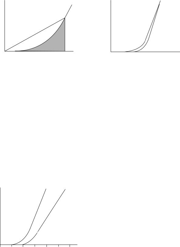
The function of tendon is to transmit the force generated by a contracting muscle to the correct point of application on a bone so as to manipulate a joint. Tendons are often preferable to direct attachment of muscle to bone because of various functional requirements. Muscles have a low tensile strength, defined as load at fracture per unit crosssectional area. This means that they must have a large cross-sectional area in order to transmit sufficient force without tearing. Around many joints (e.g., the fingers), there is insufficient space to attach many muscles. The muscle, therefore, is located further away and attachment made by a tendon, which may be tens of centimeters long in the hand and forearm. Tendons, therefore, need to be strong, so that they can be relatively slender, and stiff, so that the force developed by the muscle is transmitted to the bone without energy being wasted on stretching the tendon. A graph of force as a function of strain for a tendon is shown in Fig. 6. This type of curve is obtained by subjecting the tendon to various forces and recording the amount by which the tendon is stretched, and many of the early studies have never been bettered (6,9,18,40,65). The steepness of the stress–strain curve is a measure of the stiffness of the material. Figure 6 shows that for small strains the tendon requires very little force to stretch it but thereafter it stiffens considerably. The stress at this point is 10 MPa, which is several orders of magnitude greater than the stresses needed to shear the matrix (6). If the stiffness did not increase, a muscle would continue to stretch the tendon and the force would not be transmitted to the bone.
Tendons are believed to function in the body at strains up to 0.04 (66,67). Beyond this strain, a tendon does not return to its original length when the applied stress is removed. A tendon will break at strains of 0.1 (67). The
30
20
(N)Force 10
0
0 |
0.02 |
0.04 |
0.06 |
|
|
Strain |
|
Figure 6. Force–strain curve for tendon.
246 LIGAMENT AND TENDON, PROPERTIES OF
Stress |
Strain |
Figure 7. Schematic of stress–strain showing energy stored in a stretched tendon.
initial stages of tendon extension involve straightening the crimp of the collagen fibers described previously.
The energy stored in the stretched tendon is given by the area under the stress–strain curve, as shown in Fig. 7, and it is this energy that must be used to create a tear in the tissue. This energy is lower in a material with a J-shaped stress–strain relationship than if, for example, it were linear. Minimizing the energy available to cause a fracture in this way gives the tissue a property known as resilience, that is, a tendon does not suddenly fail if it is overloaded— unlike a steel wire. While the crimped collagen fibers are being straightened, the weak matrix must be sheared. Because of the fluid-like nature of the matrix, it tends to flow. The rate of flow depends on the force applied to it and the amount of flow is greater the longer the force is applied. The result is that tendons are ‘‘viscoelastic’’ (18,38,68). The effect of the rate at which force is applied to the tendon is shown in Fig. 8.
This time dependence of mechanical behavior leads to a phenomenon known as ‘‘creep’’. For example, when a load of 10 N was applied to a human flexor digitorum tendon, the initial strain was 0.015, but 100 s later under the same load, the strain had increased to 0.016 (69). Thus, if a
Stress
0 |
0.02 |
0.04 |
0.06 |
Strain
Figure 8. Schematic stress–strain curves for tendon to show the effects of different loading rates. These curves correspond to slow loading (continuous curve) and rapid loading (dashed curve).
stress
Strain
Figure 9. Stress–strain curves for tendon that is stretched (continuous curve) and then relaxed (dashed curve), showing hysteresis; that is, not as much energy is recovered on relaxing as was initially used to stretch the tissue.
tendon is stretched rapidly, the matrix has less chance to flow and creep in the material is less resulting in the tendon being stiffer as shown in Fig. 8. Viscous flow of the matrix also provides a mechanism for dissipating energy; more work is done in stretching a tendon than is recovered when the tendon is allowed to relax. This phenomenon is known as hysteresis and is illustrated in Fig. 9. The behavior of a tendon is therefore intermediate between that of a steel wire that stores all the energy used to stretch it, and a viscous liquid that simply flows to a new position and does not store any of the energy put in to cause it to flow.
MEASURING THE PROPERTIES OF LIGAMENTS AND TENDONS
To quantify the physical properties of ligaments and tendons, mechanical testing of bone–ligament/tendon–bone complexes is often performed (70–73). That this method is often used is partly due to the difficulty in testing isolated ligaments and tendons. Ideally, testing isolated ligaments and tendons would provide measures of the material properties of the tissue alone, but such tests are complicated by difficulties in effectively securing the cut ends (2). Putting the free ends in clamps often results in stress concentrations at the grips, which may contribute to premature failure. Although the use of bone–ligament/ tendon–bone complexes still provides more secure clamping, it also increases the difficulty of separating the properties of the ligament from those of the insertion sites. When such complexes are subjected to tensile loading, the resulting load-displacement curve represents the mechanical properties of the bone–ligament/tendon–bone complex as whole rather than specifically about the material that makes up the ligament or tendon.
In order to obtain the material properties of the ligaments, one needs to measure their length (to calculate strain from deformation divided by original length) and cross-sectional area (to calculate stress from applied force divided by original area). From stress–strain curves material properties such as Young’s modulus (slope of stress– strain curve), maximum stress, maximum strain, and energy density (area under stress–strain curve) can be
determined. Special devices such as buckle transducers (74) and Hall-effect displacement transducers (75) have been used to measure ligament strains during testing. The drawback of such devices is that they rely on direct contact with the tissue sample and that may influence the results. Optical analysers have also been employed as a noncontact method to measure ligament strains (2). However, inaccuracies may occur because the irregular dye blobs used as markers change shape on stretching making it difficult to define unique points. Another technique that has been employed to measure strain is the use of video dimension analyser (VDA) (76,77). This method requires no direct contact with the specimen, but relies on a recorded video image.
The irregular and complex shape and geometry of ligaments and tendons also make it difficult to measure their cross-sectional area. Although flexible callipers, which are able to follow contours better, have been used to measure ligament cross-sectional areas (2), they still require contact, and this results in errors in measurements. Other investigators have calculated the cross-sectional area of a known length of ligament from measurements of its density by floatation in a mixture of xylene and carbon tetrachloride (78). A number of noncontact methods, such as the use of a rotating microscope (79), use of the VDA (80) and the laser micrometer (81) have also been employed to measure the cross-sectional area.
When mechanical properties of ligaments and tendons are being determined, it is important to consider the rate at which they are loaded since they are viscoelastic, that is, their mechanical properties depend on the rate at which they are deformed (82–86). This sensitivity to strain rate means that ligaments and tendons exhibit properties of stress relaxation (decreased stress with time under constant deformation) and creep (increased deformation with time under constant load) (87,88).
Because of the complex geometry of tendons and ligaments, the orientation of the specimens during mechanical testing affects their physical properties and the manner in which they fail, and should therefore be taken into account when performing mechanical tests. Torsion has been implicated as a factor in the rupture of the ACL during sporting injuries (89–91). Cyclic loading has also been found to lower the yield point or soften the ligament by increasing its compliance (decrease the slope of the linear region of the stress–strain curve) (55). Azangwe et al. (92) showed that, when combined, tension–torsion loading affects both structural and mechanical properties of anterior cruciate ligaments.
LIGAMENT AND TENDON FAILURE MECHANISMS
Since ligaments and tendons consist of collagen fibers reinforcing a weak matrix, it is reasonable to compare their behavior under tensile loading with that of synthetic fiber-reinforced composites, since their failure mechanisms are well established. This section describes some of the modes of failure of fiber-reinforced composites and how they are related to those of the ligaments and tendons. Detailed accounts of failure modes of synthetic
LIGAMENT AND TENDON, PROPERTIES OF |
247 |
fiber-reinforced composites can be found in textbooks, for example, Agarwal and Broutman (45), and Kelly and Macmillan (50). When a material is subjected to any kind of loading, it can absorb energy by two basic mechanisms:
1.Material deformation.
2.Creation of new surfaces.
Material deformation occurs whenever a material is subjected to load. However, if the energy supplied is sufficiently large, cracks may be initiated. Whether they propagate depends on the relative amounts of energy required to create new surfaces compared with that stored in the deformed matrix. For brittle materials such as glass, the energy required to create a new fracture surface is small and though only a small amount of deformation takes place, this is elastic and the associated energy is sufficient to propagate the crack. This means that brittle materials have a low energy-absorption capability. On the other hand, for ductile materials, large plastic deformations occur, which dissipate energy, resulting in large energies being absorbed rather than being available to drive a fracture. This finding shows that the total energy-absorb- ing capability or ‘‘toughness’’ of a material can be enhanced by increasing either the length of the path of the crack during separation or the material-deformation capability. In metals, the latter mechanism frequently occurs and metallurgical processes are developed to maintain ductility. In composites, replacing low energy-absorbing constituents with greater energy-absorbing constituents can enhance the toughness.
As is the case with many materials, failure in a fiberreinforced composite emanates from small inherent defects in the material. Several failure events may occur during the fracture of a fiber reinforced composite material, such as,
1.Microcracking of the matrix.
2.Separation of fibers from the matrix (debonding and pull-out).
3.Breaking of fibers,
Forcing a crack to take a longer path is the main mechanism encountered in fiber composites to increase toughness. In fibrous materials, when a matrix crack encounters a strong fiber placed perpendicular to the direction of crack propagation, if the crack cannot cross the fiber because it is too strong, the crack is forced to branch to run parallel with the fiber (Fig. 10). If the crack goes right around the fiber, this may result in fiber debonding, and pull-out, that is, becoming detached from the matrix and pulled out leaving fiber ends showing as the material is stretched. In many cases the surface area produced by secondary cracks is much larger than the area of the primary cracks. This may increase the fracture energy many times and is an effective way of increasing the toughness of composites or the total energy absorbed during fracture. Fibers may eventually fracture when their strength is exceeded. For most synthetic fiber-reinforced composites, fibers are separated by matrix and therefore are unable to pass energy directly
248 LIGAMENT AND TENDON, PROPERTIES OF
Figure 10. Model of crack tip in a fiber-reinforced composite showing how a crack may propagate Fibers become debonded from the matrix and are pulled out as the crack widens.
from one to another, hence it is unlikely that they all fail together. Collagen fibers in ligaments are similarly separated by the matrix, and hence the transmission of stress from fiber to fiber in the ligament is indirect (7). The situation is more complicated in biological tissues because of the complexity of the components and their arrangement within the tissue and because of the possibility of repair. It seems reasonable to assume that a crack will start first in the weak matrix rather than in the strong collagen fibers. It is unlikely that the crack will spread into a fibril, as previously discussed, but it will be deflected by the fibrils
into new directions.
It appears that tendons and ligaments can continue to withstand stress long after they are damaged, but before complete fracture occurs (70,71,93). However, since damage implies an irreversible change, at least until the biological repair process begins, the tissue will not return to its original dimension when the stress is removed. When testing bone–tendon/ligament–bone complexes, an additional failure mode may occur at the ligament insertion to bone. For example, the ligamentum flavum tears at the enthesis, the junction with the bone, leaving virtually no possibility of natural healing (72). Fortunately, this injury does not appear to occur in vivo and can occur in isolation or in combination with other failure modes. As mentioned previously, most ligaments appear to be prestressed within the body so that if they are severed or avulsed they retract and the damaged ends are no longer in contact. This makes it very difficult for cells within the tissue then to bridge the gap by synthesizing new matrix.
The tearing of tendons is a fairly common injury, though rupture of the tendon tends to occur at the junction with bone. Healing of torn tendons in humans is slow, and is not always improved by surgical intervention (3). Healing starts with the invasion of cells into the damaged area that first lay down fine, poorly oriented collagen fibrils (94). These fibrils later increase in diameter and become increasingly oriented as the cell population drops and the structure becomes more akin to that of the original tendon (95).
Damage to and repair of ligaments follows a very similar pattern to that described for tendons. A point to note in all
this is that there are differences in healing characteristics between different ligaments. The ACL of the knee joint, for example, appears to have a poor healing capacity when injured prompting the need for reconstruction. At the junctions of ligaments to bone, the fibers of the ligament become more compact, then cartilaginous and finally calcified before finally merging into the bone and there are changes in cell phenotype and expressed proteins (96,97). This complex structure reflects the difficulty of attaching a tough, flexible material to a hard, brittle one.
LIGAMENT AND TENDON REPAIR
Because of the slow rate of healing of certain ligaments and tendons, their reconstruction with synthetic materials has been attempted, with varying degrees of success, for many years (98). The area of prosthetic materials and methods is so vast that only a very brief survey will be attempted here. The main approaches that have been tried are replacement using tissues taken from another part of the body, or from an animal, and complete substitution by a synthetic material. None of these approaches has so far proved entirely satisfactory. Success rates of 80–90% are reported for both techniques (99–101), but some long-term studies have shown that this success rate may fall to 40–50% after5 years (102) and there are reports of high, 50%, incidence of degenerative change (101,103). Friedman et al. (104), reviewed the autogenous reconstruction of the ACL using patellar tendon, iliotibial band, gracilis tendon, semitendinosus, or meniscus that gives some idea of the range of tissues that have been tried. Unfortunately, most repairs stretch with time resulting in loss of stability (105); this is probably due to the differences in structure and mechanical properties between the original tissue and the replacement, described earlier. If the replacement tissue is stretched too much while being fixed in position, it may be irreversibly strained.
Early attempts at the synthetic replacement of ligaments and tendons, using silk, for example, were not successful. These prostheses were intended to be permanent, but had not the strength and fatigue resistance to withstand the millions of cycle of loading imposed on them during the lifetime of the recipient. More recently polyester (106), carbon fiber (107,108), or various combinations of synthetic materials and autogenous tissue have all been tried but still seem not to overcome this particular problem (103,109,110).
Another approach that is currently being explored for augmenting or reproducing ligaments and tendons is that of tissue engineering (111,113), though this has not yet reached the stage of clinical utility. These techniques may include the development of biodegradable scaffolds, on which it is hoped to encourage cells from the patient to grow a replacement tissue (113), and growth factors (114) and their introduction into the tissue using gene transfer (112,115).
SUMMARY
Tendons and ligament are connective tissues subject primarily to tensile forces. They comprise crimped collagen
fibrils and some elastin embedded in a weak matrix. Collagen fibers in tendon, composed of bundles of fibrils, are highly aligned along the direction of applied force. The initial structural response to the application of force is straightening of the crimp, which occurs at a strain of2%, after which the tendon stiffens considerably. Higher strains are not immediately reversibly and may lead to structural damage to the tissue. The function of the crimp appears to be to minimize the energy stored in the stretched tissue thus reducing the energy available to cause fracture. Many ligaments can be stretched more than tendons, because of their more complex collagen fibril organization and, sometimes, the high proportion of elastin present. Tendons and ligaments are viscoelastic; they do not store all the energy used to stretch them, their response to load depends on the rate at which it is applied, and they continue to deform even if the applied load remains constant. Because a lot of energy is required to produce a large fracture surface, they do not break easily, that is, they are tough materials. Because some ligaments do not heal well when injured, there is a need to replace them. However, success in this area is still limited. Future replacements may include tissue engineered ligaments.
ACKNOWLEDGMENTS
We thank Professor D.W.L. Hukins for valuable discussions over many years and the many other colleagues who have contributed to some of the work we have described here. We also thank the Medical Research Council, the Engineering and Physical Sciences Research Council, and the Arthritis Research Campaign for financial support.
BIBLIOGRAPHY
1.Fank CB, Shrive NG. Ligament. In: Nigg BM, Herzog W, editors. Biomechanics of the Musculo-Skeletal System. Chichester: J Wiley; 1999.
2.Frank CB, Hart DA, Shrive NG. Molecular biology and biomechanics of normal and healing ligaments—a review. Osteoarthritis Cartilage 1999;7:130–140.
3.Maffulli N. Rupture of the Achilles tendon. J Bone Joint Surg 1999;81-A:1019–1036.
4.Woo SL, Debski RE, Zeminski J, Abramowitch SD, Saw SS, Fenwick JA. Injury and repair of ligaments and tendons. Annu Rev Biomed Eng 2000;2:83–118.
5.Nachemson AL, Evans JH. Some mechanical properties of the third human lumbar interlaminar ligament (ligamentum flavum). J Biomech 1968;1:211–220.
6.Elliott DH. Structure and function of mammalian tendon. Biol Rev 1965;40:392–421.
7.Hukins DWL, Aspden RM. Composition and properties of connective tissues. Trends Biochem Sci 1985;10:260–264.
8.Brodsky B, Ramshaw JA. The collagen triple-helix structure (Review). Matrix Biol 1997;15:545–554.
9.Kastelic J, Galeski A, Baer E. The multicomposite structure of tendon. Connect Tissue Res 1978;6:11–23.
10.Vincent JFV. Structural Biomaterials Princeton. 2nd ed. New Jersey: Princeton University Press; 1990.
11.Parry DA, Barnes GR, Craig AS. A comparison of the size distribution of collagen fibrils in connective tissues as a function of age and a possible relation between fibril size
LIGAMENT AND TENDON, PROPERTIES OF |
249 |
distribution and mechanical properties. Proc R Soc London Ser B Biol Sci 1978;203:305–321.
12.Patterson-Kane JC, Wilson AM, Firth EC, Parry DA, Goodship AE. Comparison of collagen fibril populations in the superficial digital flexor tendons of exercised and nonexercised thoroughbreds. Equine Vet J 1997;29:121–125.
13.Kadler KE, Holmes DF, Graham H, Starborg T. Tip-mediated fusion involving unipolar collagen fibrils accounts for rapid fibril elongation, the occurrence of fibrillar branched networks in skin and the paucity of collagen fibril ends in vertebrates. Matrix Biol 2000;19:359–365.
14.Buckley CP, Lloyd DW, Konopasek M. On the Deformation of Slender Filaments with Planar Crimp: Theory, Numerical Solution and Applications to Tendon Collagen and Textile Materials. Proc R Soc London Ser A, Math Phys Sci 1980; 372:33–64.
15.Diamant J, Keller A, Baer E, Litt M, Arridge RGC. Ultrastructure of collagen as a function of ageing. Proc R Soc London Ser B Biol Sci 1972;180:293–312.
16.Betsch DF, Baer E. Structure and mechanical properties of rat tail tendon. Biorheology 1980;17:83–94.
17.Haut RC, Lancaster RL, Decamp C. Mechanical properties of the canine patellar tendon—some correlations with age and the content of collagen. J Biomech 1992;25:163.
18.Woo SL. Mechanical properties of tendons and ligaments. I. Quasi-static and nonlinear viscoelastic properties. Biorheology 1982;19:385–396.
19.Woo SLY, Sites TJ. Current advances on the study of the biomechanical properties of tendons and ligaments. In: Nimni ME, editor. Collagen, Volume II, Biochemistry Biomechanics. Boca Raton: CRC Press; 1988.
20.Campbell MA, Tester AM, Handley CJ, Checkley GJ, Chow GL, Cant AE, Winter AD, Cain WE. Characterization of a large chondroitin sulfate proteoglycan present in bovine collateral ligament. Arch Biochem Biophys 1996;329:181–190.
21.Robbins JR, Vogel K. Regional expression of mRNA for proteoglycans and collagen in tendon. Eur J Cell Biol 1994;64: 264–270.
22.Thomopoulos S, Hattersley G, Rosen V, Mertens M, Galatz L, Williams GR, Soslowsky LJ. The localized expression of extracellular matrix components in healing tendon insertion sites: an in situ hybridization study. J Orthop Res 2002;20:454–463.
23.Hocking AM, Shinomura T, McQuillan DJ. Leucine-rich repeat glycoproteins of the extracellular matrix. Matrix Biol 1998;17:1–19.
24.Wilda M, Bachner D, Just W, Geerkens C, Kraus P, Vogel W, Hameister H. A comparison of the expression pattern of five genes of the family of small leucine-rich proteoglycans during mouse development. J Bone Miner Res 2000;15:2187–2196.
25.Corsi A, Xu T, Chen XD, Boyde A, Liang J, Mankani M, Sommer B, Iozzo RV, Eichstetter I, Robey PG, Bianco P, Young MF. Phenotypic effects of biglycan deficiency are linked to collagen fibril abnormalities, are synergized by decorin deficiency, and mimic Ehlers-Danlos-like changes in bone and other connective tissues. J Bone Miner Res 2002;17:1180–1189.
26.Gelman RA, Blackwell J. Collagen-mucopolysaccharide interactions at acid pH. Biochim Biophys Acta 1974;342: 254–261.
27.Lindahl U, Hook M. Glycosaminoglycans and their binding to biological macromolecules. Annu Rev Biochem 1978;47: 385–417.
28.Scott JE, Orford CR. Dermatan sulphate-rich proteoglycan associates with rat tail tendon collagen at the d band in the gap region. Biochem J 1981;197:213–216.
29.Weber IT, Harrison RW, Iozzo RV. Model structure of decorin and implications for collagen fibrillogenesis. J Biol Chem 1996;271:31767–31770.
250LIGAMENT AND TENDON, PROPERTIES OF
30.Pins GD, Christiansen DL, Patel R, Silver FH. Self-assembly of collagen fibers. Influence of fibrillar alignment and decorin on mechanical properties. Biophys J 1997;73:2164–2172.
31.Evanko SP, Vogel KG. Proteoglycan synthesis in fetal tendon is differentially regulated by cyclic compression in vitro. Arch Biochem Biophys 1993;307:153–164.
32.Koob TJ, Clark PE, Hernez DJ, Thurmond FA, Vogel KG. Compression loading in vitro regulates proteoglycan synthesis by tendon fibrocartilage. Arch Biochem Biophys 1992; 298:303–312.
33.Cockburn CG, Barnes MJ. Characterization of thrombospondin binding to collagen (type I) fibers: role of collagen telopeptides. Matrix 1991;11:168–176.
34.Amiel D, Gelberman R, Harwood F, Siegel D. Fibronectin in healing flexor tendons subjected to immobilization or early controlled passive motion. Matrix 1991;11:184–189.
35.Kannus P, Jozsa L, Jarvinen TA, Jarvinen TL, Kvist M, Natri A, Jarvinen M. Location and distribution of non-collagenous matrix proteins in musculoskeletal tissues of rat. Histochem J 1998;30:799–810.
36.Jozsa L, Lehto M, Kannus P, Kvist M, Reffy A, Vieno T, Jarvinen M, Demel S, Elek E. Fibronectin and laminin in Achilles tendon. Acta Orthop Scand 1989;60:469–471.
37.Mow VC, Mak AF, Lai WM, Rosenberg LC, Tang LH. Viscoelastic properties of proteoglycan subunits and aggregates in varying solution concentrations. J Biomech 1984;17:325–338.
38.Hooley CJ, Cohen RE. A model for the creep behaviour of tendon. Int J Biol Macromol 1979;1:123–132.
39.Kannus P. Structure of the tendon connective tissue. Scand J Med Sci Sports 2000;10:312–320.
40.Parry DA, Craig AS, Barnes GR. Tendon and ligament from the horse: an ultrastructural study of collagen fibrils and elastic fibers as a function of age. Proc R Soc London Ser B Biol Sci 1978;203:293–303.
41.Ross R. The elastic fiber. J Histochem Cytochem 1973;21: 199–208.
42.Minns RJ, Soden PD, Jackson DS. The role of the fibrous components and ground substance in the mechanical properties of biological tissues: a preliminary investigation. J Biomech 1973;6:153–165.
43.Gosline JM. The elastic properties of rubber-like proteins and highly extensible tissues. Symp Soc Exp Biol 1980;34:331–357.
44.Evans JH, Nachemson AL. Biomechanical study of human lumbar ligamentum flavum. J Anat 1969;105:188–189.
45.Agarwal BD, Broutman LJ. Analysis and performance of fiber composites New York: Wiley; 1980.
46.Gordon JE. The new science of stronmg materials. Harmondsworth: Penguin Books Ltd.; 1976.
47.Aspden RM. Fiber reinforcing by collagen in cartilage and soft connective tissues. Proc R Soc London Ser B Biol Sci 1994;B-258:195–200.
48.Goh KL, Aspden RM, Mathias KJ, Hukins DWL. Effect of fiber shape on the stresses within fibers in fiber-reinforced composite materials. Proc R Soc London Ser 1999;A-455: 3351–3361.
49.Goh KL, Mathias KJ, Aspden RM, Hukins DWL. Finite element analysis of the effect of fiber shape on stresses in an elastic fiber surrounded by a plastic matrix. J Mater Sci 2000;35:2493–2497.
50.Kelly A, Macmillan NH. Strong Solids. 3rd ed.; Oxford: Oxford University Press; 1986.
51.DeVente JE, Lester GE, Trotter JA, Dahners LE. Isolation of intact collagen fibrils from healing ligament [letter]. J Electron Microsc 1997;46:353–356.
52.Holmes DF, Chapman JA, Prockop DJ, Kadler KE. Growing tips of type-I collagen fibrils formed in vitro are near paraboloidal in shape, implying a reciprocal relationship between
accretion and diameter. Proc Natl Acad Sci USA 1992;89: 9855–9859.
53.Kadler KE, Holmes DF, Trotter JA, Chapman JA. Collagen fibril formation. Biochem J 1996;316 (Pt 1): 1–11.
54.Aspden RM. The theory of fiber reinforced composite materials applied to changes in the mechanical properties of the cervix during pregnancy. J Theor Biol 1988;130:213–221.
55.Cabaud HE. Biomechanics of the anterior cruciate ligament. Clin Orthop 1983; 26–31.
56.Dodds JA, Arnoczky SP. Anatomy of the anterior cruciate ligament: a blueprint for repair and reconstruction. Arthroscopy 1994;10:132–139.
57.Noyes FR, DeLucas JL, Torvik PJ. Biomechanics of anterior cruciate ligament failure: an analysis of strain-rate sensitivity and mechanisms of failure in primates. J Bone Joint Surg Am 1974;56:236–253.
58.Hukins DWL, Kirby MC, Sikoryn TA, Aspden RM, Cox AJ. Comparison of structure, mechanical properties and functions of lumbar spinal ligaments. Spine 1990;15:787–795.
59.Viidik A. Functional properties of collagenous tissues. Int Rev Connect Tissue Res 1973;6:127–215.
60.Claes L, Neugebauer R. In vivo and in vitro investigation of the long-term behaviour and fatigue strength of carbon fiber ligament replacement. Clin Orthop 1985;186:99–111.
61.Tkaczuk H. Tensile properties of human lumbar longitudinal ligaments. Acta Orthop Scand Suppl 1968; 115.
62.Shah JS, Jayson MIV, Hampson WGJ. Mechanical implications of crimping in collagen fibers of human spinal ligaments. Proc Instn Mech Eng [H], J Eng Med 1979;8: 95–102.
63.Kirby MC, Sikoryn TA, Hukins DWL, Aspden RM. Structures and mechanical properties of the longitudinal ligaments and ligamenta flava of the spine. J Biomed Eng 1989;11:192–196.
64.Brand RA. Knee ligaments: a new view. J Biomech Eng 1986;108:106–110.
65.Viidik A. Tensile strength properties of Achilles tendon systems in trained and untrained rabbits. Acta Orthop Scand 1969;40:261–272.
66.Rigby J, Hirai N, Spikes JD, Eyring M. The mechanical properties of rat tail tendon. J Gen Physiol 2003;43:265–283.
67.Haut RC, Little RW. A constitutive equation for collagen fibers. J Biomech 1972;5:423–430.
68.Dorrington KL. The theory of viscoelasticity in biomaterials. Symp Soc Exp Biol 1980;34:289–314.
69.Cohen RE, Hooley CJ, McCrum NG. Viscoelastic creep of collagenous tissue. J Biomech 1976;9:175–184.
70.Kennedy JC, Hawkins RJ, Willis RB, Danylchuck KD. Tension studies of human knee ligaments. Yield point, ultimate failure, and disruption of the cruciate and tibial collateral ligaments. J Bone Joint Surg Am 1976;58:350–355.
71.Neumann P, Keller TS, Ekstrom L, Perry L, Hansson TH, Spengler DM. Mechanical properties of the human lumbar anterior longitudinal ligament. J Biomech 1992;25:1185–1194.
72.Sikoryn TA, Hukins DWL. Mechanism of failure of the ligamentum flavum of the spine during in vitro tensile tests. J Orthop Res 1990;8:586–591.
73.Azangwe G, Mathias KJ, Marshall D. Macro and microscopic examination of the ruptured surfaces of anterior cruciate ligaments of rabbits. J Bone Joint Surg Br 2000;82:450–456.
74.Barry D, Ahmed AM. Design and performance of a modified buckle transducer for the measurement of ligament tension. J Biomech Eng 1986;108:149–152.
75.Beynnon B, Howe JG, Pope MH, Johnson RJ, Fleming BC. The measurement of anterior cruciate ligament strain in vivo. Int Orthop 1992;16:1–12.
76.Woo SL, Newton PO, MacKenna DA, Lyon RM. A comparative evaluation of the mechanical properties of the rabbit
medial collateral and anterior cruciate ligaments. J Biomech 1992;25:377–386.
77.Lam TC, Shrive NG, Frank CB. Variations in rupture site and surface strains at failure in the maturing rabbit medial collateral ligament. J Biomech Eng 1995;117:455–461.
78.Sikoryn TA, Hukins DWL. Failure of the longitudinal ligaments of the spine. J Mater Sci Lett 1988;7:1345–1349.
79.Gupta P, Subramanian KN, Brinker WO, Gupta AN. Tensile strength of canine cruciate ligaments inthe dog. Am J Vet Res 1971;32:183–190.
80.Njus GO, Njus NM. A non-contact method for determining cross-sectional area of soft tissue. Trans Orthopaed Res Soc 1984;32:126–131.
81.Woo SL, Danto MI, Ohland KJ, Lee TQ, Newton PO. The use of a laser micrometer system to determine the cross-sectional shape and area of ligaments: a comparative study with two existing methods. J Biomech Eng 1990;112:426–431.
82.King GJ, Pillon CL, Johnson JA. Effect of in vitro testing over extended periods on the low-load mechanical behaviour of dense connective tissues. J Orthop Res 2000;18:678–681.
83.Kwan MK, Lin TH, Woo SL. On the viscoelastic properties of the anteromedial bundle of the anterior cruciate ligament. J Biomech 1993;26:447–452.
84.Provenzano P, Lakes R, Keenan T, Vanderby, Jr. R. Nonlinear ligament viscoelasticity. Ann Biomed Eng 2001;29: 908–914.
85.Silver FH, Christiansen DL, Snowhill PB, Chen Y. Role of storage on changes in the mechanical properties of tendon and self-assembled collagen fibers. Connect Tissue Res 2000; 41:155–164.
86.Woo SL, Gomez MA, Akeson WH. The time and historydependent viscoelastic properties of the canine medical collateral ligament. J Biomech Eng 1981;103:293–298.
87.Thornton GM, Oliynyk A, Frank CB, Shrive NG. Ligament creep cannot be predicted from stress relaxation at low stress: a biomechanical study of the rabbit medial collateral ligament. J Orthop Res 1997;15:652–656.
88.Thornton GM, Shrive NG, Frank CB. Altering ligament water content affects ligament pre-stress and creep behaviour. J Orthop Res 2001;19:845–851.
89.Speer KP, Warren RF, Wickiewicz TL, Horowitz L, Henderson L. Observations on the injury mechanism of anterior cruciate ligament tears in skiers. Am J Sports Med 1995;23:77–81.
90.Fischer JF, Leyvraz PF, Bally A. A dynamic analysis of knee ligament injuries in alpine skiing. Acta Orthop Belg 1994;60:194–203.
91.Emerson RJ. Basketball knee injuries and the anterior cruciate ligament. Clin Sports Med 1993;12:317–328.
92.Azangwe G, Mathias KJ, Marshall D. The effect of torsion on the appearance of the rupture surface of the ACL of rabbits. Knee 2002;9:31–39.
93.Woo SL, Orlando CA, Gomez MA, Frank CB, Akeson WH. Tensile properties of the medial collateral ligament as a function of age. J Orthop Res 1986;4:133–141.
94.Davison PF. The organization of collagen in growing tensile tissues. Connect Tissue Res 1992;28:171.
95.Greenlee, Jr. TK, Pike D. Studies on tendon healing in the rat. J Plast Reconstr Surg 1071;48:260–270.
96.Matyas JR, Anton MG, Shrive NG, Frank CB. Stress governs tissue phenotype at the femoral insertion of the rabbit MCL. J Biomech 1995;28:147–157.
97.Moriggl B, Kumai T, Milz S, Benjamin M. The structure and histopathology of the ‘‘enthesis organ’’ at the navicular insertion of the tendon of tibialis posterior. J Rheumatol 2003;30:508–517.
98.Cotton FJ, Morrison GM. Artifical ligaments at the knee: a technique. N Engl J Med 1934;210:1331–1332.
LIGAMENT AND TENDON, PROPERTIES OF |
251 |
99.Fujikawa K, Kobayashi T, Sasazaki Y, Matsumoto H, Seedhom BB. Anterior cruciate ligament reconstruction with the Leeds-Keio artificial ligament. J Long Term Eff Med Implants 2000;10:225–238.
100.Chen CH, Chen WJ, Shih CH. Arthroscopic reconstruction of the posterior cruciate ligament: a comparison of quadriceps tendon autograft and quadruple hamstring tendon graft. Arthroscopy 2002;18:603–612.
101.Ruiz AL, Kelly M, Nutton RW. Arthroscopic ACL reconstruction: a 5-9 year follow-up. Knee 2002;9:197–200.
102.Schroven IT, Geens S, Beckers L, Lagrange W, Fabry G. Experience with the Leeds-Keio artificial ligament for anterior cruciate ligament reconstruction. Knee Surg Sports Traumatol Arthrosc 1994;2:214–218.
103.Drogset JO, Grontvedt T. Anterior cruciate ligament reconstruction with and without a ligament augmentation device: results at 8-Year follow-up. Am J Sports Med 2002;30:851–856.
104.Friedman MJ, Sherman OH, Fox JM, Del PW, Snyder SJ. Ferkel RJ. Autogeneic anterior cruciate ligament (ACL) anterior reconstruction of the knee. A review. Clin Orthop Relat Res 1985;9–14.
105.Alexander H, Weiss AB. Editorial Comment. Clin Orthop Relat Res 1985;2–3.
106.Fujikawa K, Ohtani T, Matsumoto H, Seedhom BB. Reconstruction of the extensor apparatus of the knee with the Leeds-Keio ligament. J Bone Joint Surg Br 1994;76:200–203.
107.Jenkins DH, Forster IW, McKibbin B, Ralis ZA. Induction of tendon and ligament formation by carbon implants. J Bone Joint Surg Br 1977;59:53–57.
108.Turner IG, Thomas NP. Comparative analysis of four types of synthetic anterior cruciate ligament replacement in the goat: in vivo histological and mechanical findings. Biomaterials 1990;11:321–329.
109.Marumo K, Kumagae Y, Tanaka T, Fujii K. Long-term results of anterior cruciate ligament reconstruction using semitendinosus and gracilis tendons with Kennedy ligament augmentation device compared with patellar tendon autografts. J Long Term Eff Med Implants 2000;10:251–265.
110.Fukubayashi T, Ikeda K. Follow-up study of Gore-Tex artificial ligament—special emphasis on tunnel osteolysis. J Long Term Eff Med Implants 2000;10:267–277.
111.Woo SL, Hildebrand K, Watanabe N, Fenwick JA, Papageorgiou CD, Wang JH. Tissue engineering of ligament and tendon healing. Clin Orthop 1999; S312–S323.
112.Huard J, Li Y, Peng H, Fu FH. Gene therapy and tissue engineering for sports medicine. J Gene Med 2003;5:93–108.
113.Gentleman E, Lay AN, Dickerson DA, Nauman EA, Livesay GA, Dee KC. Mechanical characterization of collagen fibers and scaffolds for tissue engineering. Biomaterials 2003;24: 3805–3813.
114.DesRosiers EA, Yahia L, Rivard CH. Proliferative and matrix synthesis response of canine anterior cruciate ligament fibroblasts submitted to combined growth factors. J Orthop Res 1996;14:200–208.
115.Martinek V, Latterman C, Usas A, Abramowitch S, Woo SL, Fu FH, Huard J. Enhancement of tendon-bone integration of anterior cruciate ligament grafts with bone morphogenetic protein-2 gene transfer: a histological and biomechanical study. J Bone Joint Surg Am 2002;84-A:1123–1131.
Further Reading
Akeson WH, Pedowitz R, O‘Connor JJ, editors. Knee Ligaments: Structure, Function, Injury and Repair. 2nd ed. New York: Lippincott Williams & Wilkins; 2003.
Mow VC, Hayes WC, editors. Basic Orthopaedic Biomechanics. New York: Raven Press; 1991.