

42 HYPERTHERMIA, SYSTEMIC
75.Kangasniemi M, et al. Thermal therapy of canine cerebral tumors using a 980 nm diode laser with MR temperaturesensitive imaging feedback. Lasers Surg Med 2004;35(1):41–50.
76.Hynynen K. The Feasibility of Interstitial Ultrasound Hyperthermia. Med Phys 1992;19(4):979–987.
77.Skinner MG, Iizuka MN, Kolios MC, Sherar MD. A theoretical comparison of energy sources—microwave, ultrasound and laser—for interstitial thermal therapy. Phys Med Biol 1998; 43(12):3535–3547.
78.Chopra R, Bronskill MJ, Foster FS. Feasibility of linear arrays for interstitial ultrasound thermal therapy. Med Phys 2000; 27(6):1281–1286.
79.Chopra R, Luginbuhl C, Foster FS, Bronskill MJ. Multifrequency ultrasound transducers for conformal interstitial thermal therapy. IEEE Trans Ultrason Ferroelectr Freq Control 2003;50(7):881–889.
80.Nau WH, et al. MRI-guided interstitial ultrasound thermal therapy of the prostate: A feasibility study in the canine model. Med Phys 2005;32(3):733–743.
81.Diederich CJ, et al. Catheter-based ultrasound applicators for selective thermal ablation: Progress towards MRI-guided applications in prostate. Int J Hyperther 2004;20(7):739–756.
82.Uchida T, et al. Transrectal high-intensity focused ultrasound for treatment of patients with stage T1b-2NOMO localized prostate cancer: A preliminary report. Urology 2002; 59(3): 394–398.
See also BRACHYTHERAPY, HIGH DOSAGE RATE; HEAT AND COLD, THERAPEUTIC; HYPERTHERMIA, SYSTEMIC; HYPERTHERMIA, ULTRASONIC; PROSTATE SEED IMPLANTS.
HYPERTHERMIA, SYSTEMIC
R. WANDA ROWE-HORWEGE
University of Texas Medical
School
Houston, Texas
INTRODUCTION
Systemic hyperthermia is deliberate heating of the whole body to achieve an elevated core temperature for therapeutic purposes. Other terms used are whole-body hyperthermia, systemic or whole body thermal therapy, and hyperpyrexia. The goal of systemic hyperthermia is to reproduce the beneficial effects of fever. Typically, core body temperatures of 41–42 8C are induced for 1–2 h, or alternatively 39–40 8C for 4–8 h. Systemic hyperthermia, by virtue of application to the whole body, aims to alleviate systemic disease conditions, in contrast to local or regional hyperthermia that treats only a specific tissue, limb, or body region.
HISTORICAL BACKGROUND
The use of heat to treat disease goes back to ancient times. Application of fire to cure a breast tumor is recorded in an ancient Egyptian papyrus, and the therapeutic value of elevated body temperature in the form of fever was appreciated by ancient Greek physicians. Hippocrates wrote, ‘‘What medicines do not heal, the lance will; what the lance does not heal, fire will,’’ while Parmenides stated,
‘‘Give me a chance to create a fever and I will cure any disease.’’ In the first century AD, Rufus (also written as Refus or Ruphos) of Ephesus advocated fever therapy for a variety of diseases. Hot baths were considered therapeutic in ancient Egypt, Greece, Rome, China, and India as they still are in many aboriginal cultures today, along with burying diseased individuals in hot sand or mud. Hot baths and saunas are an integral part of health traditions throughout the Orient, in Indian Ayurvedic medicine, as well as in Eastern European and Scandinavian countries. Following several earlier anecdotal reports, several nineteenth century German physicians observed regression or cure of sarcoma in patients who suffered prolonged, high fevers due to infectious diseases. This led to efforts to induce infectious fevers in cancer patients, for example, by applying soiled bandages or the blood of malaria patients to wounds. The late nineteenth century New York physician, William Coley, achieved cancer cures by administration of erysipelas and other bacterial endotoxins, now known as Coley’s toxins, and attempted to create standardized preparations of these pyrogens (1). At around the same time, treatment of syphilis by placing the patient in a stove-heated room, or a heat box, became commonplace. Successful hyperthermic treatment of other sexually transmitted diseases, such as gonorrhea, and neurological conditions, such as chorea minor, dementia paralytica, and multiple sclerosis along with arthritis, and asthma were widely reported. Interestingly, it was noted by Italian physicians that upon completion of the draining of the Pontine Swamps near Rome by Mussolini in the 1930s, not only was malaria eradicated, but the prevalence of cancer in the area was the same as in the rest of Italy, whereas earlier the whole malariainfected region was noted for its absence of cancer. It was concluded that the frequent fever attacks common in malaria stimulated the immune system to prevent the development of cancers.
The science of hyperthermia became grounded in the first few decades of the twentieth century when some of the biological effects of elevated body temperature were elucidated and attempts were made to understand and control the therapeutic application of heat. Numerous devices were developed to produce elevated temperatures of the body, by a variety of physical means. After a shift in focus to local and regional hyperthermia, there is now a resurgence of interest in systemic hyperthermia for treatment of cancer, as well as other systemic diseases. Whole-body hyperthermia treatment is now carried out at several university centers in the United States, and Europe (Table 1), where controlled clinical trials are being carried out. Numerous private clinics, principally in North America, Germany, Austria, Eastern Europe, Japan, and China also perform systemic hyperthermia, mostly as part of holistic, alternative, treatment regimens.
PHYSICS OF SYSTEMIC HYPERTHERMIA
As shown schematically in Fig. 1, in order to achieve body temperature elevation, there must be greater deposition of heat energy in the body than heat energy lost from

43
Table 1. Clinical Academic/Regional Systemic Hyperthermia Centers
Country |
City |
Institution |
Principal Investigator |
Heat Type, Machinea |
Protocol (time, temp) |
|
|
|
|
|
|
Asia |
|
|
|
|
|
China |
Baoding |
Second Hospital of Baoding |
Chunzhu Yin |
RF |
3.5–6 h, 40–40.5 8C |
China |
Changchun |
Jilin Tumor Hospital |
Changguo Hong |
RF |
3.5–6 h, 40–40.5 8C |
China |
Jiangmen |
Guangdong Jiangmen Renmin Hospital |
Wenping Wu |
IR |
|
China |
Shanghai |
Changhai Hospital, Second Military Medical School |
Yajie Wang |
|
|
China |
Shanghai |
Department of Tumor Hyperthermia Center |
Kai-sheng Hou |
IR, ET-Space |
1–2 h, 41.8–42.5 8C |
|
|
|
|
extracorporeal |
|
China |
Shanghai |
Shanghai Jingan Central Hospital |
Weiping Tao |
IR, ET-Space |
2 h, 41.6 8C 4 h, 42.1 8C |
|
|
|
|
extracorporeal |
|
China |
Tai’an City |
88th Hospital of PLA |
Yong Peng |
|
1–2 h, 41.8 8C 6 h, |
|
|
|
|
|
39.5–40 8C |
China |
Zhengzhou |
Modern Hospital, Zhengzhou |
Dingjiu Li |
RF |
3.5–6 h, 40–40.5 8C |
Japan |
Tokyo |
Luke Hospital |
Akira Takeuchi |
IR |
|
Europe |
|
|
|
|
|
Belarus |
Minsk |
Belarus Center for Pediatric Oncology |
Reimann Ismail-zade |
HF EM, Yakhta-5 |
2 h, 41.8–42.5 8C 1 h, |
|
|
and Hematology |
|
|
42.5–43 8C |
Germany |
Berlin |
Ludwig Maximilian University |
Bert Hildebrandt, Hanno |
IR, Iratherm |
1 h, 41.8 8C |
|
|
Charite´ Medical Center |
Riess, Peter Wust |
|
|
Germany |
Frankfurt |
Krankenhaus Nordwest |
Elke Ja¨ger, Akin Atmata |
IR, Aquatherm |
1 h, 41.8 8C |
Germany |
Munich |
Ludwig Maximilian University Hospital Clinic |
Harald Sommer |
IR, Iratherm |
1 h, 41.8 8C |
Hungary |
Kecskeme´t |
Institute of Radiology of Kecskeme´t |
Miklo´s Szu¨ cs |
IR, OncoTherm |
1 h, 41.8 8C |
Norway |
Bergen |
University of Bergen, Haukeland University Hospital |
Baard-Christian Schem |
IR, Iratherm |
1 h, 41.8 8C |
Russia |
Novosibirsk |
Siberian Scientific Research Institute of Hyperthermia |
Roman Tchervov |
Water bath |
43.5–44.0 8C |
Russia |
Obninsk |
Medical Radiological Research Center of |
Yuri Mardynsky |
HF EM, Yakhta 5 |
1–2 h, 41.0–42.3 8C |
|
|
Russian Academy |
|
|
|
|
|
of Medical Sciences, Obninsk |
|
|
|
North America |
|
|
|
|
|
United States |
Galveston, TX |
University of Texas Medical Branch |
Joseph Zwischenberger |
extracorporeal |
2 h, 42.5 8C |
United States |
Houston, TX |
University of Texas Medical School |
Joan M. Bull |
IR, Heckel |
6 h, 40 8C |
United States |
Buffalo, NY |
Roswell Park Cancer Institute |
William G. Kraybill |
IR, Heckel |
6 h, 40 8C |
United States |
Durham, NC |
Duke Comprehensive Cancer Centerb |
Zeljko Vujaskovic |
IR, Heckel |
6 h, 40 8C |
aRadio frequency ¼ RF; infrared ¼ IR. bStarting in 2006.
44 HYPERTHERMIA, SYSTEMIC
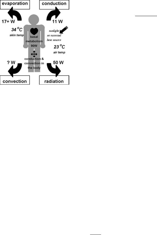
Figure 1. Schematic of heat balance mechanisms in the human body. Body temperature is determined by the balance of metabolic heat production plus heating from external sources, and heat losses by radiation, evaporation, convection, and conduction.
conduction, convection, radiation and evaporation, that is,
Q0 |
Dt > Q0 |
Dt |
1 |
Þ |
dep |
loss |
|
ð |
where Q0 ¼ DQ/Dt represents the change in heat energy, Q (measured in Joules or calories), over a time period Dt. Net heat energy deposition in a volume element DV
of tissue of density rtis results in an increase in temperature DT dependent on the specific heat of the tissue,
ctis,
Q0 |
|
Qloss0 |
!Dt ¼ ðrtisDVÞctisDT |
|
|||
dep |
|
|
|||||
DV |
DV |
|
|||||
|
|
|
DT ¼ ðQdep0 |
Qloss0 |
Þ |
Dt |
ð2Þ |
|
|
|
|
||||
|
|
|
rtisctis |
||||
Heat deposition is the sum of the absorbed power density, Pabs, from external sources and heat generated by metabolism, Qmet,
DQ0 |
|
DQmet0 |
|
|
dep |
|
|
||
|
¼ Pabsðx; y; z; tÞ þ |
|
ðx; y; z; tÞ |
ð3Þ |
DV |
DV |
|||
If the air temperature is higher than the body surface temperature, heat is absorbed from air surrounding the body by the skin, as well as during respiration. Power deposition in tissue from external electromagnetic fields depends on the coupling of the radiation field (microwave, RF, ultrasound, visible or IR light) with tissue. The body’s metabolic rate depends on the amount of muscular activity, the temperature, pressure and humidity of the environment, and the size of the body. Metabolic rate increases nonlinearly with core body temperature, in part due to the exponential increase of the rate of chemical reactions with temperature (Arrhenius equation). An empirical relationship between
basal metabolic rate and core |
temperature has been |
|||||||
determined as |
|
|
|
|
|
|
|
|
Basal MR |
¼ |
|
85 |
1:07ðTcoreÞ |
ð |
4 |
Þ |
|
0:5 |
||||||||
|
||||||||
|
|
|
|
|||||
which can be exploited to maintain elevated body temperatures
(2). At room temperature a human body produces 84 W, which increases to 162 W at a core temperature of 41.8 8C.
Heat losses from the body are often termed sensible (convective, conductive, radiative) and insensible (evaporative, latent). The primary mode of heat loss from the body is by radiation, as described by the Stefan–Boltzmann law,
Q0 |
|
4 |
|
rad |
¼ eskinsAskinðTskin TsÞ |
ð5Þ |
|
DV |
|
where Q0rad/DV is the power radiated, eskin is the emissivity of the skin (radiating material), s is Stefan’s con-
stant ¼ 5.6703 10 8 W m 2/K, Askin is the skin surface area, Tskin is the temperature of the skin (radiator), and Ts is the temperature of the surroundings (e.g., air, water,
wax). Human skin is a near perfect radiator in the IR, with an emissivity of 0.97. At room temperature, >50% of the heat generated by metabolism is lost by radiation; a clothed adult loses some 50 W at room temperature. This increases to 66% at a core temperature of 41.8 8C, as is targeted in a number of systemic hyperthermia protocols, when the skin temperature rises to 39–40 8C (3).
Direct transfer of body heat to the molecules around the body (typically air) occurs by conduction, or molecular agitation within a material without any motion of the material as a whole, which is described by Fourier’s law,
DQcond0 |
¼ kAskin |
DT |
ð6Þ |
DV |
Dx |
where DQcond is the heat energy transferred per unit volume in time Dt, k is the thermal conductivity (W mK 1)
of the material surrounding the body (air, water), and DT is the temperature difference across thickness Dx of the material. Air is a poor thermal conductor, therefore heat loss by conduction is relatively low. On the other hand, water has a thermal conductivity 20 times that of air at 0 8C, increasing further with temperature, therefore during hyperthermia it is important that any water in contact with the skin is not at a lower temperature. The relative thermal conductivity of body tissues is important in determining thermal conduction within the body from external sources of heat. For example, fat is a relative thermal insulator with a thermal conductivity one third of that of most other tissues, therefore fat bodies are slower to heat.
Convective heat transfer involves material movement and occurs principally via blood moving heat to, or from, the skin and other tissues, and air currents (respiratory and environmental) moving warm air to or from the body. Equation 7 is written for the blood,
DQ0conv ¼ rbcb½wbðx; y; z; TÞ ðT TbÞ þ Ubðx; y; z; TÞ rT&
DV
ð7Þ
where wb is the specific capillary blood flow rate, Ub is the specific blood flow through other vessels. In the context of
systemic hyperthermia, where a patient is in a closed chamber, environmental air currents can be minimized. Heat loss by respiration, however, can amount to almost 10% of metabolic heat generation.
Another route of heat loss from the body is evaporation of perspiration from the skin. Because of the very large heat of vaporization of water, cooling of the blood in skin capillaries occurs due to evaporation of sweat. Evaporation from exhaled moisture also results in cooling of the surrounding air.
DQevap0 |
Lv |
|
|
|
¼ mw |
|
ð8Þ |
DV |
Dt |
||
where mw is the mass of the water and Lv is the latent heat of vaporization (2.4 106 J kg 1 at 34 8C). In hot conditions with maximal rates of evaporation, heat loss through evaporation of sweat can be as much as 1100 W. Heat loss in the lungs is 10 W.
Combining the heat generation and heat loss terms leads to a general heat transfer equation, an extension
of the classic Pennes bioheat transfer equation. |
|
||||||||||||
|
DV |
þ Pabs |
|
|
|
|
|
|
|
||||
|
DQmet0 |
|
|
|
|
|
|
|
|
|
|
|
|
|
DV |
|
þ DV |
þ DV |
þ DV |
þ DV |
|
||||||
|
|
DQ0 |
|
DQ0 |
DQ |
DQ0 |
DQ0 |
|
|||||
|
|
rad |
|
cond |
|
conv0 |
|
resp |
|
evap |
|
||
|
|
¼ rtisctisDT |
|
|
|
|
|
|
ð9Þ() |
||||
into which the expressions given in Eqs. 2–8 may be substituted. Precise solution of this equation for temperature distribution is complex and requires a number of simplifying assumptions which have generated significant controversy in bioheat transfer circles. Modeling of temperature distributions within a body subjected to hyperthermia is also complex because of the heterogeneity of thermal characteristics between and within tissue, the directionality of power application, and the dynamic nature of thermoregulation by human body. Nonetheless, the factors governing systemic heating of the body can be appreciated.
INDUCTION OF SYSTEMIC HYPERTHERMIA
Apart from the induction of biological fever by pathogens or toxins, all methods of hyperthermia involve transfer of heat into the body from an external energy source. The required net power to raise the temperature of a 70 kg human from 37 to 41.8 8C (2) is 400 W (5.7 mW). While the heat absorption from these sources is highly nonuniform, distribution of thermal energy by the vascular system quickly results in a uniform distribution of temperature. Indeed, systemic hyperthermia is the only way to achieve uniform heating of tissues. Because physiological thermoregulation mechanisms such as vasodilation and perspiration counteract attempts to increase core body temperature, careful attention must be paid to optimizing the physical conditions for heating such that there is efficient deposition of heat energy in the body and, even more importantly, minimization of heat losses. Wrapping the body in reflective blankets, foil, or plastic film to reduce radiative and evaporative losses, or keeping the surrounding air moist to
HYPERTHERMIA, SYSTEMIC |
45 |
minimize losses by perspiration are key techniques for achieving a sustained increase in body temperature.
Noninvasive methods of heating include immersing the body in hot water or wax, wrapping the body in a blanket or suit through which heated water is pumped, placing the patient on a heated water mattress, surrounding the body with hot air, irradiating with IR energy, and applying RF or microwave electromagnetic energy. These techniques may be applied singly or in combination. For example, the Pomp–Siemens cabinet used until recently throughout Europe, as well as in the United States, a modification of a device originally developed by Siemens in the 1930s, has the patient lying on a heated water mattress under which an inductive loop generates an RF field, all inside a chamber through which hot air is circulated. The Russian Yakhta-5 system applies a high frequency (13.56 MHz) electromagnetic field through a water-filled mattress to permit whole body heating up to 43.5 8C and simultaneous deep local hyperthermia through additional applicators providing 40.6 MHz electromagnetic radiation. The majority of whole-body hyperthermia systems currently in clinical use employ IR radiation to achieve systemic heating. Invasive approaches to systemic hyperthermia are extracorporeal heating of blood, removed from the body via an arteriovenous shunt, prior to returning it to the circulation, as well as peritoneal irrigation with heated fluid (4). A useful schematic summary of whole-body hyperthermia induction techniques along with references is provided by van der Zee (5).
All of these approaches involve a period of steady temperature increase, followed by a plateau or equilibrium phase where the target temperature is maintained for anywhere from 30 min to several hours, and finally a cool-down phase. Depending on the method of hyperthermia induction, the patient may be anesthetized, consciously sedated, administered analgesia, or not given any kind of medication at all. An epidural block is sometimes given to induce or increase vasodilation. During radiant heat induction, the temperature of the skin and superficial tissues (including tumors) is higher than the core (rectal) temperature whereas during the plateau (maintenance) phase, the skin–superficial tissue temperature drops below the core temperature. As already described, heat losses due to physiological mechanisms limit the rate of heating that can be achieved. When insulation of the patient with plastic foil was added to hot air heating, the heating time to 41.8 8C was decreased from 230 to 150 min (65%), and further to 110 min (48%) by addition of a warm water perfused mattress (5). The homogeneity of the temperature distribution was also significantly increased by the addition of insulation and the water mattress. Noninvasive systemic hyperthermia methodologies typically produce heating rates of 1–10 8C h 1 with 2–3 8C h 1 being most common. More rapid heating can be achieved by the invasive techniques, at the expense of greater risk of infection and morbidity.
COMMERCIALLY AVAILABLE WHOLE-BODY HYPERTHERMIA SYSTEMS
A number of commercially available devices have resulted from the development of these initially experimental
46 HYPERTHERMIA, SYSTEMIC
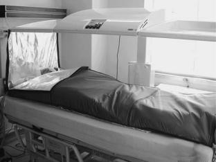
systems. The Siemens–Pomp system has already been mentioned, but is no longer commercially available. Similarly, neither the radiant heat chamber developed by Robins (3), and marketed as the Aquatherm system, nor the similar Enthermics Medical Systems RHS-7500 radiant heat device, both producing far IR radiation (IR C) in a moist air chamber, are currently being sold, though they are still in use in several centers. A close relative is the Iratherm2000 radiant heat chamber originally developed by von Ardenne and co-workers (6). In this device, waterfiltered infrared radiators at 2400 8C emit their energy from above and below the patient bed, producing near-IR (IR A) radiation that penetrates deeper into tissue than far IR radiation, causing direct heating of the subcutaneous capillary bed. Thermal isolation is ensured by reflective foils placed around the patient. However, note that significant evaporative heat loss through perspiration can be a problem with this system. Also with a significant market share is the Heckel HT 2000 radiant heat device in which patients lie on a bed enclosed within a soft-sided rectangular tent whose inner walls are coated with reflective aluminum foil that ensures that the short wavelength infrared A and B radiation emitted by four radiators within the chamber uniformly bathes the body surface. Once the target temperature is reached, the chamber walls are collapsed to wrap around the body, thereby preventing radiative and evaporative heat loss, and permitting maintenance of the elevated temperature, as shown in Fig. 2.
Another radiant heat device, used mainly in Germany, is the HOT-OncoTherm WBH-2000 whole-body hyperthermia unit which is a chamber that encloses all but the patient’s head. Special light-emitting diode (LED) radiators deliver computer-generated, alloy-filtered IR A wavelengths that penetrate the skin to deliver heat to the capillary bed. The manufacturer claims that these wavelengths also preferentially stimulate the immune system. Recently, Energy Technology, Inc. of China has released the ET-SPACE whole-body hyperthermia system, which
Figure 2. Heckel HT-2000 radiant heat whole body hyperthermia system. Unit at the University of Texas Medical School at Houston. Patient is in the heat maintenance phase of treatment, wrapped in the thermal blankets which form the sides of the chamber during active heating.
produces IR A radiation in a small patient chamber into which warm liquid is infused to help increase the air humidity and thereby reduce perspiration losses. A number of low cost, far infrared, or dry, saunas are being sold to private clinics, health clubs, and even individuals for treatment of arthritis, fibromyalgia, detoxification, and weight loss. Examples are the Smarty Hyperthermic Chamber, the TheraSauna, the Physiotherm, and the Biotherm Sauna Dome. Table 2 summarizes features of these commercially available whole-body hyperthermia devices.
BIOLOGICAL EFFECTS OF SYSTEMIC HYPERTHERMIA
An understanding of the biological effects of systemic hyperthermia is critical to both its successful induction and to its therapeutic efficacy. Systemic responses to body heating, if not counteracted, undermine efforts to raise body temperature, while cellular effects underlie both the rationale for the use of hyperthermia to treat specific diseases, and the toxicities resulting from treatment. Although improved technology has allowed easier and more effective induction of systemic hyperthermia, most of the recent clinical advances are due to better understanding and exploitation of specific biological phenomena.
Physiological Effects of Elevated Body Temperature
The sympathetic nervous system attempts to keep all parts of the body at a constant temperature, tightly controlled by a central temperature ‘set point’ in the preoptic–anterior hypothalamus and a variety of feedback mechanisms. The thermostat has a circadian rhythm and is occasionally reset, for example, during fever induced by infectious agents and endotoxins, but not in endogenously induced hyperthermia. Occasionally, it breaks down completely as in malignant hyperthermia or some neurological disorders affecting the hypothalamus. Ordinarily, when core body temperature rises, the blood vessels initially dilate, heart rate rises, and blood flow increases in an effort to transport heat to the body surface where it is lost by radiation, conduction, and convection. Heart rate increases on average by 11.7 beats min 1 8C 1 and typically remains elevated for several hours after normal body temperature is regained. Systolic blood pressure increases to drive the blood flow, but diastolic pressure decreases due to the decreased resistance of dilated vessels, thus there is an increase in cardiac output. Heart rate and blood pressure must therefore be monitored during systemic hyperthermia, and whole-body hyperthermia is contraindicated in most patients with cardiac conditions. Interestingly, hyperthermia increases cardiac tolerance to ischemia/reperfusion injury probably due to activation of manganese superoxide dismutase (Mn-SOD) and involvement of cytokines.
Respiration rate also increases and breathing becomes shallower. Perspiration results in evaporation of sweat from the skin and consequent cooling, while the respiration rate increases in order to increase cooling by evaporation of moisture from expired air. Weight loss occurs despite fluid intake. There is a decrease in urinary output and the urine has a high specific gravity, concentrating urates and

47
Table 2. Commercially Available Clinical Whole-Body Hyperthermia Devices
Manufacturer |
Website |
Device Name |
Heating Mechanism |
Temperature Range, 8C |
Application |
|
|
|
|
|
|
|
|
Energy Technology |
http://www.eti.com.cn/EN/pro/product2.htm |
ET-SPACE |
Multiple IR radiators |
39–41.8 |
Oncology |
|
|
|
|
|
(IR A) |
|
|
Heckel Medizintechnik GmbH |
http://www.heckel-medizintechnik.de/ |
HT 2000 M |
4 |
300W IR radiators |
38.5–40.5 |
Oncology, |
|
frameset_e.html |
|
|
(IR A, B) |
|
rheumatology |
Hot-Oncotherm |
http://www.hot-oncotherm.com/ |
WBH-2000 |
Multiple LED |
37–42 |
Oncology |
|
|
oncothermia.htm |
|
|
radiators (IR A) |
|
|
Von Ardenne Institut fu¨ r |
http://www.ardenne.de/med_eng/ |
Iratherm 800 |
4 |
IR radiators (IR A) |
37–38 |
Physical medicine, |
Angewandte |
|
|
|
|
|
complementary |
Medizinische Forschung, GmbH |
|
|
|
|
|
medicine, oncology |
|
|
Iratherm 1000 |
6 |
IR radiators (IR A) |
37–39 |
|
|
|
Iratherm 2000 |
10 IR radiators (IR A) |
37–42 |
|
|
|
|
|
|
|
|
|
48 HYPERTHERMIA, SYSTEMIC
phosphates. In endogenously induced hyperthermia, but not in fever, glomerular filtration, as evidenced by the creatinine clearance, decreases with increasing temperature. As already mentioned, metabolic rate increases nonlinearly with temperature, which leads to an increase in blood sugar, decreased serum potassium levels, and increased lactic acid production. All the above normal physiological effects may be enhanced or counteracted by anesthesia or sedation, as well as by disease states such as cancer because of drugs used in treatment or intrinsic pathophysiological consequences of the disease.
At 42.5 8C, the normal thermocompensatory mechanisms break down and the body displays the symptoms of advanced heat stroke, namely, lack of sweating, rapid heart beat, Cheyne–Stokes breathing, central nervous system disfunction, and loss of consciousness. Ultimately, breathing ceases despite the continuation of a heart beat.
Cellular Thermal Damage
When temperature is increased by a few degrees Celcious, there is increased efficiency of enzyme reactions (Arrhenius equation), leading to increased metabolic rates, but at temperatures > 40 8C molecular conformation changes occur that lead to destabilization of macromolecules and multimolecular structures, for example, to the side chains of amino acids in proteins, which in turn inhibit enzyme action. Small heat shock proteins (HSP) interact with the unfolding proteins to stabilize them and prevent their aggregation and precipitation. Eventually, however, at42 8C, complete denaturation of proteins begins that totally disrupts many molecular processes, including deoxyribonucleic acid (DNA) repair. Thus systemic hyperthermia can have significant effects when paired with drugs that cause DNA damage (e.g., for chemotherapy of cancer).
Membranes are known to be extremely sensitive to heat stress because of their complex molecular composition of lipids and proteins. At a certain temperature, lipids change from the tightly packed gel phase to the less tightly packed liquid crystalline phase, and permeability of the cell membrane (membrane fluidity) increases. As temperature increases further, the conformation of proteins also becomes affected, eventually resulting in disorderly rearrangement of the lipid bilayer structure and receptor inactivation or loss. Temperature changes of 5 8C are necessary to cause measurable changes in normal cell membrane permeability. Heat-induced cell membrane permeability can be exploited to increase drug delivery, for example, transdermally, or into tumor cells. Increased vascular permeability due to thermal increase of endothelial gap size also aids drug delivery into tumors. At higher temperatures, heat damage to membranes can cause cell death, but it will also interfere with therapeutic approaches that depend on membrane integrity (e.g., receptor targeted drug delivery, antibodies, etc.). Irreversible disruption of cytoplasmic microtubule organization and eventual disaggregation, as well as disruption of actin stress fibers and vimentin filaments, occur at high temperatures (43–45 8C) above those used in whole-body hyperthermia, but these cytoskeletal effects are of concern with loco-regional hyperthermia.
A variety of effects in the cell nucleus also occur at high temperatures (>41 8C) including damage to the nuclear membrane, increases in nuclear protein content, changes in the structure of nucleoli, inhibition of DNA synthesis and chromosomal damage in S-phase. These changes in nuclear structure compromise nuclear function and may cause cell death, though they are unlikely to be significant at the temperatures achieved in systemic hyperthermia. Disaggregation of the spindle apparatus of mitotic cells may be responsible for the high thermal sensitivity of cells in mitosis, as well as in S phase. Hyperthermic inactivation of polymerase b, an enzyme primarily involved in DNA repair, is sensitized by anesthetics and may have a role to play in the enhancement of the effects of ionizing radiation by systemic hyperthermia, as well as in augmenting the cytotoxic effect of drugs that cause DNA damage.
Metabolic Effects
Moderate increases in temperature lead to increased cellular reaction rates, which may be seen as increased oxygen consumption and glucose turnover. In consequence, cells may become deprived of nutrients, the intracellular ATP concentration falls, accumulation of acid metabolites increases pH, and thermal sensitivity increases. Such conditions are found in tumors and may contribute to their sensitivity to heat. Further acidifying tumor cells during hyperthermic treatment seems a promising approach as is discussed further below. At high temperatures, the citric acid cycle may be damaged leading to other acidic metabolites. Increased plasma acetate has been measured following clinical whole-body hyperthermia treatments, which reduces both release of fatty acids from adipose tissue into plasma and subsequent lipid oxidation.
Endocrine Function
Increases in plasma levels of an array of hormones have been noted after whole-body hyperthermia. Increased ACTH levels appear to be accompanied by increased levels of circulating endorphins. This may explain the sense of well-being felt by many patients after systemic hyperthermia treatment, and the palliative effect of hyperthermia treatments for cancer. Increased secretion of somatotropic hormone after systemic hyperthermia has also been measured (7).
Thermal Tolerance
Thermal tolerance is a temporary state of thermal resistance, common to virtually all mammalian cells, which develops after a prolonged exposure to moderate temperatures (40–42 8C), or a brief heat shock followed by incubation at 37 8C, and also certain chemicals. The decay of thermotolerance occurs exponentially and depends on the treatment time, the temperature, and the proliferative status of the cells. Several days are usually required for baseline levels of heat sensitivity to be regained, which has important implications for fractionated therapy. When pH is lowered, less thermal tolerance develops, and its decay is slower. Thus the long periods at moderate temperature achieved by clinical systemic hyperthermia systems should
|
2.0 |
|
uptake ratio |
1.0 |
|
41.5˚C/37˚C |
|
|
|
0.0 |
|
|
100 nm |
210 nm |
|
|
Liposome size |
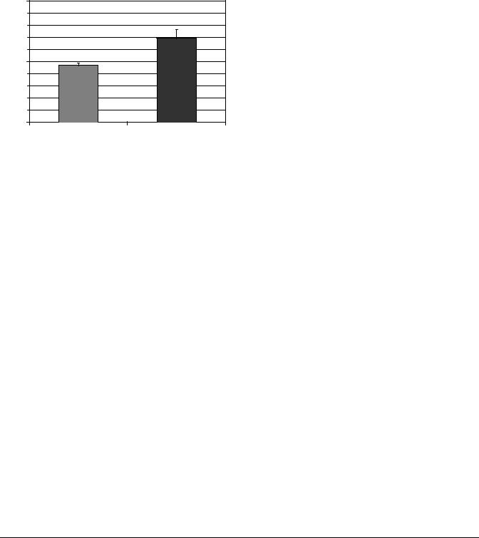
Figure 3. Increase in tumor uptake of large liposomes after 1 h of 41.5 8C whole-body hyperthermia. Systemic heat treatment increased the effective pore size from 210 to 240 nm. Because of the large pore size in MTLn3 tumors, 100 nm (average diameter) liposomes were able to pass into the tumor equally well at normal and elevated temperatures. The increased effective pore size due to hyperthermia allowed larger 200 nm liposomes, which were partially blocked at normal temperatures, to pass more effectively into the tumor.
induce thermal resistance in normal cells, while the acidic parts of tumors should be relatively unaffected. This has not, however, been studied clinically. The mechanisms involved in the induction of thermotolerance are not well understood, but there is mounting evidence that heat shock proteins are involved.
Step-Down Sensitization
Another distinct phenomenon is step-down sensitization in which an exposure of cells to temperatures >43 8C results in increased sensitivity to subsequent temperatures of 42 8C or lower. This can be important clinically for local and regional hyperthermia if there are marked variations in temperature during the course of treatment, the magnitude of the effect depending on the magnitude of the temperature change. It has been suggested that this phenomenon could be exploited clinically by administering a short, high temperature treatment prior to a prolonged treatment at a lower temperature, thereby reducing pain and discomfort. Since temperatures >43 8C cannot be tolerated systemically, a local heat boost would be required to take advantage of this effect for whole body hyperthermia. So far, there is no evidence that tumor cells are differently sensitized by step-down heating than normal cells.
HYPERTHERMIA, SYSTEMIC |
49 |
Effect of Hyperthermia on Tumors
It was initially thought that tumor cells have intrinsically higher heat sensitivity than normal cells, but this is not universally true. Although some neoplastic cells are more sensitive to heat than their normal counterparts, this appears to be the case at temperatures higher than those used in systemic hyperthermia. Tumors in vivo, on the other hand, often do have a higher thermal sensitivity than normal tissues because of abnormal vasculature (reduced blood flow), anaerobic metabolism (acidosis), and nutrient depletion. Due to their tortuous and poorly constructed vasculature, tumors have poor perfusion, thus heat dissipation by convection is reduced. At high temperatures (43 8C and up) this means that tumors become a heat reservoir with a consequent rise in temperature, which if maintained for too long damages the microcirculation and further impairs convective heat loss. Also increased fibrinogen deposition at damaged sites in the vascular wall leads to clusion of tumor microvessels. Significant heating of the tumor cells results, which may be directly cytotoxic. Additionally, the impaired blood flow brings about acidosis, increased hypoxia and energy depletion all of which increase the heat sensitivity of tumor cells (8). At lower temperatures, typical of those achieved in whole-body hyperthermia, blood flow increases (9) though the mechanism is not well understood. For these reasons, along with the historical evidence for antitumor effects of fever and the metastatic nature of malignant disease, cancer has become the main focus of systemic hyperthermia.
Systemic hyperthermia results in increased delivery of drugs to tumor sites because of increased systemic blood flow. It can also increase blood vessel permeability by increasing the effective pore size between the loosely bound endothelial cells forming tumor microvessels, permitting larger molecules, such as nanoparticles and gene therapy vectors, to pass into the interstitium (10). Figure 3 shows increased uptake of 210 nm liposomes in rat breast tumors after 1 h of 41.5 8C whole-body hyperthermia. Heat may also be toxic to endothelial cells, resulting in a transient normalization of vascular architecture and improvement in blood flow (11). Another barrier to drug delivery is the high interstitial pressure of many tumors. Since wholebody hyperthermia, even at fever-range temperatures, causes cell death (apoptosis and necrosis) within tumors it reduces the oncotic pressure allowing greater penetration of large molecules. Table 3 summarizes the interactions of systemic hyperthermia which facilitate nanoparticle delivery to tumors.
Table 3. Whole-Body Hyperthermia Facilitates Nanoparticle Therapy
Heat Interaction |
Therapeutic Effect |
|
|
" Blood flow |
" Nanoparticle delivery to tumor |
" In endothelial gap size |
" Nanoparticles in interstitium |
" Endothelial cell apoptosis/necrosis ! transient normalization of vasculature |
" Nanoparticles in interstitium |
" Tumor cell apoptosis/necrosis # oncotic pressure |
" Nanoparticles in interstitium |
Temperature-dependent " in permeability of liposome bilayer |
" And synchronization of drug release |
Cellular and molecular effects in tumor |
" Drug in tumor cell " drug efficacy |
Direct interactions with drug |
" Drug efficacy |
50 HYPERTHERMIA, SYSTEMIC
Whole-Body Hyperthermia and the Immune System
An increase in ambient temperature can serve as a natural trigger to the immune system and it appears that the thermal microenvironment plays a critical role in regulating events in the immune response. The early work of Coley on cancer therapy with infectious pyrogens implicated fever-induced immune stimulation as the mediator of tumor responses (1). While there have been numerous in vitro studies of the effect of temperature on components of the immune system, indicating that the thermal milieu regulates T lymphocytes, natural killer (NK) cells, and dendritic cells (DC), in vivo examinations of the immune effects of systemic hyperthermia are relatively few. Initial animal model studies concluded that whole-body hyperthermia resulted in immunosuppression, but high temperatures were used, tumors were mostly immunogenic, and immune response was merely inferred from the incidence of metastatic spread rather than from measurement of specific markers of immune system activation. The majority of in vivo studies in animals provide evidence of a nonspecific host reaction in response to hyperthermia in which both T and B lymphocytes, as well as macrophages, are involved (12). Although NK cells are intrinsically more sensitive in vitro to heat than B and T cells, their activation by systemic hyperthermia has been observed. Microwave induced whole-body hyperthermia of unrestrained, unanesthetized mice at 39.5–40 8C for 30 min, three or six times weekly, resulted in increased NK cell activity and reduced pulmonary metastasis in tumor-bearing mice, but none in normal mice (13). Evidence for hyperthermia-induced human tumor lysis by IL-2 stimulated NK cells activated by HSP72 expression also exists (14). Increased numbers of lymphocyte-like cells, macrophages, and granulocytes are observed in the tumor vasculature and in the tumor stroma of xenografts and syngeneic tumors in mice immediately following a mild hyperthermia exposure for 6–8 h. In the SCID mouse/human tumor system tumor cell apoptosis seen following treatment was due largely to the activity of NK cells. The investigators hypothesize heat dilatation of blood vessels and increased vessel permeability may also give immune effector cells greater access to the interior of tumors (15). In balb/C mice, fever-range whole-body hyperthermia increased lymphocyte trafficking, resulting in early responsiveness to antigen challenge (16). Thus systemic hyperthermia may be an effective, nontoxic adjuvant to immunotherapy.
A recent clinical study examined the effect of wholebody hyperthermia combined with chemotherapy on the expression up to 48 h later of a broad range of activation markers on peripheral blood lymphocytes, as well as serum cytokines and intracellular cytokine levels in T cells, and the capacity of these cells to proliferate. Immediately after treatment with 60 min of 41.8 8C WBH as an adjunct to chemotherapy, a drastic but transient, increase in peripheral NK cells and CD56þ cytotoxic T lymphocytes was observed in the patients’ peripheral blood. The number of T cells then briefly dropped below baseline levels, a phenomeonon that has also been observed by others (17). A marked, but short-lived, increase in the patients’ serum levels of interleukin-6 (IL-6) was also noted. Significantly
increased serum levels of tumor necrosis factor-alpha (TNF-alpha) were found at 0, 3, 5 and 24 h posttreatment. Further immunological consequences of the treatment consisted of an increase in the percentage of peripheral cytotoxic T lymphocytes expressing CD56, reaching a maximum at 48 h post-WBH. Furthermore, the percentage of CD4+ T cells expressing the T cell activation marker CD69 increased nearly twofold over time, reaching its maximum at 48 h. Since similar changes were not observed in patients receiving chemotherapy alone, this study provided strong evidence for prolonged activation of human T cells induced by whole-body hyperthermia combined with chemotherapy (18).
Activation of monocytes has been observed following hot water bath immersion such that response to endotoxin stimulation is enhanced with concomitant release of TNF-a. Macrophage activation and subsequent lysosomal exocytosis were observed in the case of a patient treated for liver metastases by hyperthermia. Lysosomal exocytosis induced by heat may be an important basic reaction of the body against bacteria, viruses, and tumor growth and was proposed as a new mechanism of thermally induced tumor cell death mediated by an immune reaction (19).
Several investigators have suggested that the immune changes seen during in vivo whole-body hyperthermia are mediated by elevations in the plasma concentrations of either catecholamines, growth hormone, or beta-endorphins. In volunteers immersed in a heated water bath, neither recruitment of NK cells to the blood, nor the percentages or concentrations of any other subpopulations of blood mononuclear cells were altered by hormone blockade. However, somatostatin partly abolished the hyperthermia induced increase in neutrophil number. Based on these data and previous results showing that growth hormone infusion increases the concentration of neutrophils in the blood, it was suggested that growth hormone is at least partly responsible for hyperthermia induced neutrophil increase. A similar study suggested that hyperthermic induction of T lymphocytes and NK cells is due to increased secretion of somatotropic hormone (7).
The peripheral blood level of prostaglandin E2 (PGE2), which may act as an angiogenic switch, transforming a localized tumor into an invasive one by stimulating new blood vessel growth, and which also has an immunosuppressive effect, is elevated in patients with tumors compared to healthy control subjects. In a clinical study of cancer patients receiving 1–2 h of 41.8–42.5 8C whole-body hyperthermia, or extracorporeal hyperthermia, blood levels of PGE2 decreased markedly after treatment and correlated with tumor response (20).
In addition to their role as protectors of unfolding proteins, extracellular heat shock proteins (HSP) can act simultaneously as a source of antigen due to their ability to chaperone peptides and as a maturation signal for dendritic cells, thereby inducing dendritic cells to cross-present antigens to CD8þ T cells (21). Heat shock proteins can also act independently from associated peptides, stimulating the innate immune system by eliciting potent proinflammatory responses in innate immune cells. The heat shock response also inhibits cyclooxygenase-2 gene expression at the transcriptional level by preventing the activation of
nuclear factor-kappaB (NFkB) (22). Thermal upregulation of HSPs (HSP70 and HSP110) is strongest in lymphoid tissues and may relate to the enhanced immune responses that are observed during febrile temperatures. It has been proposed that local necrosis induced by hyperthermic treatment induces the release of HSPs, followed by uptake, processing and presentation of associated peptides by dendritic cells. By acting as chaperones and as a signal for dendritic cell maturation, HSP70 might efficiently prime circulating T cells. Therefore, upregulating HSP70 and causing local necrosis in tumor tissue by hyperthermia offers great potential as a new approach to directly activate the immune system, as well as to enhance other immunotherapies (23,24).
CLINICAL TOXICITIES OF WHOLE-BODY HYPERTHERMIA TREATMENT
At fever-range temperatures, adverse effects of systemic hyperthermia treatment are minimal however, at higher temperatures they can be significant, even fatal. On the other hand, the teratogenic effects (birth defects, still births, spontaneous abortions) and 8Cular damage (cataract induction) resulting from electromagnetic fields used in local hyperthermia are not seen in systemic hyperthermia. The transient cardiorespiratory effects of elevated temperature can, however, lead to severe toxicity. Elevated heart rate, especially at high temperatures may result in arrythmias or ischemic heart failure, consequently patients have to be very carefully screened with regard to their cardiac status. Beta blockade has generally been found to be deleterious although infusion of esmolol has been safely carried out (25). Pulmonary hypertension and edema due to capillary leak may also be seen, but like the cardiac effects, these return to baseline a few hours after treatment. Increased serum hepatic enzymes have been noted, but these may be cancer related. All these toxicities are less prevalent or less severe with radiant heat systems, particularly at lower temperatures, and when light conscious sedation is used rather than general anesthesia. For example, decreased platelet count, decreased plasma fibrinogen, and other factors leading to increased blood clotting have been noted, particularly in extra-corporeal hyperthermia, but also with other methods of heating carried out under inhalation-administered anesthesia drugs. On the other hand, with whole-body hyperthermia under conscious sedation there is no evidence of platelet drops (26) and animal studies even show platelet stimulation providing protection against radiation induced thrombocytopenia.
Since systemic hyperthermia is almost never used as a single treatment modality, it is important to recognize that whole-body hyperthermia combined with radiation and chemotherapy can enhance some of the toxicities associated with these modalities. For example, the cardiotoxicity of doxorubicin and both the renal toxicity and hematological toxicity of platinum agents may increase under hyperthermia (27), while the muscle and peripheral nervous system effects of radiation and some drugs can also be enhanced (28). Bone marrow suppression is the limiting
HYPERTHERMIA, SYSTEMIC |
51 |
toxicity of many chemotherapy drugs but there is little data to suggest that whole body hyperthermia exacerbates this effect. On the contrary, the synergy of hyperthermia with several chemotherapy agents may mean that lower doses can be used, resulting in less toxicity. For example, systemic hyperthermia combined with carboplatin achieves therapeutic results without elevation of myelosuppression and responses have occurred at lower than normal doses (29). Pressure sores can easily develop at elevated temperatures thus care must be taken not only in patient placement and support, but also with application of monitoring devices. If heat dissipation is locally impaired, for example, at pressure points, hot spots occur that can lead to burns. This is rarely a problem with fever-range wholebody hyperthermia, but in anesthetized patients undergoing high heat regimens burns are not uncommon.
Following systemic hyperthermia treatments, malaise and lethargy are almost universally experienced although these may be counteracted by pain relief and a sense of well-being due to released endorphins. However, the faster the target temperature is reached, the less the exhaustion (6), thus attention to minimizing heat dissipation during the heat-up phase and using efficient heating devices, such as those that generate heat by several mechanisms (e.g., radiant heat and EM fields), add a regional heat boost, or produce near-IR radiation that is preferentially absorbed, is advantageous to patient well being. Fever after treatment in the absence of infectious disease is not uncommon and may be associated with an inflammatory response to tumor regression. Nausea and vomiting during the first couple of days after treatment are also common. Outbreaks of herpes simplex (cold sores) in susceptible individuals have also been noted, but are easily resolved with acyclovir.
THERMAL DOSE
The definition of dose for systemic hyperthermia is problematic. An applied dose would be the amount of heat energy generated or delivered to the body but even if it can be measured, this quantity does not predict biological effects. By analogy with ionizing radiation, the absorbed dose would be amount of thermal energy absorbed per unit mass of tissue (J kg 1), however, this is not a quantity that can be readily measured, or controlled, neither would it necessarily predict biological effects. As indicated in the previous sections, the effects of systemic hyperthermia depend on (1) the temperature, and (2) the duration of heating, but not on the energy required to produce the temperature rise. This leads to the concept of time at a given temperature as a practical measure of dose. In reality, however, temperature is seldom constant throughout a treatment, even in the plateau phase of systemic hyperthermia, so time at temperature is at best a crude measure. Nonetheless, it is the one that is used most often clinically for whole-body hyperthermia because of its simplicity. Ideally, the dose parameter should allow for comparison of treatments at different temperatures. Based on the Arrhenius relationship and measured cell growth inhibition curves, the heating time at a given temperature relative to the heating time at a standard temperature or

52 HYPERTHERMIA, SYSTEMIC
thermal dose equivalent (TDE), was defined empirically as,
T1 ¼ t2 RðT1 T2Þ |
ð10Þ |
A discontinuity occurs in the temperature-time curves between 42 and 43 8C for both cells in culture and heated tissues, thus the value of R changes for temperatures above the transition: R 2 < 42.5 8C and R 5 > 42.5 8C in vitro while for in vivo heating studies, R ¼ 2.1 below the transition temperature and 6.4 above 42.5 8C. In practice, a finite time is required for the body or tissue of interest to reach the target temperature, temperature fluctuates even after the target temperature is reached, and there is a cooling period after heating ceases. If the temperature is measured frequently throughout treatment, the temperature–time curves can be integrated to provide the accumulated thermal dose that produces an equivalent effect to that resulting from holding the cells–tissue at a constant reference temperature for a given a period of time:
t f |
|
t43 ¼ Z R43 TðtÞdt |
ð11Þ |
ti
where ti and tf are the initial and final times of the heating procedure (30). This thermal isoeffect dose (TID) is usually expressed in minutes is sometimes known as the tdm43 or the cumulative equivalent minutes (CEM 43 8C). While a biological factor has now been built in to the dose measure, and the integrated TID allows for temperature variations during heat-up and cool-down phases, it does not take into account thermal tolerance and step-down sensitization. Nor is it particularly relevant to clinical whole-body hyperthermia where multiple physical and biological effects combine in a complex manner although for a given patient, time– temperature profiles are generally reproducible from one treatment to another. A further modification attempts to take into account temperature inhomogeneity through the measurement of temperature at multiple sites and defining T90, namely, that temperature exceeded by 90% of the measurements (or correspondingly 20%: T20; or 50%: T50). The TID is then expressed as cumulative equivalent minutes that T90 is equal to 43 8C (CEM 43 8C T90) (31).
The efficiency of adjuvant hyperthermia in enhancing the biological effectiveness of other treatments is often reported in terms of the thermal enhancement factor (TEF) or thermal enhancement ratio (TER). This quantity is defined in terms of the isoeffect dose as,
dose of treatment to achieve a given endpoint
TER ¼ dose of treatment with heat ð12Þ to achieve the same endpoint
In clinical and laboratory studies, the TER is often computed on the basis of isodose rather than isoeffect, for example, in the case of hyperthermia plus drug induced arrest of tumor growth, TER ¼ TGDHT/TGTRT, where TGDHT is the tumor growth delay due to hyperthermia plus chemotherapy, and TGTRT is the tumor growth delay resulting from chemotherapy at room temperature. Similarly, the enhancing effect of hyperther-
mia on radiation treatment may be expressed through
TER ¼ D0HT/D0RT or TER ¼ LD50HT/LD50RT, where D0 is the time required to reduce survival to 1/e of its initial
value, and LD50 is the lethal dose to 50% of cells.
TEMPERATURE MEASUREMENT
Since systemic hyperthermia achieves a uniform temperature distribution, except for possible partial sanctuary sites, thermometry for systemic hyperthermia is much less challenging than for regional or intracavitary hyperthermia, but it is still important to prevent adverse effects, especially burns. Also, convection can induce steep thermal gradients, especially around major blood vessels, so that careful placement of temperature probes is required. Most practitioners of whole-body hyperthermia measure temperature in several locations, typically the rectum, the esophagus, and at several skin sites. During heat-up, the esophageal temperature is usually 1–2 8C higher than the rectal temperature, but during plateau phase it drops to 0.5–1.5 8C below the rectal temperature. Continuous and accurate temperature measurement is particularly important when temperatures >418C are to be achieved, as critical, life-threatening changes can occur in minutes or even seconds and over changes in temperature of as little as 0.1–0.2 8C because of the nonlinear response to temperature. For moderate temperature systemic hyperthermia, temperature measurement to within 0.1 8C is usually adequate, but a precision of 0.01 8C is desirable when heating to >41 8C and also allows determination of the specific absorption rate from the slope of the temperature versus time curve. The temperature measuring device must be insensitive to all other influences, such as ambient temperature, moisture, nearby electromagnetic fields, and so on and satisfying this criterion can be difficult. Frequent calibration of thermometers in the working range of temperatures is important since some thermometers appear fine at 30 8C, but drift substantially at 40 8C and above. Stringent quality control of any thermometry system is required to monitor accuracy, precision, stability, and response time.
Table 4 summarizes the different types of thermometer probes available for internal and external body temperature measurements, and their relative merits and disadvantages for systemic hyperthermia. Thermistors are most often used for standard temperature monitoring sites while thermocouples are used for tumor or other intra-tissue measurements. Recently, noninvasive methods of temperature measurement have been developed that are beginning to see application in hyperthermia. Thermography provides a two-dimensional (2D) map of surface temperature by measurement of infrared emission from the body, though deep-seated hot structures may be visualized because of heat carried by blood flow from the interior heat source to the skin. It is useful to detect skin hotspots and therefore in burn prevention. Since temperature-induced changes in the mechanical properties of tissue lead to altered ultrasound propagation velocity, mapping of ultrasound velocity can also provide a visual map of temperature. Tomographic reconstruction of 2D or 3D temperature is theoretically possible, but it is difficult in practice because of the heterogeneity of tissue characteristics. A

53
Table 4. Temperatures Probes for Systemic Hyperthermia
Probe Type |
Measurement Principle |
Accuracy |
Sensitivity |
Stability |
Advantages or Disadvantages |
|
|
|
|
|
|
Clinical |
Expansion of mercury or |
Moderate 0.1 8C |
Low |
High |
Large size, inflexible. Slow response. |
|
alcohol in glass |
High 0.02 8C |
|
|
|
Platinum resistance |
Linear resistance change |
|
Used as standard for |
Expensive. Difficult to calibrate. Large size. |
|
thermometer |
with temperature |
|
|
calibration |
Sensitive to shock. |
|
|
|
|
of other types |
|
|
|
Moderate 0.1 8C |
|
of thermometers. |
|
Thermocouple |
Seebeck effect: temperature |
Moderate to |
Moderate |
Small sensor. Nonlinear voltage |
|
|
dependent voltage |
|
high |
|
change with temp. Sensitive to EM |
|
difference between two |
|
|
|
fields. Can’t handle steep temp. gradients. |
|
conductors made of different |
|
|
|
|
|
metals |
|
|
|
|
Thermistor (e.g., |
Inverse relationship between |
High < 0.05 8C |
High |
Poor Require frequent |
Short time constant. Not interchangeable. |
Bowman |
temperature |
|
|
recalibration |
Sensitive to EM fields. |
Loop Larsen probe) |
and semiconductor resistance |
|
|
|
|
GaAs |
Temperature specific absorption |
Moderate |
Low |
|
Small size |
Optical (fiber optic probe): |
Change w/temp.: |
|
|
|
Not sensitive to EM fields. Small size. |
LCD birefringent crystal |
Color reflectance |
|
Low |
Low |
Unstable |
fluorescent phosphor |
|
|
|
|
|
|
Refraction of polarized light |
|
Low |
|
|
|
Decay of fluorescence |
Very high |
|
|
|
|
|
|
|
|
|

54
Table 5. Summary of Clinical Trials of Whole-Body Hyperthermiaa
|
Public |
Study |
Number of |
Disease |
|
|
Reference, |
First Author |
Year |
Type |
Patients |
|
Protocol |
Result of WBH |
PMID |
|
|
|
|
|
|
|
|
WBH Alone |
|
|
|
|
|
|
|
Kraybill, W.G. |
2002 |
Phase I |
|
Advanced solid tumors |
3–6 h at 39.5–40.0 8C |
Well tolerated No significant |
16, 12028640 |
|
|
|
|
|
|
adverse events # in circulating |
|
|
|
|
|
|
|
lymphocytes |
|
Steinhausen, D. |
1994 |
Phase I |
103 |
Advanced refractory or |
1 h at 41.8 8C |
Minimal side effects 52 |
8023241 |
|
|
|
|
recurrent cancers |
þ hyperglycemia |
responses (50%) |
|
WBH þ Chemotherapy |
|
|
|
|
þ hyperoxemia |
|
|
|
|
|
|
1 h at 41.8 8C þ ifosfamide |
|
|
|
Bakshandeh, A. |
2003 |
Phase II |
25 |
Nonmetastatic malignant |
Grade III/IV neutropenia and |
12609573 |
|
|
|
|
|
pleural mesothelioma |
þ carboplatin þ etoposide |
thrombocytopenia 5 partial |
|
|
|
|
|
|
2 h at 41.8–42.0 8C þ BCNU |
remissions (20%) |
|
Bull, J.M. |
1992 |
Phase II |
17 |
Advanced metastatic |
Limiting toxicity |
33, 1607734 |
|
|
|
|
|
sarcoma |
|
¼ thrombocytopenia |
|
|
|
|
|
|
|
7 responses/SD (41%) |
|
|
|
|
|
|
6 h at 40.0 8C þ doxil þ 5-FU |
" survival |
|
Bull, J.M. |
2002 |
Phase I |
13 |
Various chemotherapy |
Grade III toxicities |
60 |
|
|
|
|
|
resistant cancers |
þ metronomic interferon-a |
9 responses/SD |
|
|
|
|
|
|
6 h at 40.0 8C þ cisplatin |
(69%) |
|
Bull, J.M. |
2004 |
Phase I |
33 |
Advanced metastatic |
20 responses/SD (66%) " |
35 |
|
|
|
|
|
cancers (GI, breast, |
þ gemcitabine |
survival " quality of life |
|
|
|
|
|
head and neck, sarcoma, |
þ interferon-a |
|
|
|
|
|
|
neuroendocrine) |
|
18 responses/SD (86%) " |
|
Douwes, F. |
2004 |
Pilot |
21 |
Ovarian cancer |
1-2 h at 41.5–42.0 8C |
15108039 |
|
|
|
|
|
|
þ cisplatin |
quality of life |
|
|
|
|
|
|
or carboplatin |
|
|
|
|
|
|
|
þ hyperglycemia |
Slight " in myelotoxicity 10 |
|
Engelhardt, R. |
1990 |
Pilot |
23 |
Advance metastatic |
1 h at 41.0 8C þ cisplatin |
52, 2198312 |
|
|
|
|
|
melanoma |
þ doxorubicin |
responses/SD Response rate |
|
|
|
|
|
|
|
¼ that in literature for |
|
|
|
|
|
|
|
chemo alone |
|
Guan, J. |
2005 |
Phase II |
32 |
Advanced cancers |
|
94% responses/SD Pain |
65 |
|
|
|
|
|
|
reduction in all pts. |
|
|
|
|
|
|
|
Increased KPS |
|
|
|
|
|
|
1 h at 41.8 þ oxaliplatin |
Decreased tumor markers |
|
Hegewisch-Becker, S. |
2002 |
Phase II |
41 |
Pretreated advanced |
No excess toxicity 31 |
55, 12181242 |
|
|
|
|
|
metastatic |
þ leucovorin þ 5FU |
responses/SD (76%) |
|
|
|
|
|
colorectal cancer |
|
|
|
Hildebrandt, B. |
2004 |
Phase I/II |
28 |
Metastatic colorectal |
1 h at 41.8–42.1 8C |
Grade III/IV toxicities 11 |
44, 15204528 |
|
|
|
|
cancer |
þ hyperglycemia |
responses/SD (39%) |
|
|
|
|
|
|
þ hyperoxemia |
|
|
|
|
|
|
|
þ folinic acid þ 5-FU |
|
|
|
|
|
|
|
þ mitomycin C |
|
|
55
Hou, K. |
2004 |
Phase II |
54 |
Advanced cancers |
1–2 h at 41.8–42.5 8C, |
75.3% responses/SD 72.6% # |
|
|
|
|
|
extracorporeal |
tumor markers 70% pain |
|
|
|
|
|
þ chemotherapy |
relief improved sleep " weight, |
|
|
|
|
|
vs. chemotherapy alone |
appetite, KPS All signifly > control |
|
|
|
|
|
|
Reversible toxicities |
Ismael-Zade, R.S. |
2005 |
Pilot |
5 |
Pediatric renal cell |
3 h at 41.8–42.5 8C |
No complications 5 responses (100%) |
|
|
|
|
carcinoma |
þ doxorubicin |
|
|
|
|
|
|
þ interferon-a |
|
Kurpeshev, O.K. |
2005 |
Phase II |
42 |
Various disseminated |
1–2 h at 41.0–42.3 8C |
Regression of metastases. |
|
|
|
|
cancers |
þ poly-chemotherapy |
Pain reduction |
Richel, O. |
2004 |
Phase II |
21 |
Metastatic and recurrent |
1 h at 41.8 8C þ carboplatin |
Grade III/IV leucopenia, |
|
|
|
|
cervical cancer |
|
thrombopenia, |
|
|
|
|
|
|
anemia, renal toxicity 16 |
|
|
|
|
|
1 h at 41.8 8C þ carboplatin |
responses/SD (76%) |
Robins, H.I. |
1993 |
Phase I |
30 |
Various refractory cancers |
Myelotoxicity 9 responses (30%) |
|
Robins, H.I. |
1997 |
Phase I |
16 |
Various refractory cancers |
1 h at 41.8 8C þ L-PAM |
Lower platelet nadir |
|
|
|
|
|
|
myelosuppression 8 |
|
|
|
|
|
1 h at 41.5–41.8 8C þ paclitaxel |
responses/SD (50%) |
Strobl, B. |
2004 |
Phase II |
7 |
Metastatic cervical cancer |
Grade II alopecia Grade III/IV |
|
|
|
|
|
|
þ carboplatin |
thrombopenia, neutropenia |
|
|
|
|
|
1 h at 41.8 8C þ carboplatin |
" survival |
Westermann A.M. |
2001 |
Phase II |
14 |
Platinum resistant |
Grade IV thrombocytopenia, |
|
|
|
|
|
ovarian cancer |
|
grade III neutropenia 9 |
|
|
|
|
|
1 h at 41.8 8C þ ifosfamide |
responses/SD (64%) |
Westermann A.M. |
2003 |
Phase II |
95 |
Metastatic sarcoma |
Neutropenia, thrombocytopenia, |
|
WBH þ Radiation |
|
|
|
|
þ carboplatin þ etoposide |
infection 58 responses/SD (61%) |
|
|
|
|
1 h at 43 8C þ fractionated RT |
|
|
Overgaard, J. |
1995 |
Randomized |
70 |
Metastatic melanoma |
Improved local tumor control, |
|
|
|
multicenter |
|
|
vs. FRT alone |
" survival |
Robins, H.I. |
1990 |
Pilot |
8 |
Nodular lymphoma, chronic |
41.8 8C þ TBI vs. LON þ TBI |
8 responses/SD " survival |
|
|
|
|
lymphocytic leukemia |
|
|
68
15700247
66
57, 15581981
53, 8355046 48, 8996137
58
11378341
50, 12759526
41, 7776772
24, 2182581
aPublished since 1990.

56 HYPERTHERMIA, SYSTEMIC
number of magnetic resonance (MR) techniques have been used for thermal mapping and BSD Medical and SIEMENS Medical Systems have collaborated to develop a hybrid hyperthermia/MRI system, although it is not a whole-body hyperthermia machine. Currently, the most widely accepted MR technique is the proton resonance frequency (PRF) method that exploits the temperature dependence of the chemical shift of water. Unlike the value of the water spin-lattice relaxation time or the molecular diffusion coefficient, both of which have been used for MRI temperature measurements, the thermal coefficient relating temperature to the water chemical shift has been shown to be essentially independent of tissue type and physiological changes induced by temperature (32). Recently an interleaved gradient echo–echo planar imaging (iGE-EPI) method for rapid, multiplanar temperature imaging was introduced that provided increased temperature contrast- to-noise and lipid suppression without compromising spa- tio-temporal resolution (33).
CLINICAL EXPERIENCE
Cancer
Systemic hyperthermia has been used mostly for treatment of cancer because of its potential to treat metastatic disease. Initial treatments aimed to produce direct killing of tumor cells based on the premise, now understood not to be universally true, that cancer cells are more susceptible to elevated temperatures than normal cells, and the higher the temperature the greater the tumor cell kill. Maximally tolerated temperatures of 41.5–42 8C were therefore maintained for 1–2 h as the sole treatment. Response rates were, however, disappointing. Tumor regressions were observed in less than half the cases, no tumor cures were achieved, and remissions were of short duration. It became apparent that the heterogeneity of cell populations within tumors, along with micro-environmental factors, such as blood/ nutrient supply, pH, and oxygen tension prevent the thermotoxic results achieved in the laboratory. Consequently, the focus of research on systemic hyperthermia shifted to using hyperthermia as an adjunct to other cancer therapies, principally chemotherapy and radiotherapy. It is important to note that because of the experimental status of systemic hyperthermia treatment for cancer, almost all clinical trials, summarized in Table 5, have been performed on patients with advanced disease for whom whole-body hyperthermia, either as a sole therapy, or as an adjunct, is a treatment of last resort. In these cases, any response whatsoever is often remarkable. Nonetheless, a number of hyperthermia centers in Europe have discontinued systemic hyperthermia because the high temperature protocols required intensive patient care and led to unacceptable toxicities, especially in light of the efficacy and reduced toxicities of newer generation chemotherapies. Large, randomized, multicenter, Phase III trials are, however, needed to firmly establish the benefits of systemic hyperthermia in conjunction with chemotherapy and radiation. Also, validation and optimization of fever-range temperature protocols are much needed.
Systemic Hyperthermia and Chemotherapy. The bene-
ficial interaction of hyperthermia with several classes of chemotherapy agents, acting via several mechanisms as summarized in Table 6, has spurred a variety of thermochemotherapy regimens and several clinical trials of systemic hyperthermia and chemotherapy are ongoing. While the results have been mixed, elevated response rates were recorded in the treatment of sarcoma when systemic hyperthermia was combined with doxorubicin and cyclophosphamide (54) or BCNU (34). Systemic hyperthermia is the only way to heat the lung uniformly, and impressive response rates and increased durations of response have been achieved in both small cell and nonsmall cell lung cancer treated with the combination of whole body hyperthermia at 41 8C for 1 h with adriamycin, cyclophosphamide, and vincristine (ACO protocol) (34). Neuroendocrine tumors also appear to have increased sensitivity to systemic hyperthermia and multidrug chemotherapy (51).
Optimal combination of whole-body hyperthermia with chemotherapy requires an understanding of the mechanisms of interaction of heat with individual drugs or drugs in combination. Preclinical data is consistent with the concept that the timing of chemotherapy during whole-body hyperthermia should affect therapeutic index. For example, Fig. 4 shows the effect on tumor cures in mammary carcinoma bearing rats of 6 h of 40 8C whole-body hyperthermia administered with, or 24 or 48 h after gemcitabine. A synergistic response was obtained when hyperthermia was begun with gemcitabine administration or 48 h later. The effect of gemcitabine was completely negated, however, when hyperthermia was administered 24 h after the start of heating, perhaps due to cell cycle effects. With cisplatin, the greatest therapeutic index is achieved if the drug is given 24 h before the start of wholebody hyperthermia, thereby preventing thermal augmentation of cisplatin induced nephrotoxicity (55). In a clinical investigation of multiple cycles of radiant heat whole-body hyperthermia combined with carboplatin, Ifosfamide, etoposide, and granulocyte colony stimulating factor, it was found that toxicity was minimized when carboplatin was
|
|
GEM+WBH(0) |
|
GEM+WBH(+24) |
|
GEM+WBH(+48) |
|
|
|
|
|||
|
|
|
|
cures |
5.0 |
|
|
|
|
|
|
|
|
3.57 |
|
|
|
|
|||
4.0 |
|
|
|
|
|
|||
|
|
|
2.98 |
|
|
|||
tumor |
3.0 |
|
|
|
|
|
||
|
|
|
|
|
|
|
||
|
|
|
|
|
|
|
||
2.0 |
|
|
|
|
|
|
|
|
for |
|
|
|
|
|
|
|
|
1.0 |
|
|
|
|
|
|
|
|
TER |
|
|
|
|
|
|
|
|
|
|
|
0.06 |
|
|
|
||
0.0 |
|
|
|
|
|
|
||
|
|
|
|
|
|
|
|
|
|
|
GEM+WBH(0 h) GEM+WBH(+24 h) GEM+WBH(+48 h) |
||||||
|
|
|
||||||
GEM+WBH schedule
Figure 4. Schedule dependence of fever range whole-body hyperthermia enhanced gemcitabine tumor cures. A supraadditive cure rate occurred when whole-body hyperthermia (WBH) was given at the same time as gemcitabine administration or 48 h later. When hyperthermia followed gemcitabine by 24 h the number of cures dropped to almost zero, well below the number achieved with gemcitabine alone.

|
|
|
HYPERTHERMIA, SYSTEMIC |
57 |
Table 6. Chemotherapy Agents Used with Whole-Body Hyperthermia |
|
|
||
|
|
|
|
|
|
Likely Mechanism |
Drugs Used with |
Investigator |
|
Class of Agent |
of Heat Interaction |
WBH in Clinical Studies |
Referencesa |
|
|
|
|
|
|
Alkylating agents |
Impaired DNA repair |
Cyclophosphamide (CTX) |
Parks, 1983 (4) |
|
|
Improved pharmacokinetics |
|
Engelhardt, 1988 (34) |
|
|
|
Dacarbazine (DTIC) |
Lange, 1983 (35) |
|
|
|
Melphalan (L-PAM) |
Robins, 1997 (36) |
|
|
|
Ifosfamide (IFO) |
Engelhardt, 1988 (34) |
|
|
|
|
Issels, 1990 (37) |
|
|
|
|
Westermann, 2003 (38) |
|
Nitrosoureas |
Impaired DNA repair |
BCNU |
Parks, 1983 (4) |
|
|
Improved pharmacokinetics |
|
Bull, 1992 (39) |
|
|
|
Me-CCNU |
Bull, 1992 (39) |
|
Platinum agents |
Impaired DNA repair |
Cisplatin (CDDP) |
Parks, 1983 (4) |
|
|
Altered plasma protein binding |
|
Herman, 1982 (40) |
|
|
|
|
Engelhardt, 1990 (41) |
|
|
|
|
Robins, 1993 (42) |
|
|
|
|
Douwes, 2004 (43) |
|
|
|
Carboplatin (CBDCA) |
Westermann, 2003 (38) |
|
|
|
|
Hegewisch-Becker, 2002 (44) |
|
|
|
|
Hegewisch-Becker, 2003 (45) |
|
|
|
|
Douwes, 2004 (43) |
|
|
|
|
Richel, 2004 (46) |
|
|
|
|
Strobl, 2004 (47) |
|
|
|
Oxaliplatin |
Elias, 2004 (48) |
|
|
|
|
Hegewisch-Becker, 2002 (44) |
|
Anthracyline antibiotics |
Impaired DNA repair |
Adriamycin |
Engelhardt, 1990 (41) |
|
|
Enzyme activation |
|
Bull, 2002 (49) |
|
|
|
Bleomycin |
Herman, 1982 (40) |
|
Antimetabolites |
Increased drug transport |
5-FU |
Lange, 1983 (35) |
|
|
Cell cycle arrest |
|
Larkin, 1979 (50) |
|
|
Impaired DNA repair |
|
Bull, 2002 (49) |
|
|
|
|
Hegewisch-Becker, 2002 (44) |
|
|
|
Gemcitabine |
Bull, 2004 (51) |
|
Antiproliferatives |
Impaired DNA repair |
Etoposide (VP-16) |
Barlogie, 1979 (52) |
|
|
|
|
Issels, 1990 (37) |
|
|
|
|
Westermann, 2003 (42) |
|
Topoisomerase inhibitors |
Impaired DNA repair |
Irinotecan (CPT-11) |
Hegewisch-Becker, 2003 (45) |
|
|
|
|
Elias, 2004 (48) |
|
Taxanes |
Microtubule disruption |
Paclitacel |
Strobl, 2004 (49) |
|
|
Apoptosis |
Docetaxel |
Strobl, 2004 (49) |
|
Biological response |
Increased anti-viral and |
Interferon |
Robins, 1989 (53) |
|
modifiers |
antiproliferative activity |
|
|
|
|
|
|
Bull, 2002, 2004 (34,49) |
|
aReferences prior to 1980, or not in English, are not provided in the Bibliography at the end of this article.
given during the plateau phase of WBH, 10 min after target temperature was reached (56).
A major rationale for whole-body hyperthermia in cancer treatment is the ability to treat metastases, but this is actually a controversial issue. There have been no clinical studies specifically designed to study the effect of systemic hyperthermia on either the efficacy against metastatic disease or prevention of development of metastases. Increased survival in advanced malignancies is often interpreted to mean a reduction in metastatic disease, but direct measurement of the incidence and response of metastases is rare. Based on some animal studies, it has been suggested that systemic hyperthermia could actually promote the metastatic spread of tumor cells, but this has not been confirmed. One clinical study found an increase of tumor cells in blood 24 h after 41.8 8C WBH, but there was no
evidence that this caused metastatic spread of disease (57). Several animal experiments do support the efficacy of whole-body hyperthermia against metastases. In mouse models of lung cancer and melanoma, the number of lung metastases was scored after repeated systemic microwave heating. It was found that the number of lung metastases was significantly reduced, and NK-cell activity was higher, in treated animals. The authors hypothesized that WBH interferes with the spread of organ metastases, possibly through a mechanism involving NK cells (13). Another study of mouse Lewis lung carcinoma in which the animals were treated with 60 min of systemic hyperthermia at 42 8C, demonstrated a reduction in the number and percentage of large metastases (>3 mm) on day 20 posttumor implantation. Addition of radiation led to a reduction to 50% of control of the number of lung metastases as
58 HYPERTHERMIA, SYSTEMIC
well as the percent of large metastases on day 20 (58). In a breast cancer ocult metastasis model in rats, 6 h of 40 8C whole-body hyperthermia combined with daily, low dose, metronomic irinotecan resulted in delayed onset, and reduced incidence, of axillary lymph node metastases compared to control in rats, as did treatment with 40 8C WBH alone. The combination therapy also reduced axillary metastasis volume. Interestingly, none of the therapies significantly affected inguinal lymph node metastases, but lung metastases were decreased in both the combination therapy and WBH alone groups. Rats treated with fever-range whole-body hyperthermia and metronomic irinotecan also survived significantly longer (36%) than control animals (59).
Systemic Hyperthermia and Radiotherapy. The augmentation of ionizing radiation induced tumor kill by hyperthermia is well documented for local hyperthermia and has led to numerous protocols combining whole-body hyperthermia with radiation therapy (60,61). Hyperthermia is complementary to radiation in several regards: ionizing radiation acts predominantly in the M and G1 phases of the cell cycle while hyperthermia acts largely in S phase; radiation is most effective in alkaline tissues whereas hyperthermic cytotoxicity is enhanced under acidic conditions; radiation is not effective in hypoxic regions yet hyperthermia is most toxic to hypoxic cells. Thus when hyperthermia is combined with radiotherapy, both the hypoxic, low pH core of the tumor is treated as well as the relatively well perfused outer layers of the tumor. Furthermore, because of its vascular effects, hyperthermia enhances tumor oxygenation thus potentiating radiation cell kill. Hyperthermia also increases the production of oxygen radicals by radiation, and reduces the repair of DNA damage caused by ionizing radiation. Thus hyperthermia and radiotherapy together often have a synergistic effect, and this combination is now well accepted for treatment of a number of tumors.
Fever-Range WBH. Like systemic hyperthermia alone, combined modality treatments were initially aimed to achieve maximally tolerated temperatures. Such regimens, however, carry significant risk to the patient, require general anesthesia, and necessitate experienced, specialist personnel to provide careful monitoring of vital signs and patient care during the treatment. More recently, it has been appreciated that lower core body temperatures (39–40 8C) maintained for a longer time (4–8 h), much like fever, can indirectly result in tumor regression through effects on tumor vasculature, the immune response, and waste removal (detoxification). The optimum duration and frequency of mild hyperthermia treatment has, however, not yet been determined. Protocols range from single treatments of 4–6 h, or similar long duration treatments given once during each cycle of chemotherapy, to daily treatments of only 1 h. Several studies of mild, fever-range, whole-body hyperthermia with chemotherapy have demonstrated efficacy against a broad range of cancers (34,17) and clinical trials are currently being conducted at the University of Texas Health Science Center at Houston, Roswell Park Cancer Institute,
New York, and by the German Interdisciplinary Working Group on Hyperthermia (62).
Systemic Hyperthermia and Metabolic Therapy.
Increased rates of metabolic reactions lead to rapid turnover of metabolites, causing cellular energy depletion, acidosis, and consequent metabolic disregulation. Tumors, which have increased metabolic rates [glucose, adenomine triphosphate (ATP)] compared to normal cells, may be particularly sensitive to thermally induced energy depletion and this has been exploited in the Cancer Multistep Therapy developed by von Ardenne, which is a combined hyperthermia–chemotherapy–metabolic therapy approach to cancer (63). The core of this approach is systemic hyperthermia at 40–42 8C, sometimes with added local hyperthermia to achieve high temperatures within the tumor. A 10% solution of glucose is infused into the patient to achieve a high accumulation of lactic acid within the tumor that cannot be cleared because of sluggish blood flow and confers an increased sensitivity to heat to the tumor cells. Administration of oxygen increases the arterial oxygen pressure and stimulates lysozymal cytolysis. Finally low dose chemotherapy is added.
Palliation. Pain relief is reported by many patients receiving systemic hyperthermia treatment, whether with chemotherapy or radiation. Indeed, almost all patients undergoing thermoradiotherapy report pain relief. Immediate pain relief following treatment is likely to stem from an increased level of circulating b-endorphins, while longer term pain relief may be due to increased blood flow, direct neurological action, and disease resolution, for example, tumor regression in cancer patients, or detoxification. Meaningful improvements in quality of life typically result from such pain relief. Localized infrared therapy using lamps radiating at 2–25 mm is used for the treatment and relief of pain in numerous medical institutes in China and Japan.
Diseases Other than Cancer. Therapeutic use of heat lamps emitting IR radiation is commonplace throughout the Orient for rheumatic, neurological and musculoskeletal conditions, as well as skin diseases, wound healing, and burns. The improvements reported appear to be largely due to increased blood flow bringing nutrients to areas of ischemia or blood vessel damage, and removing waste products. Scientific reports of these treatments are, however, difficult to find. Application of heat via hot baths or ultrasound has long been standard in physical therapy for arthritis and musculoskeletal conditions, though ice packs are also used to counter inflammatory responses. Heat decreases stiffness in tendons and ligaments, relaxes the muscles, decreases muscle spasm, and lessens pain. Unfortunately, few clinical trials of efficacy have been performed, and methodological differences or lack of rigor in the studies hinder comparisons (64). A clinical trial in Japan reported a supposedly successful solution for seven out of seven cases of rheumatoid arthritis treated with whole-body IR therapy, and it is reported that the King of Belgium was cured of his rheumatoid arthritis in three months due IR treatments. Systemic hyperthermia with
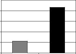
whole-body radiant heat units is being carried out in clinical centers as well as many private clinics in Germany for the purpose of alleviating rheumatoid arthritis. It has been proposed that the induction of TNF receptors by WBH may induce a remission in patients with active rheumatoid arthritis. The use of heat packs has long been standard to relieve the pain of fibromyalgia. Again, the therapeutic effect is believed to be due to increased circulation flushing out toxins and speeding the healing process. Whole-body hyperthermia treatment for fibromyalgia and chronic fatigue syndrome (CFS) is to be found in a number of private clinics. Hyperthermia increases the number and activity of white blood cells, stimulating the depressed immune system of the CFS patient.
Because of its immune stimulating effects, whole-body hyperthermia is a strong candidate for treatment of chronic progressive viral infections, such as HIV and hepatitis C. A clinical trial at the University Medical Center Utrecht, The Netherlands has evaluated extracorporeal heating to induce systemic hyperthermia of 41.8 8C for 120 min under propofol anesthesia for treatment of hepatitis C (65). Human immunodeficiency virus (HIV)-infected T cells are more sensitive to heat than healthy lymphocytes, and susceptibility increases when the cells are presensitized by exposure to tumor necrosis factor. Thus, induction of whole-body hyperthermia or hyperthermia specifically limited to tissues having a high viral load is a potential antiviral therapy for acquired immunodeficiency syndrome (AIDS). An Italian study has found treatment of AIDS with beta-carotene and hyperthermia to be synergistic, preventing progression of early disease and also increasing the survival time in patients with severe AIDS. A single treatment of low flow extracorporeal hyperthermia was found effective against AIDS associated Kaposi’s sarcoma, though there was significant toxicity. Core temperature was raised to 42 8C and held for 1 h with extracorporeal perfusion and ex vivo blood heating to 49 8C. Complete or partial regressions were seen in 20/29 of those treated at 30 days post-treat- ment, with regressions persisting in 14/29 of those treated at 120 days post-treatment. At 360 days, 4/29 maintained tumor regressions with 1 patient being in complete remission still at 26 months (66).
THE FUTURE OF SYSTEMIC HYPERTHERMIA
While there is a resurgence of interest in systemic hyperthermia, this modality has not yet been adopted as a mainstream therapy, and optimal clinical trials have not yet been carried out. Well-designed, well-controlled, multicenter clinical trials need to be conducted. In order to unequivocally demonstrate the utility of whole-body hyperthermia inthe treatmentof cancer aswellasother diseases,it will be important to accrue a sufficiently large number of patients who do not have end-stage disease. Thanks to the commercial availability of systemic hyperthermia systems, the variability between induction techniques at different institutions can be removed. Newer instrumentation, particularly near-IR radiant heat devices, along with treatment at lower temperatures (fever-range thermal therapy) should lead to significantly reduced toxicity. Better exploitation of
|
|
HYPERTHERMIA, SYSTEMIC |
59 |
||
|
5 |
|
|
|
|
40˚C/37˚C ratio of HSVtk molecules per cell |
grp78-HSVtk |
|
|
|
|
4 |
|
|
|
|
|
3 |
|
|
|
|
|
2 |
|
|
|
|
|
1 |
|
|
|
|
|
|
|
|
|
|
|
|
0 |
Liver |
Tumor |
|
|
|
|
|
|
||
Figure |
5. Fever |
range whole-body |
hyperthermia |
increases |
|
therapeutic gene (grp78-HSVtk) delivery in tumor.
the narrow window of effective temperatures within which the cellular effects of heat can be exploited yet damage remains minimal, and improved understanding of the biological interactions in vivo of systemic heat with chemotherapy and radiation will be essential to optimize therapy.
The effects of systemic hyperthermia on tumor blood flow and vascular permeability have the potential to increase delivery of various small molecules, nanoparticles, and gene therapy vectors to tumors. Ferromagnetic nanoparticles can be heated by external magnetic fields and offer the potential for internal hyperthermia, both locally and systemically. Thermally sensitive liposomes that release their contents at designated temperatures are also of interest. The ability of systemic hyperthermia to aid in systemic delivery of gene therapy vectors (the holy grail of gene therapy) and enhance transfection of cells with therapeutic gene plasmids is under investigation in several laboratories (67,68), and shows potential along with targeted gene therapy via the heat shock response. For example, Fig. 5 shows a fourfold hyperthermic increase of therapeutic gene delivery to tumor when plasmid DNA was injected intravenously into mammary carcinoma bearing rats immediately after 6 h of whole-body hyperthermia at 40 8C. Thus systemic hyperthermia is likely to see increasing application as an enhancer of drug delivery.
There is a great deal of interest in the immunological consequences of whole-body hyperthermia, and as they become better understood, the combination of systemic hyperthermia with specific immunotherapies will undoubtedly be pioneered, not just for cancer but also, by analogy with fever, in a broad range of diseases.
SUMMARY
Systemic hyperthermia is founded on solid physical and biological principles and shows promise in the treatment of a number of diseases. Modern whole-body hyperthermia devices use IR-A radiation sources together with effective heat loss techniques to achieve a controlled, uniform temperature distribution throughout the body with minimal patient toxicity. A shift in paradigm has occurred away from achieving direct cell killing with short
60 HYPERTHERMIA, SYSTEMIC
bouts of maximally tolerated temperatures, to inducing indirect curative effects through longer duration treatments at lower temperatures, and synergy with other modalities, such as radiotherapy. Better understanding of the interactions of elevated temperature with metabolic and genetic pathways will allow thermally driven targeted therapies. Of particular promise is the use of systemic hyperthermia as an immune system stimulator and adjunct to immunotherapy. Application of systemic hyperthermia to nanoparticle delivery and gene therapy is emerging. Whole-body hyperthermia is moving from being dubbed an alternative therapy to becoming a standard treatment and clinical hyperthermia centers are to be found all over the world.
Useful Websites |
|
http://www.ncbi.nlm.nih. |
Techniques and |
gov/books/bv.fcgi?rid=hstat6. |
Devices Used |
section.40680 |
to Produce |
|
Hyperthermia |
http://www.ncbi.nlm.nih.gov/ |
Physics and |
books/bv.fcgi?rid=cmed. |
Physiology of |
section.7813 |
Heating |
http://www.duke.edu/~dr3/ |
Clinical Hyperthermia |
hyperthermia_general. |
Background |
html |
|
http://www.eurekah.com/isbn. |
Online book: |
php?isbn=1-58706- |
Locoregional |
248-8&bookid=143 |
Radiofrequency-perfusional |
&catid=50 |
and Whole Body |
|
Hyperthermia in |
|
Cancer Treatment: |
|
New Clinical Aspects, |
|
E.F. Baronzio and |
|
A. Gramaglia (eds.), |
|
Eurekah Bioscience |
|
Database |
http://www.esho.info/ |
European Society for |
professionals/ |
Hyperthermic |
|
Oncology |
http://www.hyperthermie.org/ |
German |
index2.html |
Interdisciplinary |
|
Working group on |
|
hyperthermia |
http://www.uth.tmc.edu/ |
Systemic Thermal |
thermaltherapy/ |
Therapy at the |
|
University of Texas |
BIBLIOGRAPHY |
|
1.Nauts HC. Bacterial pyrogens: beneficial effects on cancer patients. In: Gautherie M, Albert E, editors. Biomedical Thermology, Progress in Clinical Biological Research. New York: Alan R. Liss; 1982. p 687–696.
2.Law HT, Pettigrew RT. Heat transfer in whole body hyperthermia. Ann NY Acad Sci 1980;335:298–310.
3.Robins HI, et al. A non-toxic system for 41.8 8C whole body hyperthermia: results of a phase I study using a radiant heat device. Cancer Res 1985;45:3937–3944.
4.Parks LC, Smith GV. Systemic hyperthermia by extracorporeal induction techniques and results. In: Storm FK, editor.
Hyperthermia in Cancer Therapy. Boston: Hall; 1983.
pp407–446.
5.van der Zee J, van Rhoon GC, Faithfull NS, van den Berg AP. Clinical hyperthermic practice: whole body hyperthermia. In: Field SB, Hand JW, editors. An Introduction to the Practical Aspects of Clinical Hyperthermia. London: Taylor & Francis; 1990.
6.Wust P, et al. Feasibility and analysis of thermal parameters for the whole-body-hyperthermia system IRATHERM-2000. Int J Hyperthermia 2000 Jul–Aug; 16(4):325–339.
7.Blazickova S, Rovensky J, Koska J, Vigas M. Effect of hyperthermic water bath on parameters of cellular immunity. Int J Clin Pharmacol Res 2000;20(1–2):41–46.
8.Vaupel P, Kallinowski F. Physiological effects of hyperthermia. In: Streffer C, editor. Hyperthermia and the Therapy of Malignant Tumors. Berlin/Heidelberg, Germany: Springer-Verlag; 1987.
9.Dudar TE, Jain RK. Differential response of normal and tumor microcirculation to hyperthermia. Cancer Res 1984;44(2):605– 612.
10.Kong G, Braun RD, Dewhirst MW. Characterization of the effect of hyperthermia on nanoparticle extravasation from tumor vasculature. Cancer Res 2001;61:3027–3032.
11.Jain RK. Normalization of tumor vasculature: an emerging concept in antiangiogenic therapy. Science 2005;307(5706): 58–62.
12.Urano M, et al. Effect of whole body hyperthermia on cell survival, metastasis frequency, and host immunity in moderately and weakly immunogenic murine tumors. Cancer Res 1983;43:1039–1043.
13.Shen RN, et al. Whole-body hyperthermia decreases lung metastases in lung tumor-bearing mice, possibly via a mechanism involving natural killer cells. J Clin Immunol 1987;7(3):246–253.
14.Roigas J, Wallen ES, Loening SA, Moseley PL. Heat shock protein (HSP72) surface expression enhances the lysis of a human renal cell carcinoma by IL-2 stimulated NK cells. Adv Exp Med Biol 1998;451:225–229.
15.Burd R, et al. Tumor cell apoptosis, lymphocyte recruitment and tumor vascular changes are induced by low temperature, long duration (fever-like) whole body hyperthermia. J Cell Physiol 1998;177(1):137–147.
16.Ostberg JR, Gellin C, Patel R, Repasky EA. Regulatory potential of fever-range whole body hyperthermia on Langerhans cells and lymphocytes in an antigen-dependent cellular immune response. J Immunol 2001;167(5):2666–2670.
17.Kraybill WG, et al. A phase I study of low temperature, long duration (fever-range) whole body hyperthermia (WBH) in patients with advanced solid tumors: correlation with mouse models. Int J Hyperthermia 2002;18:253–266.
18.Atanackovic D, et al. 41.8 degrees C whole body hyperthermia as an adjunct to chemotherapy induces prolonged T cell activation in patients with various malignant diseases. Cancer Immunol Immunother Epub 2002 Oct 18 2002;51(11–12):603– 613.
19.Barni S, et al. Lysosomal exocytosis induced by hyperthermia:
anew model of cancer death. III. effect on liver metastasis. Biomed Pharmacother 1996;50:79–84.
20.Hou K. The level changes of peripheral blood PGE2 and clinical significance in patients with malignant tumor before and after whole body hyperthermia. Proceedings of the 26th Congress of the International Clinical Hyperthermia Society, September 9–12, 2004, Shenzhen, China, 2004; p 66.
21.Manjili MH, et al. Subjeck, Cancer immunotherapy: stress proteins and hyperthermia. Int J Hyperthermia 2002;18(6): 506–520.
22.Ialenti A, et al. Inhibition of cyclooxygenase-2 gene expression by the heat shock response in J774 murine macrophages. Eur J Pharmacol 2005;509(2–3):89–96.
23.Repasky EA, Issels R. Physiological consequences of hyperthermia: Heat, heat shock proteins and the immune response. Int J Hyperthermia 2002;18:486–489.
24.Ostberg JR, Repasky EA. Emerging evidence indicates that physiologically relevant thermal stress regulates dendritic cell function. Cancer Immunol Immunother [Epub ahead of print]; Apr 28, 2005.
25.Berry JM, Michalsen A, Nagle V, Bull JM. The use of esmolol in whole-body hyperthermia: cardiovascular effects. Int J Hyperthermia 1997;13(3):261–268.
26.Robins HI, et al. Adjunctive therapy (whole body hyperthermia versus Ionidamine) to total body irradiation for the treatment of favorable B-cell neoplasms: a report of two pilot clinical trials and laboratory investigations. Int J Radiat Onco Biophys 1990;18:909–920.
27.Ohno S, et al. Haematological toxicity of carboplatin and cisplatin combined with whole body hyperthermia in rats. Br J Cancer 1993;68:469–474.
28.Haveman J, et al. Effects of hyperthermia on the peripheral nervous system: a review. Int J Hyperthermia 2004;20(4):371–391.
29.Calvert AH, et al. Early clinical studies with cis-diammine-1,1- cyclobutane dicarboylate platinum II. Cancer Chemother Pharmacol 1982;9:140–147.
30.Sapareto SA, Dewey WC. Thermal dose determination in cancer therapy. Int J Radiat Oncol Biol Phys 1984;10(6):787–800.
31.Thrall DE, et al. Using units of CEM 43 degrees C T90, local hyperthermia thermal dose can be delivered as prescribed. Int J Hyperthermia 2000;16(5):415–428.
32.Webb AG. Temperature measurement using nuclear magnetic resonance. Ann Reports NMR Spectrosc 2001;45:1–67.
33.Stafford RJ, Hazle JD, Glover GH. Monitoring of high-inten- sity focused ultrasound-induced temperature changes in vitro using an interleaved spiral acquisition. Magn Reson Med 2000;43(6):909–912.
34.Engelhardt R. Summary of recent clinical experience in wholebody hyperthermia combined with chemotherapy. Recent Results Cancer Res 1988;107:200–224.
35.Lange J, Zanker KS, Siewert JR, Eisler K, Landauer B, Kolb E, Blumel G. and Remy, W. Extracorporeally induced wholebody hyperthermia in conventionally incurable malignant tumor patients Med Wochenschr. 1983;108(13):504–509.
36.Robins HI, et al. Phase I clinical trial of melphalan and 41.8 degrees C whole-body hyperthermia in cancer patients. J Clin Oncol 1997;15:158–164.
37.Issels RD, Wilmanns W, editors. Recent Results in Cancer Research, Vol. 107: Application of Hyperthermia in the Treatment of Cancer. Berlin/Heidelberg: Springer Verlag; 1988.
38.Westermann AM, et al. Systemic Hyperthermia Oncologic Working Group, A Systemic Hyperthermia Oncologic Working Group trial, Ifosfamide, carboplatin, and etoposide combined with 41.8 degrees C whole-body hyperthermia for metastatic soft tissue sarcoma. Oncology 2003;64(4):312–321.
39.Bull JM, et al. Chemotherapy resistant sarcoma treated with whole body hyperthermia (WBH) combined with 1-3-bis(2- chloroethyl)-1-nitrosourea (BCNU). Int J Hyperthermia 1992; 8(3):297–304.
40.Herman TS, Sweets CC, White DM, Gerner EW. Effect of heating on lethality due to hyperthermia and selected chemotherapeutic drugs. J Natl Cancer Inst 1982;68(3):487– 491.
41.Engelhardt R, et al. Treatment of disseminated malignant melanoma with cisplatin in combination with whole-body hyperthermia and doxorubicin. Int J Hyperthermia 1990;6(3): 511–515.
42.Robins HI, et al. Phase I clinical trial of carboplatin and 41.8 degrees C whole-body hyperthermia in cancer patients. J Clin Oncol 1993;11:1787–1794.
HYPERTHERMIA, SYSTEMIC |
61 |
43.Douwes F, et al. Whole-body hyperthermia in combination with platinum-containing drugs in patients with recurrent ovarian cancer. Int J Clin Oncol 2004;9(2):85–91.
44.Hegewisch-Becker S, et al. Whole-body hyperthermia (41.8 8C) combined with bimonthly oxaliplatin, high-dose leucovorin and 5-fluorouracil 48-hour continuous infusion in pretreated metastatic colorectal cancer: a phase II study. Ann Onc 2002;13(8):1197–1204.
45.Hegewisch-Becker S, et al. Whole body hyperthermia (WBH, 41.8 8C)combined with carboplatin and etoposide in advanced biliary tract cancer. Proc Am Soc Clin Oncol 2003; (abstr. 1247), 22:311.
46.Richel O, et al. Phase II study of carboplatin and whole body hyperthermia (WBH) in recurrent and metastatic cervical cancer. Gynecol Oncol 2004;95(3):680–685.
47.Strobl B, et al. Whole body hyperthermia combined with carboplatin/paclitaxel in patients with ovarian carcinoma–Phase-II- study. J Clin Oncol -Proc Am Soc Clin Oncol 2004;22(14S).
48.Elias D, et al. Heated intra-operative intraperitoneal oxaliplatin plus irinotecan after complete resection of peritoneal carcinomatosis: pharmacokinetics, tissue distribution and tolerance. Ann Oncol 2004;15:1558–1565.
49.Bull JM, et al. Phase I study of long-duration, low-tempera- ture whole-body hyperthermia (LL-WBH) with liposomal doxorubicin (Doxil), 5-fluorouracil (5-FU), & interferon-a (IFN-a), Proc Amer. Soc Clin Oncol 2002;(Abst. 2126).
50.Larkin JM, A clinical investigation of total-body hyperthermia as cancer therapy. Cancer Res, 1979;39(6 Pt 2):2252–2254.
51.Bull JM, et al. Update of a phase I clinical trial using feverrange whole-body hyperthermia (FR-WBH) þ cisplatin (CIS) þ gemcitabine (GEM) þ metronomic, low-dose interferon-alpha (IFN-alpha), Proceedings of the International Congress on Hyperthermic Oncology, 20.-24.04., St. Louis, (Session 21 and Poster 689); 2004.
52.Barlogie B, Corry PM, Lip E, Lippman L, Johnston DA, Tenczynski TF, Reilly E, Lawson R, Dosik G, Rigor B, Hankenson R, Freireich EJ Total-body hyperthermia with and without chemotherapy for advanced human neoplasms. Cancer Res 1979;39(5):1481–1489.
53.Robins HI, et al. Phase I trial of human lymphoblastoid interferon with whole body hyperthermia in advanced cancer. Cancer Res 1989;49(6):1609–1615.
54.Gerad H, van Echo DA, Whitacre M, Ashman M, Helrich M, Foy J, Ostrow S, Wiernik PH, Aisner J. Doxorubicin, cyclophosphamide, and whole body hyperthermia for treatment of advanced soft tissue sarcoma. Cancer. 1984 Jun 15;53(12):2585–91.
55.Baba H, et al. Increased therapeutic gain of combined cisdiamminedichloroplatinum (II) and whole body hyperthermia therapy by optimal heat/drug scheduling. Cancer Res 1989;49(24 Pt. 1):7041–7044.
56.Katschinski DM, et al. Optimization of chemotherapy administration for clinical 41.8 degrees C whole body hyperthermia. Cancer Lett 1997;115(2):195–199.
57.Hegewisch-Becker S, et al. Effects of whole body hyperthermia (41.8 degrees C) on the frequency of tumor cells in the peripheral blood of patients with advanced malignancies. Clin Cancer Res 2003;9(6):2079–2084.
58.Teicher BA, Holden SA, Ara G, Menon K. Whole-body hyperthermia and lonidamine as adjuvant therapy to treatment with cisplatin with or without local radiation in mouse bearing the Lewis lung carcinoma. Int J Hyperthermia 1995;11(5):637–645.
59.Sumiyoshi K, Strebel FR, Rowe RW, Bull JM. The effect of whole-body hyperthermia combined with ‘metronomic’ che-
motherapy on rat mammary adenocarcinoma metastases. Int J Hyperthermia 2003;19(2):103–118.
60. Overgaard J, et al. Randomised trial of hyperthermia as adjuvant to radiotherapy for recurrent or metastatic