

Ghai Essential Pediatrics8th
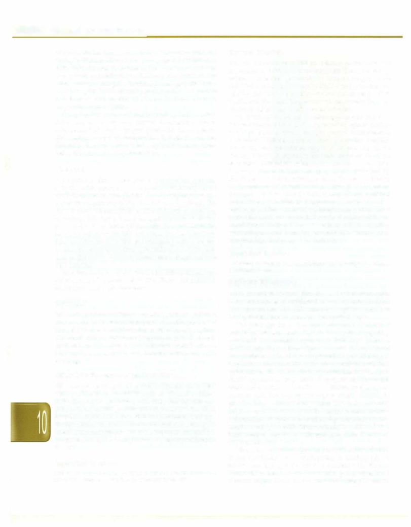
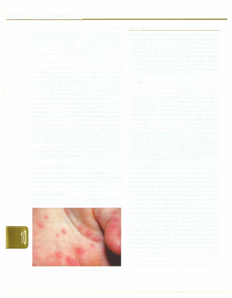
.pdfFig. 10.5: Polymorphic rash of chickenpox
leading to stroke, autoimmune thrombocytopenic purpura and Reye syndrome.
The progressive varicella syndrome is a dreaded complication in immunocompromised individuals, neo nates, pregnant women and even healthy adults and adolescents. This syndrome is characterized by continued development oflesions, hemorrhagiclesions,coagulopathy and visceral organ involvement including hepatitis, pneumonia and encephalitis. Mortality rate are high despite therapy.
Chickenpox in pregnancy is associated with an increased risk of severe disease in the mother. Congenital varicella syndrome may occur following infection in the first and second trimester at a frequency of 0.4-2%. It is characterized by skin scarring, malformed extremities, cataracts and brain abnormalities (e.g. aplasia, calci fications). Finally, if the disease occurs in the mother 5 days before to 2 days after delivery, severe and often fatal neonatal disease may result.
Herpes zoster in children is characterized by a mild vesicular rash with dermatomal distribution. Unlike in adults, pain is less and postherpetic neuralgia unusual. The risk of herpes zosteris more in immunocompromised children, children who acquire chickenpox in infancy and those whose mothers developed varicella in the third trimester.
Diagnosis
The diagnosis is clinical and usually not difficult. Chickenpox should be differentiated from other vesicular exanthematasuch asherpes simplex, enteroviral infections (hand-foot-mouth disease), insect bites and drug reactions. In atypical cases, the diagnosis is made on Tzanck smear of the lesions, which shows multinucleated cells and by demonstrating IgM antibodies to varicella.
Treatment
Management is symptomatic and includes antipyretics, antipruritic agents and good hygiene. Aspirin is contra-
Infections and Infestations -
indicated due to risk of Reye syndrome. The child should not attend school until new lesions stop appearing and all lesions have crusted. Administration of oral acyclovir (20 mg/kg/dose four times a day for 5 days) within 24 hr of onset of rash in healthy children reduces the duration of rash by one day and lesions by 25%. IV acyclovir (10 mg/ kg every 8 hr for 7 days) is given to patients with compli cated varicella and for illness in high risk patients (neo nates, immunocompromised children, pregnant women).
Prevention
Prevention against varicella with the live attenuated varicella vaccine and use of varicella zoster immune globulin (YZIG)forpostexposure prophylaxisaredetailed in Chapter 9. YZIG is fairly expensive and not always available.
Suggested Reading
Gershon AA: Varicella-zoster virus infections. Pediatr Rev 2008;29(1): 5-10
Whitley RJ. Therapy ofherpes virus infections in children. Adv Exp Med Biol 2008;609:216-32
Infectious Mononucleosis
Infectious mononucleosis, a syndrome characterized by fever, fatigue, sore throat and lymphadenopathy, is most often caused by a herpes virus, Epstein-Barr virus (EBY). Infectious mononucleosis-like illness can also be caused by toxoplasma, CMY, adenoviruses and primary HIV infection.
Epidemiology
The EBY virus, a DNA virus of the herpes virus family, is transmitted in oral secretions by closeintimatecontactlike kissing or exchange of saliva from close child contact. The virus replicates in the oral epithelial cells then spreads to salivary glands with eventual viremia to the B lympho cytes in the blood and lymphoreticular system including liver andspleen. The CDS lymphocytes proliferate to check this replication of virus in the B lymphocytes and represent the atypical lymphocytes seen in EBY infection. Like other herpes viruses, EBY establishes lifelong latent infection after the primary infection with frequent asymptomatic reactivations.
The epidemiology is related to the age of primary acquisition of EBYinfection. In developing countries, most of EBY infection occurs in infancy and early childhood, whenitiseitherasymptomatic orsimilar tootherchildhood infections. For this reason, infectious mononucleosis is uncommonly seen or reported in India. In developed countries, the age of acquisition of EBY infection shifts upwards and thus the illness is seen more commonly.
Clinical Features
Symptomatic EBY infections in older children and adults are characterized by insidious onset of malaise, fatigue,
- Essential Pediatrics
fever, headache, nausea, sore throat, abdominal pain and myalgia. Examination shows pharyngeal inflammation with exudates and petechiae at the junction of soft and hard palate, generalized lymphadenopathy (cervical, less often axillaryand inguinal), mildsplenomegaly (50%) and hepatomegaly (10%). Maculopapular rashes are seen in 3-15% cases and in 80% of those who have received ampicillin or amoxicillin.
Complications are rare and include splenic rupture following minor trauma, airway obstruction due to enlargement of oropharyngeal lymphoid tissue, menin gitis, seizures, ataxia, myocarditis, hemolytic anemia, thrombocytopenia, neutropenia, aplastic anemia, inter stitial pneumonitis and pancreatitis.
Diagnosis
Most patients show leukocytosis and absolute lympho cytosis, with presence of atypical lymphocytes on peripiheral smear. The platelet count is slightly low and hepatic transaminasesmildlyelevatedin50% patients. The Paul Bunnel (heterophile antibody) test is used for screening. This test is based on agglutination of sheep/ horse red cells by heterophile antibodies present in the serum of patients with EBV infection. This test may have false negative rates of 10% and remains positive for few months to 2 yr afterinfection.IgMantibody to viralcapsid antigen (lgM VCA) is aconfirmatorytest todiagnoseacute EBV infection.
Infectious mononucleosis should be differentiated from other causes of mononucleosis like illness, streptococcal pharyngitis and acute leukemia.
Treatment
Rest and symptomatic therapy are advised. Participation in strenuous activities andcontactsportsshouldbe prohibited in the first 2-3 weeksof illness due to risk of splenic rupture. Treatment with prednisolone (1 mg/kg/day for 7 days) is advisedfor complications suchashemolyticanemia, airway obstruction, meningitis and thrombocytopenia with bleeding.
Other Manifestations of EBV Infections
EBV has oncogenic potential and is causally associated with proliferative disorders such as virus associated hemophagocytic syndrome, oral hairy leukoplakia and lymphoid interstitial pneumonitis in patients with AIDS, nasopharyngeal carcinoma, Burkitt lymphoma, Hodgkin disease, tumors in immunocompromised patients (e.g. X linked lymphoproliferative disease, leiomyosarcoma, CNS lymphoma) and post-transplantation lymphoproliferative disease.
Suggested Reading
Bell AT, Fortune B, Sheeler R. What is the best test for diagnosing infectious mononucleosis? J Fam Pract 2006;55(9):799-802
Roseola lnfantum
Roseola infantum or exanthem subitum or Sixth disease is a common childhood exanthematous illness caused by primary infection primarily by human herpes virus (HHV)-6and less commonly by HHV-7and echovirus 16. HHV-6 and HHV-7 are DNA viruses that target the CD4 T cells, and like other herpes viruses, can remain latent in the body for several yr after acute infection.
The peak age for roseola is between 6 months and 3 yr. The prodromal period is characterized by upper respira tory signs such as rhinorrhea, pharyngeal inflammation, conjunctiva! redness, mild cervical or occipital lympha denopathy and sometimes, palpebral edema. The classic clinical illness is heralded by high fever of 38-40°C, associated with febrile seizures in 5-10% cases, and lasts 3-4 days. Fever declines abruptly and is followed by development of a rashwithin 12-24 hr. The rash is discrete erythematous and maculopapular which first appears on the trunk and then spreads to the face, neck and proximal extremities. The rash is nonpruritic, rarely becomes confluent and fades in 3-4 days. Infectiousness is low and outbreaks have not occurred. Roseola should be differen tiated from childhood illnesses such as rubella, measles, enteroviruses and drug hypersensitivity. Treatment is symptomatic and prognosis excellent.
Suggested Reading
Caserta MT, Mock DJ, Dewhurst S. Human herpes virus 6. Clin Infect Dis 2001;33:829-33
Erythema lnfectiosum
Erythema infectiosum or Fifth disease is a common exan thematous illness of childhood caused by a small DNA virus, parvovirus B19. This virus has tropism for cells of the erythroid lineage at the pronormoblast stage.
The peak age for erythema infectiosum is between 5 and 15 yr. Transmission of infection is by the respiratory route and the incubation period is 4-28 days (average 16-17 days). Theprodromal phase is mild and consists of low-grade fever, headache and symptoms of mild upper respiratory tract infection. The characteristic rash first appears as erythematous flushing ontheface in a 'slapped cheek' appearance (Fig. 10.6). It spreads rapidly to the trunk and proximal extremities as a diffuse erythematous macular rash that rapidly undergoes central clearing to give it a lacy or reticulated pattern. The rash gradually fades over a 1-3 week period. Complications include arthropathy, idiopathic thrombocytopenic purpura and aseptic meningitis. Fifth disease should be differentiated from measles, roseola, rubella and drug rash. Treatment is symptomatic.
Other seriousmanifestationsof parvovirus B19 infection include arthralgia and arthropathy in adolescents or adults, transient aplastic crises in patients with chronic hemolytic anemias, chronic anemia, pancytopenia or marrow suppression, virus associated hemophagocytic
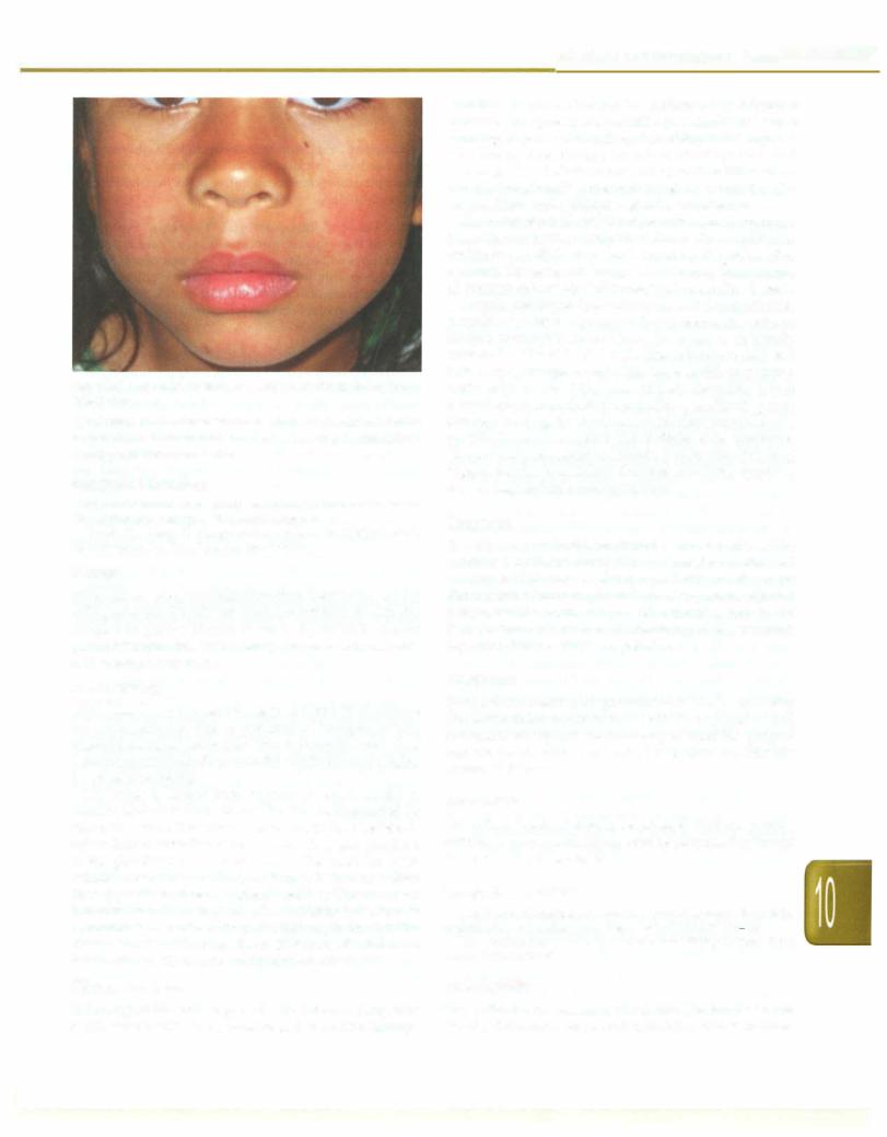
Fig. 10.6: The rash resembles a 'slapped cheek' in erythema infectiosum
syndrome in the immunocompromised, hydrops fetalis in pregnant women and rare episodes of myocarditis in healthy children or adults.
Suggested Reading
Servant-Delmas A, Lefrere JJ, Morine! F, Pille! S. Advances in human B19 erythrovirusbiology. J Virol 2010;84:9658-65
Servey JT, Reamy BV, Hodge J. Clinical presentations of parvovirus B19 infections. Am Fam Physician 2007;75:373-6
Mumps
Mumps is an acute viral infection characterized by painful enlargement of the salivary glands, most characteristically the parotid glands. Mumps is caused by an RNA virus of genus Paramyxovirus in the family Paramyxoviridae; only one serotype is known.
Epidemiology
Most cases occur between the ages of 5 and 15 yr; infants are rarely affected due to presence of transplacentally acquired maternal antibodies. Man is the only reservoir of infection; carrier statedoesnot exist. The incidence is higher in winter and spring.
The virus is spread from human reservoir by direct contact, air-borne droplets and fomites contaminated by saliva and urine. The virus proliferates in the respiratory epithelium to enter the circulation and then gets localized to the glandular and neural tissue. The virus has been isolated from saliva as long as 6 days before to 9 days afterappearanceof salivaryglandswelling.The secondary infection rate is as high as 80%. Mumps infection or immunization is believed to confer lifelong immunity. The disease is mild in the majority; in 10% cases, the infection is associated with aseptic meningitis or encephalitis.
Clinical Features
Infections and Infestations -
infection is characterized by unilateral or bilateral parotitis. This presents as earache, jaw tenderness while chewing, dryness of mouth and swelling at the angle of jaw. The ear lobe may appear to be pushed upwards and outwards. The defervescence and resolution takes about a week. Occasionally, other salivary glands, including the submaxillary and sublingual glands, are affected.
The occurrence of epididymoorchitis is more common in adolescent boys or postpubertal men. The condition is unilateral in 85% cases and occurs 1-2 weeks after parotitis. Thetestes are enlarged and tender. Some degree of atrophy follows the inflammation but sterility is rare.
Aseptic meningitis is seen in about 1-10% patients with parotitis. Mumps is perhaps the commonest cause of aseptic meningitis in children. Recovery is generally uneventful. The risk of encephalitis is between 0.02 and 0.3% cases. Mumps encephalitis has a satisfactory prog nosis with a mortality rate of less than 2%. Other neurological manifestations include auditory nerve damage leading to deafness, cerebellar ataxia, facial neuritis, transverse myelitisand Guillain-Barre syndrome. Uncommon presentations include pancreatitis (5% may trigger insulin dependent diabetes mellitus), mastitis, oophoritis, nephritis and myocarditis.
Diagnosis
The diagnosis is based on clinical features and may be confirmed by ELISA for IgM. Serum amylase is elevated in almost 90% cases. Mumps parotitis needs to be differentiated from suppurative parotitis, submandibular lymphadenitis, recurrent juvenile parotitis, calculus in Stensen duct and other viral infections causing parotitis, e.g. coxsackie A and cytomegalovirus.
Treatment
Symptomatictreatment is given in the form of antipyretics and warm saline mouthwashes. Orchitis is treated by bed rest and local support. Steroids may be used for sympto matic relief of orchitis and arthritis but does not alter the course of disease.
Prevention
The affected patient should be isolated until the parotid swelling has subsided. Mumps can be prevented by timely immunization (Chapter 9).
Suggested Reading
MacDonald N, Hatchette T, Elkout L, Sarwal S. Mumps isback: Why is eradication not working, Adv Exp Med biol 2011;697:197-220
WHO position paper on mumps and vaccines. Weekly Epidemiologic Record 2001;76:345-56
Poliomyelitis
Following an incubation period of 2-4 weeks, symptoms begin acutely with fever, malaise and headache. Mumps
The polioviruses belong to the genus Enterovirus in the family Picornaviridae and comprise three related
- Essential Pediatrics
serotypes:types 1, 2and3, all of which cancauseparalysis. Type 1 is most frequently responsible, type 3 is less commonly causative and type 2 is only rarely implicated.
Epidemiology
The disease is seasonal, occurring more commonly in summer and early autumn in temperate climates. In tropical countries, seasonalityis less clearly defined; some areas experience increase in incidence during the rainy season. Feco-oral route is the predominant mode of transmission indeveloping countries with poor sanitation, whereas oral-pharyngeal transmission predominates in industrialized countries and during outbreaks. The average incubation period of disease is 7-10 days, ranging from 4 to 35 days. The virus is shed in the stools for 6-8 weeks after infection.
Humans are the only reservoir of poliovirus and infec tion is spread from person-to-person. The virus spreads rapidly to nonimmune persons. Transmission is usually widespread in the community by the time of onset of paralysis in a child. Infants born to mothers with antibodies areprotected naturally againstparalytic disease for a few weeks. Immunity is acquired through infection with the wild virus and through immunization.
The Global Polio Eradication initiative was launched in 1988 using oral polio vaccine (OPV) as the eradication tool and employing a four pronged strategy comprising
(i)maintaining high routine immunization coverage,
(ii)supplementary immunization activities (SIAs)/pulse immunization, (iii) acute flaccid paralysis (AFP) surveil lance, and (iv) outbreak response immunization. The initiative was hugely successful with reduction of polio cases from 350,000 worldwide in 1988 to 650 in 2011 and only 215 cases in 2012 (as of 25th December 2012). Only 3 countries,Afghanistan,Nigeria andPakistanremain polio endemic; 210 of the 215 cases in 2012 were reported from thesecountries. Thelastwildpolio case wasreportedfrom India on 13January, 2011 and India is no more considered endemic for poliovirus.
Pathogenesis
The mouth is the usual portal of entry. The virus is usually present in the pharynx and stools before the onset of paralytic illness. It invades local lymphoid tissue, enters the bloodstream to invade certain nerve cells and may damage or destroy these cells.
Clinical Features
In 90-95% of individuals, poliovirus infection is inappa rent. In the remaining 5-10% of individuals, one of the following syndromes may occur.
Abortive polio occurs in 4-8% of infections and is characterizedby a minorillness with low grade fever, sore throat, vomiting, abdominal pain, loss of appetite and malaise. Recovery is rapid and complete; there is no
paralysis. It cannot be distinguished from other viral infections.
Nonparalytic asepticmeningitisoccursin1-2% of infections, with headache, neck, back and leg stiffness several days afteraprodromesimilartoabortivepolio. Recoveryoccurs within 2-10 days.
Paralytic poliomyelitisoccurs in 0.5-1% of cases. Symptoms occur in two phases, minor and major, separated by several dayswithoutsymptoms.The minorphase consists of symptoms similar to those of abortive poliomyelitis. Themajorphaseofillnessbeginswithmusclepain, spasms and the return of fever. This is followed by rapid onset of flaccid paralysis that is usually complete within 72 hr.
Spinal paralytic poliomyelitis is the most common form of paralyticpoliomyelitis, accountingfor approximately 80% cases. It results from a lower motor neuron lesion of the anterior horn cells of the spinal cord and affects the mus cles of the legs, arms and/ortrunk.Severe cases may deve lop quadriplegia and paralysis of the trunk, abdominal and thoracic muscles. The affected muscles are floppy and reflexes are diminished. The sense of pain and touch are normal. Paralysis is often asymmetrical, affecting legs more often than arms. Paralysis in extremities begins proximally andprogresses toinvolvedistalmusclegroups (i.e. descending paralysis). Residual flaccid paralysis is usually present after 60 days. Bu/bar polio accounts for 2% cases and results from a cranial nerve lesion, resulting in respiratory insufficiency and difficulty in swallowing, eating or speaking. Bulbospinalpolio accounts for 20% cases and is a combination of spinal paralytic and bulbar polio.
Polio encephalitis is characterized by irritability, delirium and loss of consciousness; seizures may occur. The paralysis may be of the upper motor neuron type.
Depending on the strain ofpoliovirus, the ratio between subclinical and clinical cases is estimatedto range between 100:1 and 1000:l. Older children and adults run a greater risk of developing paralytic illness. The case fatality rate among persons who develop the paralytic form of the disease is 2-20%. However, the case-fatality rate may be as high as 40% in those with bulbar or respiratory involvement.
Residual Paralysis
Following an acute phase of illness lasting 1--4 weeks, the recovery of paralyzed muscles begins. The extent of recoveryis variablerangingfrom mild to severe residual paresis at 60 days, depending upon the extent of damage caused to the neurons by the virus. Maximum neuro logical recovery takes place in the first 6 months of the illness; slow recovery continues up to two yr. After two year, no more recovery is expected and the child is said to have postpolio residual paralysis, which persists throughout life.
Diagnosis
Thediagnosisisbased onthe history andthecharacteristic clinical manifestations of asymmetric flaccid paralysis. Stool examination is recommended in every case of acute flaccid paralysis (AFP). Virus can be detected from onset to 8 or more weeks after paralysis; the highest probability of detection is during the first 2 weeks after onset of paralysis. Examination of the cerebrospinalfluid (cell count, Gram stain, protein and glucose) is useful in eliminating other conditions that cause AFP. Current serologic tests cannot differentiate between wild and vaccine virus strains. Collection of blood specimens for culture or serology is not recommended.
Differential Diagnosis
The two diseases most commonly confused with polio are Guillain-Barre syndrome and transverse myelitis. Other conditions with apresentation similar to those of paralytic poliomyelitis include traumatic neuritis and less fre quently, meningitis, encephalitis and illnesses produced by toxins (diphtheria, botulism) (see Chapter 19).
Treatment
Treatmentshouldbeearlyandappropriatetothestageand degree of paralysis. Children with bulbospinal polio and respiratory paralysis require hospitalization. In the acute stage, children with isolated paralysis of one or more limbs can bemanagedathome. They should be advised complete rest, proper positioning of the affected limb and passive range of movement at the joints. Massage and intra muscular injection should be avoided during acute phase of illness. Frequent change of the posture of the patient is must. The child should be made to lie on a firm bed and maintainlimbsinneutralposition. Thechildshouldliewith trunk and hip straight with slight flexion (5-10°) at knees and feet at right angle at the ankle joint. This position can be maintained with pillows, rolled towels or sand bags. Warm moist fomentations can be given with soft towels, dipped in warm water to relieve pain and spasms. Analgesics can also be given to relieve pain and fever. All the joints of affected limb/limbsshould bemovedthrough their passive range of movements, 2-3 times a day for 10 times at each joint, to prevent joint stiffness. This helps to stimulate proprioceptive impulses from muscles and tendons, helping improve muscle power.
As the acute phase of illness subsides, recovery in musclepoweris helped bygivingphysiotherapy, helping ambulation and prevention of deformities. Some children require orthosis atsome stage for ambulation. Others with fixed deformities and contractures require orthopedic intervention.
Prevention of Poliomyelitis
The available vaccines and the recommended schedule are discussed in Chapter 9.
Infections and Infestations -
Eradication of Polio
Eradication is possible because polio affects only man, immunity is lifelong, a safe vaccine is available and there are no carriers or reservoirs of the infection. The strategies for achieving this goal are:
Attaining high rntes ofroutine immunization. Every child less than 1-yr-old should be immunized with at least three doses of oral poliovirus vaccine (OPV).
National immunization days (NIDs). On these days, under the pulse polio immunization (PPI) program, additional OPV doses are administered to every child <5-yr-old. The aim of NIDs/PPI is to 'flood' the community with OPV within a very short period, thereby interrupting trans mission of virus throughout the community. Intensi fication of the PPI program is accomplished by the addition of extraimmunization rounds, adding a house to-house 'search and vaccinate' component in addition to providing vaccine at a fixed post. The number of PPI rounds conducted during any particular yr is determined by the extent of poliovirus transmission in the state or district.
Mopping-up immunization. When poliovirus transmission is reduced to well-defined and focal geographic areas, intensive house-to-house, child-to-child immunization campaigns are conducted over a period of days to break the final chains of virus transmission.
Acute flaccid paralysis surveillance. Under the global polio eradication initiative, surveillance for polio is conducted throughinvestigationofpatientswithAFP.AFPsurveillance helps to detect reliable areas where poliovirus transmission is occurring. Acute flaccid paralysis (AFP) is defined as sudden onset of weakness and floppiness in any part of the bodyinachild <15-yr-oldorparalysis inapersonofany age in whom polio is suspected. In other parts of the world, at least one case of AFP (excluding polio) occurs annually for every 100,000 children less than 15 yr of age (background AFP rate). The nonpolio causes of AFP account for this background rate. Sensitive surveillance will detect a background AFP rate of 1 per 100,000 children. In our country, wheretheincidenceof conditions suchastraumatic neuritis and AFP caused by other nonpolio enteroviruses is very high, the background nonpolio AFP rate is higher.
Details on the AFP surveillance are mentioned in Chapter 19.
Suggested Reading
National Polio Survei.llance Project, at: http://www.npspindia.org/ index.asp. accessed on May 21,2012
WHO position paper. Poliovaccines and immunization. Weekly epidemiologic record 2010;85:213-28
Hand-Foot-Mouth Disease
Hand-foot-mouth disease is a common viral illness primarily affecting children below 5 yr. It is caused by
- Essential Pediatrics
viruses of the genus Enterovirus belonging to family Picornaviridae, including polio, ECHO, coxsackie virus and enteroviruses. The most common causes of hand foot mouth disease are coxsackie virus A16 and enterovirus 71. The disease usually presents as outbreaks, often in preschool children and transmission is by direct contact with an affected patient or infected fomites.
Clinical Features
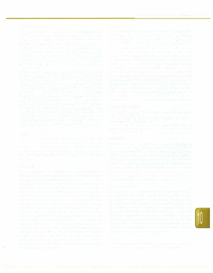
The onset is with a prodrome characterized by low grade fever, feeling of being unwell and sore throat. This is followed by development of ulcers or blisters in the oral cavity, mostly on the posterior aspect and then a papulovesicular skin rash on the palms and soles and less commonly, on buttocks, knees, elbows and genital area (Fig. 10.7). All manifestations may not be present in all patients. The illness resolves quickly over 4-5 days.
Complications include temporary loss of toe nails or finger nails about 4 weeks after onset of disease. Rare complications include aseptic meningitis, encephalitis, polio like paralysis, myocarditis and respiratory distress syndrome. Outbreaks, particularly due to enterovirus 71, are reported from China, Vietnam, Taiwan and Malaysia, in which neurologic complications are common and mortality significant. Some experts believe that entero viruses cause hand-foot-mouth disease now occupy the ecologic niche vacated by eradication of polioviruses.
Diagnosis is clinical and requires differentiation from other illnessescausing oral ulcers like herpangina,herpetic gingivostomatitis and aphthous ulcers and from chicken pox.
Treatment and Prevention
Treatment is mainly symptomatic and includes analgesics and soft diet. Isolation of affected children at home and promotion of hand hygiene to prevent disease spread is important. No vaccine is available against this disease.
Suggested Reading
Wong 55, Yip CC, Lau SK, Yuen KY. Human enterovirus 71 and hand, foot and mouth disease. Epidem.iol Infect 2010;138:1071---89
Fig. 10.7: The vesicular rash of hand-foot-mouth disease
VIRAL HEPATITIS
Hepatitis, meaning inflammation of the liver, can be caused by a variety of different hepatotropic viruses such as hepatitis A, B, C, D and E. Hepatitis A and E are responsible for most of the water-borne (community acquired) hepatitis while B, C and D are responsible for post-transfusion hepatitis. Since a considerable number of cases of post-transfusion and community-acquired hepatitis are not identified as being caused by hepatitis A-E, other hepatotropic viruses are also incriminated, including hepatitis G, TI virus and SEN virus.
Hepatitis A
Hepatitis A is caused by infection with the hepatitis A virus (HAV), a nonenveloped RNA virus. A single serotype of HAV infects humans and infection induces lifelong immunity. HAV is extremely resistant to degra dation by environmental conditions. Hence, it spreads readily by the feco-oral route through contaminated food and water and from person-to-person living with poor sanitation.Diseaseseverity increases withage at infection; children below 5 yr age have asymptomatic infection or presentwithanacuteundifferentiated febrile illness, while older children, adolescents and adults suffer from classic hepatitis. Symptomatic disease is uncommon and outbreaks rare indeveloping countries with poor hygiene sincemostindividualsareinfectedinchildhood. Inregions with intermediate endemicity like India, a significant proportion of people escapes infection in childhood and may develop symptomatic disease as adults.
Clinical Features
During an incubation or preclinical period of average 30 (range 10-50) days, the virus replicates actively. This is followed by ashortprodromalphaselastingup to a week, which is characterized by loss of appetite, fatigue, abdominal pain, nausea and vomiting, fever, diarrhea, dark urine and pale stools. Older individuals then have an icteric phase, duringwhichjaundicedevelops, with total bilirubinlevelsexceeding2-4 mg/dl. Fever improvesafter the first few days ofjaundice.In thesubsequentfew weeks of convalescence, patients show complete recovery.
In around 0.1-1% of patients, extensive necrosis of the liver occurs during the first 6-8 weeks of illness. In this case, high fever, marked abdominal pain, vomiting, jaundice and the development of hepatic encephalopathy associated with coma and seizures occur. These are the signs of fulminant hepatitis which is more common as age advances and leads to death in 70-90% of the patients. In patients who survive, neither functional nor pathologic sequelae are common despite the widespread necrosis. Infection with HAV does not lead to chronic or persistent hepatitis. Relapsing hepatitis may occur in 3-20% of patients 4 to 15 weeks after the initial symptoms have resolved.
Diagnosis
The specific diagnosis of acute hepatitis A is made by detecting serum anti-HAV IgM. Anti-HAV IgM is detectable about 3 weeks after exposure, by which time symptoms have already appeared. Its titer increases over 4-6 weeks, then declines to nondetectable levels within 6 months of infection. As IgG anti-HAV persists lifelong after acute infection, detection of IgG anti-HAV alone indicates past infection. Laboratory evaluation of liver function includes estimation of total and direct bilirubin, transaminases, alkaline phosphatase, prothrombin time, total protein and albumin.
Treatment
Therapy is supportive and is aimed at maintaining adequate nutrition. There is no evidence to suggest that restriction of fats has any beneficial effect on the course of the disease. Eggs, milk and butter may actually help provide an appropriate caloric intake. Antiviral agents have no role because the hepatic injury appears to be immunologically mediated. Referral to a liver transplant center is appropriate for patients with fulminant hepatitis.
Prevention
The ideal preventivestrategy is improvement in sanitation, hygiene and water supply. Immunization is very effective and discussed further in Chapter 9. Immunoglobulin G may be used for postexposure prophylaxis. If adminis tered within two weeks of exposure it either prevents development of disease or reduces its severity.
Suggested Reading
Mathur P, Arora NK. Epidemiological transition of hepatitis A in India: issues for vaccination in developing countries. Indian J Med Res 2008;128:699-704
Hepatitis B
Hepatitis B virus is a 3.2 kb, circular, partially double stranded DNA virus. HBV contains four open reading frames, which encode major structural and nonstructural proteins for HBV.
Epidemiology
HBV infection is prevalent in Asia, Africa, Southern Europe and Latin America, where the HBsAg sero positivity ranges from 2 to 20%. In hyperendemic areas, HBV infections occur mainly during infancy and early childhood. In Asia, perinatal transmission from HBsAg carrier mothers to their infants is an important route of transmission leading to chronicity. Approximately 90% of infants of HBeAg seropositive carrier mothers become HBsAg carriers, irrespective of a high or low HBsAg carrier rate in the population. In areas of low endemicity, horizontal infection is the main route of transmission.
Infections and Infestations -
Pathogenesis and Natural Course
HBV has an incubation period of 2--6 months. Following primary HBV infection, an acute, fulminant or chronic course may be noted.
Acute and fulminant hepatitis. Acute hepatitis is marked by symptoms similar to other acute hepatitis illnesses, i.e. fever, vomiting, jaundice and anorexia. Recovery is marked by hepatitis B surface antibody (anti-HBs) seroconversion. Fulminant hepatitis is heralded by pathologic mental status changes within 2 to 8 weeks after the initial symptoms in an otherwise healthy child. About two-thirds of children with fulminant hepatitis B present in infancy.
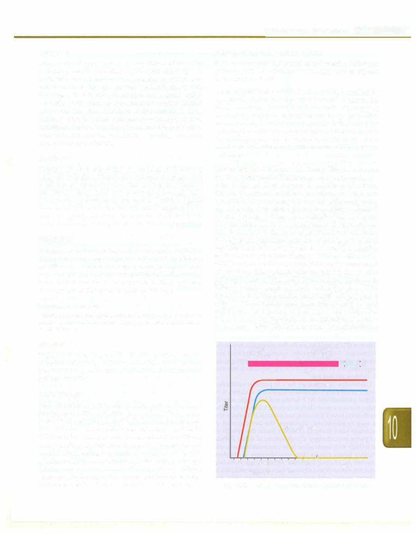
Chronic infection. Children with chronic HBV infection are mostly asymptomatic. They are generally active and grow well. Although liver damage is usually mild during childhood, serious sequelae, including cirrhosis and hepatocellular carcinoma, may develop insidiously at any age.An immune-mediated process is themain mechanism for cell damage. During acute exacerbations of chronic HBV infections, CD8+ T lymphocytes are the predominant cells in the liver in the areas of piecemeal necrosis. Since HBeAg is an important marker reflecting active viral replication and infectivity, its clearance is used as a marker for the success of antiviral therapy. Children with chronic HBV infection are HBeAg seropositive at the initial stage of infection; this antigenemia can persist for yr after primary infection (Fig. 10.8). Spontaneous clearance of HBeAg occurs gradually with increasing age. Viral replication is reduced during this process. This process of HBeAg seroconversion takes place subclinically in most individuals for a period of 2 to 7 yr (Table 10.3). This process is usually preceded by an elevation of amino transferases. After HBeAg clearance, aminotransferase
Acute |
Chronic |
(6 months) |
(years) |
HBeAg |
l·MUl#i=tl |
HBsAg
Total anti-HBc
lgM anti-HBc
/1 |
I |
|
O 4 8 12 16 20 24 28 32 36 II 52 |
/I |
Years |
Weeks after exposure
Fig. 10.8: Serological response in hepatitis B virus infection
- Essential Pediatrics
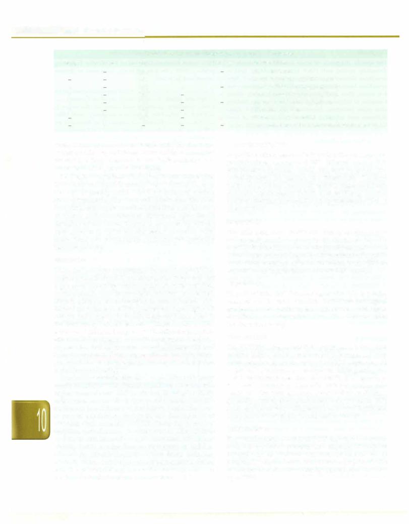
Table 10.3: Common seropatterns of hepatitis B infection
HBsAg |
Anti-HBs |
Anti-HBc |
+ |
|
IgM |
|
|
IgM |
+ |
|
IgG |
+ |
|
IgG |
+ |
|
+/ |
+ |
|
IgG |
|
+ |
IgG |
|
+ |
|
HBeAg
+
+/
+
Anti-HBe Interpretation
Acute infection
+/- Acute infection; anti-HBc window Chronic infection; high infectivity
+Chronic infection; low infectivity
|
Precore/Core mutant infection |
+/ |
Hepatitis B carrier |
+/- |
Recovery from infection |
|
Immunization; false positive; infection inremote past |
levels return to normal levels and anti-HBe develops. Longtermfollowup of HBsAg carrier children showsthat the rate of HBsAg clearance is low (0.6% annually), and occurs only after clearance of HBeAg.
During the early phase of infection, the amount of virus in the liver and blood is usually large, whereas the liver damage is mostly mild. The host immune system gradually recognizes the virus and startsto clear the virus. It results in active inflammation of the liver and elevation of serum aminotransferases. Repeated episodes of elevation of aminotransferases is followed by HBeAg seroconversion. After HBeAg seroconversion, viral replication declines andthe liver inflammation gradually becomes inactive.
Treatment
There are two approvedtherapiesfor chronic hepatitis Bin children: interferon (IFN) and lamivudine. Interferons are a group of naturally occurring agents with antiviral, antineoplastic and immunomodulatory properties. IFNa2a achieves seroconversion in approximately one third of cases; those most likelyto respond have high ALT activity and a greater histological activity index score in the liver biopsy before treatment. Children younger than 6 yr have an enhanced response toIFNa2b treatment. Side effects of IFN inchildren are flu-like symptoms, headache, depression, loss of appetite, anemia, leukopenia and thrombocytopenia. Promising results are emerging using pegylated IFN in adults with chronic hepatitis B, but data in children are lacking.
Lamivudine monotherapy for 1 yr provides satisfactory results in children with chronic hepatitis B. Children with higher pretreatment ALT levels and histologic activity indexscoresare mostlikelytorespondtolamivudine. The medication is well tolerated, has minimal side effects and is easy to administer. However, the development of resistant viral mutants (YMDD) limits the benefit of longterm monotherapy. Combinations of either INFa2a or INFa2b with lamivudine have comparable effects and slightly better results than monotherapy in children affected by chronic hepatitis. Other drugs including adefovir, entecaviranddipivoxilhave documentedclinical activity against wild and lamivudine resistant HBV, but needs to be further evaluated in children.
lmmunoprophylaxis
Hepatitis B imrnunoglobulin is used in the postexposure prophylaxis of newborns of HBV infected women. It is administered intramuscularly and may be given con currently with HBV vaccine, at a different site. The dose for infants is 0.5 ml. Combination of the imrnunoglobulin and HBV vaccination in infants born to HBsAg positive mothers prevents transmission in approximately 95% of those at risk.
Hepatitis D
Hepatitis delta virus (HOV) was first detected as a new nuclearantigeninthe hepatocytes ofpatientsinfectedwith hepatitisB virus (HBV)andwasfrequentlyassociatedwith severe acute orchronichepatitis.Transmissionofhepatitis delta virus requires either coinfection with HBV or superinfection in individuals who are HBV carriers.
Hepatitis C
Hepatitis Cvirus (HCV) was recognizedin 1989 as a major cause of non-A, non-B hepatitis. HCV is an enveloped, single-stranded, positive-sense ribonucleic acid virus, classified as an independent genus (Hepacivirus) within the Flavivirus family.
Viral Variants
The HCV RNA-dependent RNA polymerase lacks proof reading ability, which results in HCV being genetically heterogeneous. Based on analysis of HCV sequences, six major HCV genotypes are recognized. HCV genotypes 1 and 2 are the most prevalent worldwide. HCV genotype 3 is most common in Australia and the Indian sub continent. The viral genotypic distribution in children generally parallelsthatreported regionallyin adults. HCV genotype 1 correlates with higher serum viral levels and a less favorable response to antiviral treatment.
Epidemiology
The worldwide prevalence of HCV infection is approxi mately 3%, which represents an estimated 170 million infected persons. Children who received transfusions of potentially contaminated blood products prior to the institution of routine screening have seroprevalence rates up to 95%.
Clinical Features
The mean incubation period of post-transfusion acute HCY infection is 7to 8 weeks,witha rangeof 2 to 26 weeks. Acute HCY is usually anicteric or subclinical and only one-third of patients develop jaundice or symptoms. Fulminant hepatic failure due to HCY is rare. In adults, 85% of patients exposed to HCY will develop chronic infection, of which approximately 10 to 20% develop cirrhosis. In children, the course of HCY infection is generally benign. Most children withacutehepatitis C are asymptomatic.
When symptoms are present, they are often nonspecific (malaise, anorexia) or mild; jaundice is present in 25%. Most children exposed to HCY are at risk to become chronically infected based on persistently detectable serum anti-HCY antibodiesandHCY RNA Children with chronic HCY infection may also remain asymptomatic. Progression to decompensated liver disease in children is rare. Biochemical markers such as serum alanine aminotransferase typically fluctuate in HCY patients. Normal or only minimally increased transaminase levels are reported with chronic HCY infection and these can remain elevated despite anti-HCY seronegativity. Liver histology shows portal lymphoid aggregates, bile duct injury and steatosis; necroinflammatory activity is mild.
Perinatal Transmission
The rate of vertical transmission is approximately 5-6%, which is low compared to that for hepatitis B virus and human immunodeficiency virus. High-titer maternal viremia correlates with higher transmission rates. Breastfeeding is permitted unless the motherhas bleeding nipples.
Diagnosis
The diagnosis of HCY infection is based on detection of antibodies against recombinant HCY antigens by enzyme immunoassay or recombinant immunoblot assay or by detection of HCY RNA using nucleic acid tests. Enzyme immunoassay is limited by frequent false-positive results, particularly in patients with elevated globulin levels such as those with autoimmune hepatitis. Recombinant immunoblot assays are less sensitive but more specific than enzyme immunoassay in detecting anti-HCY antibodies. Recombinant immunoblot assay is, therefore, not recommended for initial HCY screening and are useful to confirm viral infection. Nucleic acid tests identify the presence of HCY very early in the course of infection and therefore, are used to diagnose infection even before the anti-HCY antibodies have appeared. These tests are also necessary to detect HCY in infants born to infected mothers, in whom HCY antibodies may be of maternal origin and in immuno compromised patients whose ability to produce HCY antibodies may be impaired.
Infections and Infestations -
Therapy
Sustained virologic responses are achieved in only 8-35% of patients given recombinant interferon monotherapy. However,significantlyhigher sustained virologic respon ses are attained (30-40%) by combining interferon with ribavirin at 15mg/kg/day. Longer-actingpegylatedinter ferons have been subsequently developed based on the premise that more sustained drug levels would result in greater antiviral activity. Several randomized clinical trials in adults verify considerably better virologic responses (50-60%) with the use of pegylated interferons, particu larly when given in conjunction with ribavirin. However, in general, sustained virologic response rates in children treated with interferon alone (30-60%) appear to be two to three-fold higher than in similarly treated adults. Importantly, biochemical and virologic responses have been accompanied by significant histologic improvement in all treated patients included in these trials, and interferon has been well tolerated in children.
Suggested Reading
Heller S, Valencia-Mayoral P. Treatment of viral hepatitis in children. Arch Med Res 2007;38:702-10
Hsu EK, Murray KF. Hepatitis B and C in children. Nat Clin Pract Gastroenterol Hepatol 2008;5:311-20
Price N, Boxall EH. Treatment of children persistently infected with hepatitis B virus: seroconversion or suppression. J Antimicrob Chemother 2007;60:1189-92
Hepatitis E
Hepatitis E virus was first described in 1978 after an epidemic affecting 52,000 individuals in Kashmir. Hepatitis E is caused by infection with thehepatitis E virus (HEY), a single-stranded RNA virus. Just like hepatitis A virus, HEY is transmitted via the fecal oral route. It is usually transmittedthrough contaminateddrinking water. Hepatitis E virus causes acute sporadic and epidemic viral hepatitis. Symptomatic HEY infection is most common in young adults aged 15-40 yr and is uncommon in children since it is mostly asymptomatic and anicteric.
Clinical Features
The incubation periodfollowing exposure to HEY ranges from 3 to 8 weeks, with a mean of 40 days. The clinical presentation of hepatitis E is similar to hepatitis A The severity of an HEY infection is generally greater than the severity of an HAY infection. In pregnant women,the disease is particularly severe where mortality approaches 20% with infections in the third trimester. Premature deliveries with high infant mortality up to 33% are observed. No evidence of chronic inflammation or of a healthy chronic carrier state has been detected and no recurrence of hepatitis E has been reported.
Diagnosis
Laboratory evaluation of HEY is similar to that of HAY and is based on detection of IgM antibodies. These

- Essential Pediatrics
antibodies (IgM and IgG) develop at the time symptoms occur, usually before the development of jaundice. IgM anti-HEY titer declines rapidly during early convale scence,whileIgG anti-HEYpersistsfor long duration and provides protection against subsequent infections.
Treatment
As no specific therapy is capable of altering the course of acute hepatitis E infection, prevention is the most effective approach against the disease.
Prevention
Good personal hygiene, high quality standards for public water supplies and proper disposal of wastehave resulted in a low prevalence of HEV infections in developed countries. At present,thereare no commercially available vaccines for the prevention of hepatitis E.
Hepatitis due to other Viruses
Certain cases of post-transfusion (10%) and community acquired hepatitis (20%) are of unknown origin. Three viruses are potentially associated with liver disease but no conclusive evidence exists to support them as a cause for these cases. These viruses are HGV/GB virus C, TT virus and SEN virus.
Suggested Reading
Aggarwal R, Jameel S. Hepatitis E. Hepatology 2011 Dec;54:2218-26
Dengue Infections
Dengue fever is an acute illness characterized by fever, myalgia, arthralgia and rash. Severe dengue infection is characterized by abnormalities in hemostasis and by marked leakage of plasma from the capillaries; the latter may lead to shock (dengue shock syndrome).
Epidemiology
The global prevalence of dengue has grown dramatically in recent decades. The disease is now endemic in more than 100 countries in Africa, the Americas, the Eastern Mediterranean, South-East Asia and the Western Pacific. WHO currently estimates there may be 50 million cases of dengue infection worldwide every year. During epidemics of dengue, attack rates among susceptible are often40-50%,but mayreach80-90%. An estimated 500,000 cases of severe dengue infection require hospitalization each year, of which very large proportions are children. Without proper treatment in severe dengue infection [earlier called dengue hemorrhagic fever (DHF) dengue shock syndrome (DSS)] case fatality rates can exceed 20%.
The spread of dengue is attributed to expanding geographic distribution of the four dengue viruses andof their mosquito vectors, the most important of which is the predominantly urban species Aedes aegypti. A rapid rise in urbanpopulationsisbringing evergreaternumbers
of people into contact with this vector, especially in areas that are favorable for mosquito breeding, e.g. where household water storage is common and where solid waste disposal services are inadequate.
Virus. Dengue fever is causedbyinfectiondue to any of the four serotypes of dengue viruses. Dengue viruses are arboviruses that belong to the family Flaviviridae. The envelop protein bears epitopes that are unique to the serotypes;theantibodiestotheseuniqueepitopesneutralize by interfering with the entry of the virus into the cells.
Transmission. Dengue viruses are transmitted to humans through the bites of infected female Aedes mosquitoes. Mosquitoes generally acquire the virus while feeding on the blood of an infected person. After incubation for 8-10 days, an infected mosquito is capable, during probing and blood feeding, of transmitting the virus, to susceptible individuals for the rest of its life. Infected female mosquitoes may also transmit the virus to their offspring by transovarial transmission, but the role of this in sustaining transmission of virus to humans has not yet been delineated.Humans are the main amplifying host of the virus, although studies have shown that in some parts of the world monkeys may become infected and perhaps serve as a source of virus for mosquitoes. The virus circulates inthebloodof infected humans fortwoto seven days, at approximately the same time as they have fever; Aedes mosquitoes may acquire the virus when they feed on an individual during this period.
Pathophysiology
The major pathophysiologic changes that determine the severity of disease in severe dengue infection and differentiate it from dengue fever are plasma leakage and abnormalhemostasisleading to rising hematocrit values, moderate to marked thrombocytopenia and varying degrees ofbleeding manifestations. Thecause of abnormal leakage of plasma is not entirely understood. However, rapid recovery without residual abnormality in vessels suggests it to be the result of release and interaction of biological mediators, which are capable of producing severe illness with minimal structural injury.
It has been observed that sequential infection with any two of the four serotypes of dengue virus results in severe dengue infections in an endemic area. How a second dengue infection causes severe disease andwhyonlysome patients get severe disease remains unclear.It is suggested that the residual antibodies produced during the first infection areabletoneutralize a secondviralinfection with the same serotype. However, when no neutralizing antibodies are present (i.e. infection due to another serotype of dengue virus), the second infection is under the influence of enhancing antibodies and the resulting infection and disease are severe. An alternative expla nation is that certain strains (South-East Asian) of the dengue virus may be inherently capable of supporting