

Ghai Essential Pediatrics8th
.pdfTreatment
Indications for inpatient treatment Most cases of enteric fever can be managed at home with oral antibiotics and advice to seek medical followup in case of failure to respond to therapy or development of complications. Children with persistent vomiting, inability to take orally, severe diarrhea or abdominal distension usually require intravenous antibiotics therapy and intravenous fluids, necessitating admission to hospital.
Antimicrobial susceptibility Though resistance to chloramphenicol was first noted soon after its first use in the 1940s, it was not until 1972 that chloramphenicol resistant typhoid fever became a major problem. Multi drug resistant typhoid fever (MDRTF) became a common occurrence by the end of 1990s, with emergence of S. typhi simultaneously resistant to all the drugs that were used as first-line treatment (chloramphenicol, trimethoprim, sulfamethoxazole and ampicillin). Fluoroquinolones, introduced in the late 1980s and early 1990s, produced very good results initially, but the past decade has seen a progressive increase in the minimum inhibitory concen trations of ciprofloxacin in S. typhi and paratyphi. Since minimum inhibitory concentrations are below the stan dard susceptibility breakpoint, laboratories report bacteria as sensitive to fluoroquinolones; but the use of fluoro quinolones is associated with a high incidence of clinical failure because drug levels needed to kill organisms are not achieved with standard doses, and often, even with highest tolerated doses. Now that the susceptibility breakpoints have been revised downwards, this discor dance between in vitro and in vivo susceptibility will be resort. Resistance to nalidixic acid has been suggested as a marker of fluoroquinolone failure.
Currently, third-generation cephalosporins such as ceftriaxone and cefixime are the first-line agents for therapy of enteric fever. Azithromycin is a new drug that is being used as an alternative agent.
Choice for empirical therapy For uncomplicated ente ric fever, oral cefixime at a dose of 20 mg/kg/day is the drug of choice, both for sensitive and multidrug resistant S. typhi. In areas where quinolone resistance is infrequent (rare at the moment in India), fluoroquinolones may still be considered the drugs of choice; however, if both quinolone resistance and resistance to other drugs (like amoxicillin, chloramphenicol, cotrimoxazole) are wide spread, the only options are oral cefixime or azithromycin.
Azithromycin (10-20 mg/kg/day) is a good second choiceagent; chloramphenicol (50mg/kg/day), amoxicillin and cotrimoxazole are other second-line agents. The choice of medication depends on individual preference, experience and level of comfort and cost considerations. Once culture results are available, therapy can be modified. There is no data at present to support use of combination therapy in enteric fever.
Infections and Infestations -
For severe illness and where complications are present, intravenous ceftriaxone and cefotaxime are used at a dose of 100 mg/kg/day. In patients with history of penicillin or cephalosporin allergy, aztreonam, chloramphenicol (in higher than usual doses) or cotrimoxazole (in higher than usual doses) are used as second-line agents. Parenteral treatment is continued until defervescence has occurred, oral intake has improved and complications resolved. Thereafter, therapy can be switched to oral cefixime to complete a total duration of 14 days. Other oral drugs that may be used for switch over therapy include cefpodoxime, azithromycin, cotrimoxazole and amoxicillin. However, the experience with cefpodoxime is limited and the other agents require switch to a different class of antimicrobials than cephalosporins.
If cultures are positive and show quinolone sensitivity, therapy should be changed to ciprofloxacin at a dose of 20 mg/kg/day as quinolones are associated with faster defervescence and lower relapse rates as compared to ceftriaxone. If cultures are positive and show quinolone resistance as well as sensitivity to other drugs ampicillin, chloramphenicol and cotrimoxazole), it is prudent to continue with ceftriaxone alone rather than change because the older drugs do not offer any advantage over ceftriaxone. If cultures are negative and defervescence has not occurred by day 7, a thorough search for alternative etiology for fever should be made and ceftriaxone continued. There is no role for changing the antimicrobial agent or adding another drug, since ceftriaxone resistance is still anecdotal.
Therapy of relapses Relapse rates vary with the type of drug and are most common withbeta lactams (ceftriaxone, cefixime) especially if shorter duration of therapy is used. Usually relapses may be satisfactorily treated with the same drug as used for primary therapy but at appropriate dose and duration. However, if the isolate is quinolone sensitive and fluoroquinolones were not used for primary therapy, they should be used for treatment of the relapse. Therapy of carriers The carrier state is uncommon in children and testing for chronic carriage 3 months after an episode of enteric fever is not recommended. However, if chronic carriage is demonstrated, treatment with amoxicillin (100 mg/kg/day) with probenecid (30 mg/ kg/day) or cotrimoxazole (10 mg/kg/day) for 6-12 weeks is recommended. If the strain is nalidixic acid sensitive, quinolones for 28 days is a better option.
Prevention
The most effective and desirable method for preventing enteric fever is by improving hygiene and sanitation. This will yield additional dividends of reduction in the burden of other water-borne illnesses as well. Vaccination is a major preventive strategy.
___E_s_s_e_n_t.aii_P_e _d.iat .ric s _________________________________ _
Suggested Reading
Kundu R, Ganguly N, Ghosh TK, Yewale VN, Shah RC, Shah NK; IAP Task Force. Report: diagnosis of enteric fever in children. Indian Pediatr 2006;43:875-83
Kundu R, Ganguly N, Ghosh TK, Yewale VN, Shah RC, Shah NK; lAP Task Force. Report: management of enteric fever in children. Indian Pediatr 2006;43:884-7
Parry CM, Hien TT, Dougan G, White NJ, Farrar JJ. Typhoid fever. N Eng J Med 2002;347:1770-82
Leptospirosis
Leptospirosis is a zoonotic disease of worldwide distri bution, caused by spirochetes. Most cases occur in tropical and subtropical countries. While rats are the principal source of human infection, dogs, cats, livestock and wild animals are otherreservoirs.Infectedanimals mayexcrete spirochete in urine for several weeks. The survival of excreted organisms depends on the moisture content and temperature of the soil. Humans acquire infection after getting exposure to water or soil contaminated with rat urine. Agricultural workers, veterinarians,meat handlers, rodent control workers and laboratory personnel are at risk of getting infected because of occupational exposure. Infectionis common inthe monsoons and during flooding.
Pathogenesis
Leptospira enter the body through abrasions and cuts in skin or through mucous membranes and spread to all organs hematogenously. The organisms damage the endothelial lining of smallbloodvessels, with leakage and extravasation of blood cells, hemorrhage and ischemic damage to various organs including liver, kidneys, meninges and muscles.
Clinical Features
Human infection ranges from asymptomatic infection to a severe multiorgan involvement which is often fatal. Symptomatic infection is a relatively mild as an anicteric febrile illness in over 70% of patients; about 20% present as aseptic meningitis, while severe leptospirosis with hepatorenal dysfunction (Weil disease)developsin5-10% ofindividuals. The incubation period is usually1-2 weeks.
The illness is often biphasic. In the initial or septicemic phase lasting 2-7 days, the onset is abruptwithhigh grade fever with rigors and chills, lethargy, severe myalgia, headache, nausea, vomiting. There may be conjunctiva! suffusion with photophobia and orbital pain, generalized lymphadenopathy and hepatosplenomegaly. Transient maculopapular rash may be seen in <10% cases. Hypo tension with bradycardia and circulatory collapse is rare. Some patients develop acute respiratory distress syndrome with respiratory failure. Most patients are asymptomatic within one week.
In some patients, after a brief asymptomatic phase, the second phase, called the immune or leptospiruric phase, becomes manifest where Leptospira localize to tissues to cause specific signs and symptoms. In this phase,
circulating autoantibodies to Leptospira are present; organisms canno more be isolated from blood or CSF but persist in tissues like kidneysandaqueous humor. During the immune phase, some children develop aseptic meningitisoruveitis withrecurrenceoffever. Encephalitis, cranial nerve palsies, paralysis and papilledema are rare. Central nervous system abnormalities usually normalize within 1 week; mortality is rare.
In icteric Ieptospirosis (Weil syndrome) after the initial phase of fever patients develop severe hepatic and renal dysfunction. Jaundice and hepatomegaly are usually detected; splenomegaly is found in20%. Renal failuremay develop, often during the second week of illness. All patients have abnormal urinary finding on urinalysis in the form of hematuria,proteinuria and casts. Azotemia is common, often associated with oliguria or anuria. Hemorrhagic manifestations are rare but when present, may include epistaxis, hemoptysis and gastrointestinal and adrenal hemorrhage. Transient thrombocytopenia may occur, and mortality is 5-15%.
Diagnosis
Complete blood count shows anemia, leukocytosis with polymorph predominance and thrombocytopenia. The CRP is elevated and liver enzymes are mildly elevated with SGOT more than SGPT. The CPK is high. In patients with Weil disease there is elevated serum creatinine, deranged coagulation parameters and direct hyper bilirubinemia with raised transaminases.
Specific diagnosis is established by serologic testing, microscopicdemonstrationof the organism or culture. The gold standard for serologic diagnosis is the microscopic agglutination test (MAT), which is only available in reference centers. Commercial kits for serologic diagnosis includerapidtestsandIgM ELISA. These tests arepositive after 5 days of illness. Cross reactivity and false positivity is seen with other infections like enteric fever and malaria. Demonstration of organism in tissues or urine by dark field microscopy or immunoflorescence and cultures are not routinely available.
Leptospirosis should be differentiated from other febrile illnesses commonly seen in the monsoon season like malaria, dengue, enteric fever, acute viral hepatitis and hantavirus infections.
Treatment
Treatment should be initiated as early as possible. For a severe case of leptospirosis, parenteral penicillin G (6-8 million U/m2/24 hr q 4 hr IV) for 7 days is the drug of choice. Ceftriaxone and IV tetracycline are also acceptable alternatives. Fororaltreatmentamoxicillinanddoxycycline (in children above 8 yr) are the drugs of choice.
Prevention
Prevention entails avoidanceof exposure to contaminated water. Single dose doxycyline or amoxicillin following
exposure can prevent illness but is not routinely recommended.
Suggested Reading
Tullu MS, Karande S. Leptospirosis in children: a review for family physicians. Indian J Med Sci 2009;63:368--78
Tetanus
Tetanus is caused by the bacterium Clostridium tetani, a spore forming, anerobic, gram-positive motile bacillus, found in human and animal feces. Its spores are widespread in the environment. Tetanus commonly occurs in areas where soil is cultivated, in rural areas, in warm climates and during summer months. According to WHO estimates, it contributes to 8% of vaccine preventable deaths.
Pathogenesis
C. tetani is a noninvasive organism. The spores of the organism remain nonpathogenic in soil or contaminated tissues until conditions are favorable for transformation into vegetative form. Transformation occurs in the presence of locally decreased oxygen reduction potential, typically in devitalized tissue, in the presence of a foreign body, trauma and crush injury and suppurative infections. Two types of toxins are produced by the organism, tetanolysin and tetanospasmin. Tetanospasmin, is the main toxin responsible for the manifestations of the disease. It binds to the neuromuscular junction at the site of injury, and undergoes retrograde axonal transport to reach the presynaptic nerve terminal where it prevents the release ofinhibitory neurotransmitters glycine and GABAleading to uncontrolled contraction of muscles.
Clinical Features
Tetanus mainly affects the unimmunized and partly immunized individuals. The disease may occur in various forms: neonatal, generalized, localized and cephalic. The most commonforms are generalized andneonatal tetanus.
Generalized tetanus has an incubation period of approxi mately 8 days (range 2-14 days). However, the disease may occur months after the initial injury. The incubation period depends on the distance of the site of injury from the central nervous system. The faster is the onset of symptoms, the poorer is the prognosis. Characteristically, there is descending paralysis, with initial involvement of the jaw muscles. There is spasm of the masseters leading totrismusorlockjaw.Subsequentinvolvement of the neck, back and abdominal muscles occurs, soon involving the whole body. As the disease progresses, minimal stimuli may lead to generalized spasms, which are the hallmark of the disease and contribute to serious complications and death. Typically, thesensoriumof the patient is preserved. There is difficulty in swallowing. Autonomic instability
Infections and Infestations -
may occur, with blood pressure fluctuations in the form of hypertension or hypotension, diaphoresis and arrhythmias. Recovery usually begins after 3 weeks and approximately takes four weeks. Recovery from tetanus occurs bysproutingnewnerveterminals in the spinal cord leading to relaxation of the contracted muscles.
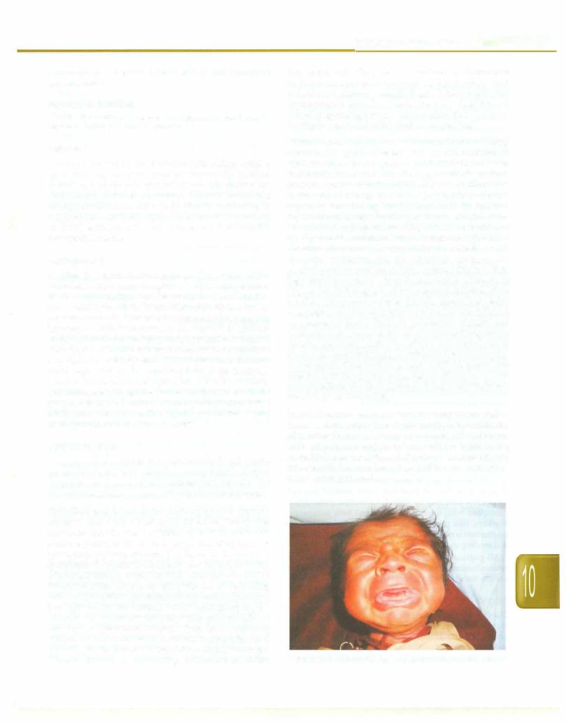
Neonatal tetanus is a major cause of mortalityin developing countries. Pregnant women who are not immunized against tetanus do not pass on protective antibodies to theirbabies. Infectionresultsof unhygienicbirthpractices, most commonly when the umbilical cord is contaminated at the time of cutting after delivery. Symptoms usually appear by the third day afterbirth, never in the first two days of life and rarely after the age of two weeks. Excessive unexplained crying followed by refusal of feeds and apathy are the common initial symptoms. The baby develops progressive feeding difficulty, becomes rigid, develops paralysis and may develop opisthotonic posturing and experience painful spasms. The mouth is kept slightly open due to pull and spasm of the neck (Fig. 10.13). Reflex spasm of the masseter makes feeding painful. Pharyngeal muscles go into spasm and cause dysphagia andchoking,lockjaw or reflextrismusfollowed by spasms of limbs. There is generalized rigidity and opisthotonusin extension. Spasm of larynx and respiratory muscles are characteristically induced by stimuli such as touch, noise andbrightlight, resulting inepisodes of apnea and cyanosis. Constipation persists until the spasms are relieved. In.tercurrent infections, dehydration and acidosis may complicate the clinical picture. It has a very high case fatality rate of 70 to 100%.
Localized tetanus is less severe in comparison and is characterized byrigidity and painconfined to the muscles adjacent to the wound. It may lead to generalized tetanus later. In patients with isolated localized tetanus, the mortality is lessthan 1%. Cephalic tetanus is aform of local tetanus, which occurs due to injury of the bulbar muscles. It has a poor prognosis.
Fig. 10.13: Neonatal tetanus. Courtesy: Dr Amarjeet Mehta, Jaipur
--E•s•s•e•n•t•ia• l•P•e•d• i•a ·.·tncs. . |
---------------------- |
- |
---------- |
|
|
Treatment
Most patients require intensive care management and good supportive care. The aims of treatment are airway maintenance, prevention of further toxin absorption, relieving clinical features, e.g. spasms, controlling autonomic instability and antibiotics. Airway manage ment may require intubation and mechanical ventilation, especially in severe cases and if the infant gets frequent episodes of largyngeal spasms, apneic attacks with cyanosis or central respiratory failure. Neutralization of free toxin is done by administering human tetanus immunoglobulin (TIG); however, antitoxin cannot dislodge the toxin already fixed to the nerve roots. The route of administration is intramuscular or intrathecal. The usual dose is 500 to 1000 IU. Antibiotic therapy is needed to abolish the bacteria from the wound site. The commonly used antibiotics are crystallinepenicillin or metronidazole.
Spasms are precipitated by minimal stimuli, therefore, efforts should be made to avoid noxious stimuli including bright lights, pain and loud noises. Patient should be kept in a dark, quiet and isolated room, which should be lighted well to permit observation of the child; handling should be minimum. Intramuscular injections must be avoided. Temperature should be maintained within normal limits. Relief of spasms is done by using benzodiazepines. The most commonly used agent is diazepam, either as an intermittentIV bolus or as continuous infusion. Diazepam prevents further spasms by causing GABA-mediated central inhibition. It also helps by reducing anxiety and promoting muscle relaxation. Other agents used for severe spasms include pancuronium bromide.
Supportive care includes adequate hydration, early detection of myoglobinuria and prevention of renal shutdown. Oropharyngeal secretions should be sucked periodically. Maintenance of oxygen is important. Oral feeding should be stopped and an IV line should be established for providing adequate fluids, calories and electrolytes and for administration of medications. After three to four days of treatment, milk feeding through nasogastric tube may be started. Autonomic instability is controlled with the use of alpha and beta adrenergic blockers, like propranolol and labetalol. Intravenous magnesium is effective in decreasing autonomic instability and treating muscle spasms.
All patients should receive a complete course of immunization with tetanus toxoid once recovered, as the disease does not induce protective antibodies.
Prognosis
The disease has high mortality rate in spite of adequate supportive care, which may reach up to 50% in severe generalized tetanus and 90% in neonatal form. The outcome depends on the incubation period, the site of injury, the rate of progression of illness and presence of
autonomic instability. Survivors do not manifest any neurological sequelae, except when apneic episodes are unduly prolonged and unattended. The prognosis in neonatal tetanus isworse if the (i) onset of symptoms occurs within the first weeks of life, (ii) interval between lockjaw and onset of spasms is less than 48 hr, (iii) high fever and tachycardia are present, and (iv) spasms, especially of larynx resulting in apnea are severe and frequent.
Prevention
Immunization with tetanus toxoid leads to induction of protective antibodies (Chapter 9). Maternal and neonatal tetanus can be effectively prevented by immunizing the mother during pregnancy, and ensuring clean delivery and cord care.
Suggested Reading
Okoromah CN, Lesi FE. Diazepam for treating tetanus. Cochrane Database Syst Rev 2004; CD003954
Roper MH, Vandelaer JH, Gasse FL. Maternal and neonatal tetanus. Lancet 2007;370:1947-59
Singhi S, Jain V, Subramanian C. Post-neonatal tetanus: issues in intensive care management. Indian J Pediatr 2001; 68:267-72
Rickettsial Infections
Rickettsial diseases are a group of febrile illnesses caused by obligate intracellular gram-negative bacilli and transmitted to man by arthropod vectors.
Etiopathogenesis and Epidemiology
Rickettsia are a group of motile, gram-negative, nonspore forming highlypleomorphic bacteria that present as cocci, rods or thread like obligate, intracellular parasites. Scrub typhus caused by R.tsutsugamushi, Indian spotted fever caused by R.conorii and Q fever caused by C. burnetti are the rickettsial infections prevalent in India. Cases have been reported from all states chiefly from rural and forested areas and occasionally also from urban areas.
Scrub typhus is transmitted by bite of the trombiculid mite and Indian spotted fever by ticks. Rickettsial disease is due to invasion of the endothelial region of the vas culature and subsequent microvasculitis. This process especially affects the brain, cardiac and skeletal muscle, skin, liver, lungs and kidneys.
Clinical Manifestations
Incubation period in children varies from 2 to 14 days. A history of exposure to tick, history of origin from an endemic area or a similar illness in family members may be forthcoming. Severity of manifestations varies from a mild, self limiting illness to a life-threatening disease.
Initially the illness appears to be nonspecific and patients present with unrelenting headache, very high fever, anorexia, myalgias, restlessness, calf muscle pain and tenderness. Gastrointestinal symptoms include abdominal pain, nausea, vomiting and diarrhea. Skin rash
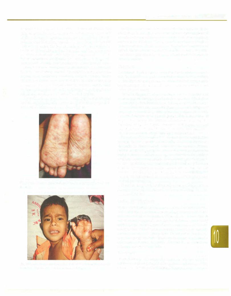
is usually not present until after 2-4 days of illness. The triad of fever, headache and rash is observed in only half of the patients. In spotted fever, rash is initially discrete pale rose red blanching macules or maculopapules on the extremities. Later, the rash spreads rapidly to involve the entire body including palms and soles and becomes more petechial sometimes with palpable purpura (Fig. 10.14). In severe form of the disease, petechiae may enlarge into ecchymosis, which can become necrotic. Severe vaso occlusive disease secondary to rickettsial vasculitis and thrombosis is infrequent but can result in gangrene of the digits, toes, earlobes, scrotum, nose or entire limbs (Fig. 10.15). In scrub typhus, rash isseen initially on trunk or may not be present at all. Painless eschar, may be seen at the initial site of tick attachment and regional lymphadenopathy and is seen in scrub typhus.
Complications may involve any organ system and include encephalopathy, pulmonary edema, myocarditis, acute renal failure and vascular collapse.
Fig. 10.14: Maculopapular rash on the soles. Courtesy, Dr Atul Kulkarni,
Sholapur
Fig. 10.15: Necrotic rash in spotted fever. Note the gangrene of ear
lobes and fingers. Courtesy: Dr Atul Kulkarni, Sholapur
Infections and Infestations -
In Q fever no vector is involved. Transmission is by inhalation ofinfecteddustfromsoil previouslycontaminated by urine or feces of diseased animals. This fever is rarely reported in children. This presents in acute as well as chronic forms. Rash which is typical of variants of rickettsial fever isnot seen inQfever. Pneumonia, hepatitis and meningitis are other features. Endocarditis is seen in chronic variety.
Diagnosis
Laboratoryfindings are nonspecific. Totalleukocytecount may be initially normal or low but leukocytosis develops as the disease progresses. Anemia, thrombocytopenia, hyponatrernia and elevated serum aminotransferases are some other features.
Specific diagnosis of a rickettsial illness is confirmed by serological testing. Serological evidence of infection occurs not earlier than the second week of illness in any of the rickettsial diseases and hence a specific diagnosis may not be available until after the patient has fully recovered or worsened. Serodiagnosis ofrickettsialdisease is possible using the immunoflourescence assay for detection of IgG and IgM. However, it is expensive and not widely available. ELISA is specific and sensitive allowing detection of IgG and IgM antibodies.
Detection of IgM antibodies to scrub typhus and Indian spotted fever is available in India. The Weil-Felix test depends on detection of antibodies to various Proteus species containing antigen with cross reacting epitopes to antigens from members of the genus Rickettsia. It is widely available but unacceptable for accurate diagnosis because of low sensitivity and specificity. Weil-Felix test can be used in developing countries where other tests are not available for diagnosis of rickettsia infection but the test should be interpreted in conjunction with history and clinical presentation.
If clinical suspicion for rickettsia is high, then empirical therapy should be started without waiting for any confirmatory test.
Differential Diagnosis
Spottedfever can mimic a great numberof febrile illnesses. Most important of these are meningococcernia, measles and enteroviral exanthemas. Other diseases included in differential diagnosis are typhoid fever, secondary syphilis, leptospirosis, toxic shock syndrome, scarlet fever, rubella, Kawasaki disease, parvoviral infection, idiopathic thrombocytopenic purpura, thrombotic thrombo cytopenic purpura, hemolytic uremic syndrome, Henoch Schonlein purpura, hepatitis, dengue fever, infectious mononucleosis and malaria.
Treatment
Doxycycline is the drug of choice for all age groups. Chloramphenicol is reservedforpatientswithdoxycycline allergy and for pregnant women. Doxycycline can be used
__E_s_s_e_n_t.ia•l•P•e•d-ia.ri.tc s __________________________________
safely in young children because tooth discoloration is dose dependent and children are unlikely to require multiple courses. Furthermore, the increased mortality with chloramphenicol compared with tetracycline, when other factors such as severity are considered, have led to preference for doxycycline even in young children. The therapy should be continued for a minimum of 5-7 days and for at least 3 days until the patient is afebrile in order to avoid relapse. Patients treated with one of these regimens usually become afebrile within 48 hr and thus the entire therapy lasts for less than 10 days. In patients with severe disease, admission to an intensive care unit and appropriate supportive therapy may be required.
Prevention
Known tick infested areas should be avoided. Daily inspection of body for ticks is particularly important. Disinfection of dogs will minimize the tick population. Health education of people about modeof transmission by ticksandmeansofpersonalprotectionisequallyimportant. Prophylactic antimicrobial therapy is not recommended.
Epidemiology and Pathogenesis
Agent. All patients of pulmonary tuberculosis and most cases of extrapulmonary disease are caused by human type strain of Mycobacterium tuberculosis. A few cases of extrapulmonary illness particularly the tubercular lymphadenitis may be due to the bovine strain.
Reservoir of infection. The infection is spread by the tuberculous patient, who discharges tubercle bacilli in his sputum or nasopharyngeal secretions during bouts of coughing or sneezing, etc. Such patients are open or infective cases. In the pediatric age groups, few infections may also occur by the transplacental route (congenital tuberculosis).
Mode of infection. The usual mode of infection is through inhalation of droplets of infected secretions. The infected sputum spitted carelessly by open cases of tuberculosis dries up and the tubercle bacilli are resuspended in the dust and air. This may be a source of infection through breathing. Infection through ingestion of infected material is rare. Rarely infection may be transmitted through skin, mucous membrane or transplacentally.
TUBERCULOSIS
Tuberculosis is a chronic infectious disease caused by Mycobacterium tuberculosis. Tuberculosis still is one of the deadliest diseases in the world killing nearly 2 million people every yr. More than ninety percent of all tuber culosis cases occur in the developing countries, where limited resources are available for optimal treatment. Tuberculosis continues to be an important cause of morbidity and mortality for children worldwide.
Magnitude
Most children acquire the organism from adults in their surroundings. Several estimates make use of an arbitrary calculation assigning 10% of the tuberculosis burden to children. Tuberculosisinfectionanddiseaseamongchildren are much more prevalent in developing countries, where resources for control are scarce. The annual risk of tuberculosis infection in developing countries in children is 2-5%. The estimated lifetime risk of developing tuber culosisdiseaseforayoungchildinfectedwith M. tuberculosis as indicated by positive tuberculin test isabout10%. About 5% of those infected are likely to develop disease in the first yr after infection and the remaining 5% during their lifetime. These rates increase about six-fold in HIV infected individuals. Nearly 8-20% of the deaths caused by tuberculosis occur in children. The age of the child at acquisition of infection has a great effect on the occurrence of tuberculosis disease. Approximately 40% of infected children less than 1 yr of age if left untreated develop radiologically significant lymphadenopathy or segmental lesions compared with 24% of childrenbetween 1 and 10 yr and16% ofchildren11and15 yrofage. InIndia,over100,000 children die from h1berculosis every yr.
Host Factors
Age. No age is exempt from tuberculosis. Tubercle bacilli are nottransferredacross the healthyplacentabut the fetus may be infected from the infected placenta. Frequency of infection with tubercle bacilli increases progressively as the child grows in age. An infant is more likely to develop disease after an infection compared to an older child.
Sex. Adolescent children, especially girls, are prone to develop active tuberculosis disease during puberty.
Malnutrition. Undernourishedchildren are moresusceptible to develop tuberculosis, probably due to depressed immunological defenses. Tuberculosis may precipitate kwashiorkor or marasmus in an infant with borderline undemutrition. A malnourished patient, who does not respond to the dietary therapy should be promptly investigated for tuberculosis.
Immunodeficiency. Children with primary or secondary immune deficiencies (including HIV) are more likely to develop disseminated disease. The diseases thataffect the cell mediated immw1ity are more likely to increase the susceptibility.
Intercurrent infections. A quiescent tuberculous infection may flare up after an attack of measles or pertussis, that suppresses cell mediated immune response.
Environment. The risk of acquiring infection has been associated consistently with the extent of contact with the index case, the burden of organisms in the sputum and the frequency of cough in the index case. Patients with smear positive pulmonary tuberculosis are more likely to transmit infection. An increased risk of developing

infection has been seen in institutional settings, including nursing homes, correctional institutions and homeless shelters.
Pathology
The inhaled tubercle bacilli may lodge in the pulmonary alveoli and cause inflammation with hyperemia and congestion. Initially, the polymorphonuclear leukocytes infiltrate at the site oflesion. The phagocyticabilityofthese cells is poor and they are soon eliminated.
Further course of the infection depends on the immune response of the host. If the host resistance is good, the inflammatory exudate around the primary focus is absorbed and the caseous areainspissated. Healing occurs by fibrosis and calcification. When the cell mediated immune response is weak, thebacillicontinue to multiply and the inflammatory process extends to the contiguous areas. Progressive primary disease is a serious compli cation of the pulmonary primary complex (PPC) in which the PPC, insteadof resolving/calcifying, enlarges steadily and develops large caseous center. The center then liquefies; this may empty into an adjacent bronchus leading to formation of a cavity. This is associated with large numbers of tubercle bacilli. From this stage, the bacilli may spread to other parts of the lobe or the entire lung. This may lead to consolidation of area of lung or bronchopneumonia. Cavitary disease is uncommon in children. The enlarged lymph nodes may compress the neighboring airway. Ball-valve effect due to incomplete obstructionmay leadtotrapping ofair distal to obstruction (emphysema). Enlarged paratracheal nodes may cause stridor and respiratory distress. Subcarinal nodes may impinge on the esophagus and may cause dysphagia. If the obstruction ofbronchus is complete, atelectasis occurs.
Outcome of bronchial obstruction may include:
(i) complete expansion and resolution of chest X-ray findings; (ii) disappearance of the segmental lesions; and (iii) Scarring and progressive compression of the lobe or segment leading tobronchiectasis. A caseated lymph node may erode through the wall of the bronchus, leading to tuberculous bronchitis/endobronchial tuberculosis. Fibrosis and bronchiectatic changes may supervene. Discharge of the bacteria into the lumen may lead to its bronchial dissemination.
Hematogenous dissemination of M. tuberculosis occurs early in the course of the disease; this results when the bacilli find their way into bloodstream through lymph nodes. This may result in foci of infection in various organs. If the host immunesystemis good, thenthesefoci arecontainedand disease does not occur. Seeding of apex of lungs leads to development of Simon focus. Lowering ofhost immunity may leadto activation of these metastatic foci and development of disease. This is especially seen in young infants, severely malnourished children and children with immunodeficiency. Massive seeding of bloodstream with M. tuberculosis leads to miliary
Infections and Infestations -
tuberculosis, where all lesions are of similar size. This usually occurs within 3-6 months after initial infection.
Pulmonary tuberculosis resulting from endogenous reactivation of foci of infection is uncommon in children; butmaybeseeninadolescents.Thecommonestsiteforthis typeofdiseaseisthe apexofthelung (Puhllesion),because the blood flow is sluggish at apex. Regional lymph nodes are usually not involved. Miliary and meningeal tuber culosisusuallyoccurwithin a yr of the primary lesion.
Clinical Features
The incubation period varies between 4 and 8 weeks.
lntrathoracic Tuberculosis
The onset of symptoms is generally insidious, but may be relatively acute in miliary tuberculosis.
Primary infection usually passes off unrecognized. Asymptomatic infection is defined as infection associated with tuberculinhypersensitivity and a positive tuberculin test but with no striking clinical or roentgenographic manifestations.
Most symptoms in children with pulmonary primary complex (PPC) are constitutional in the form of mildfever, anorexia, weight loss, decreased activity. Cough is an inconsistent symptom and may be absent even in advanced disease. Irritating dry cough can be a symptom of bronchial and tracheal compression due to enlarged lymph nodes. In somechildren, the lymph nodes continue to enlarge even after resolution of parenchymal infiltrate. This may lead to compression of neighboring regional bronchus. The PPC may bepicked-up accidentally during evaluation of intercurrent infections.
Progressive primary disease (PPD) is the result of the progression of primary disease. Children with PPD may present with high-grade fever and cough. Expectoration of sputum and hemoptysis are usually associated with advanced disease anddevelopmentof cavity or ulceration of the bronchus. Abnormal chest signs consist mainly of dullness, decreased air entry and crepitations. Cavitating pulmonary tuberculosis is uncommon in children.
Children with endobronchial tuberculosis may present with fever and troublesome cough (with or without expectoration). Dyspnea, wheezing and cyanosis may be present. Occasionally, the child not responding to bronchodilators may be misdiagnosed as asthma. In a wheezing child not responding to bronchodilators less than2-yr-old, the possibility of endobronchial tuberculosis should always be considered. Partial compression of the airway can lead to emphysema. Features of collapse may be present if a large airway is completely compressed.
Miliary tuberculosis is characterized by hematogenous spread and progressive development of innumerable small foci throughout the body. The disease is most common in infantsandyoung children. The onset of illness is often sudden. The clinical manifestations depend on the
__E_s_s_e_n_t.ia•l•P•e•d-ia.t._rics __________________________________
numbers of disseminated organisms and the involved organs. The child may have high-grade fever, which is quite unlike other forms of tuberculosis. The child may also have dyspnea and cyanosis. There are hardly any pulmonary findings but fine crepitations and rhonchi may be present. These findings may occasionally be confused with other acute respiratory infections of childhood. The illness may be severe, with the child having high fever, rigors and alteration of sensorium. In addition, these children may have lymphadenopathy and hepato splenomegaly. Theotherpresentationofmiliarytuberculosis may be insidious with the child appearing unwell, febrile and losing weight. Choroid tubercles may be seen in about 50% patients. Meningitis may occur in 20-30% cases.
Pleural effusion follows the rupture of a subpleural focus into the pleural cavity. The pleura may also be infected by hematogenous spread from the primary focus. The effusion usually occurs because of hypersensitivity to tubercular proteins. If the sensitivity is high, there is significant pleural effusion along with fever and chest pain on affected side. Minor effusions associated with the rupture of primary foci are usually not detected. Tuber culous effusion is uncommon in children younger than 5 yr of age and is rarely associated with segmental lesion and miliary tuberculosis. The onset may be insidious or acute with rise in temperature, cough, dyspnea and pleuritic pain on the affected side. There is usually no expectoration. Pain in chest may disappear once the fluid separates the inflamed pleural surfaces; this may be replaced by some discomfort. Increase in effusion may make breathingshallowanddifficult. The clinical findings depend on the amount of fluid in the pleural cavity. In early stages, a pleural rub may be present. Early signs include decreased chest wall movement, impairment of percussion note and diminished air entry on the affected side. As the fluid collection increases, the signs of pleural effusion become more definite.
Extrathoracic Tuberculosis
The most common forms of extrathoracic disease in children include tuberculosis of the superficial lymph nodes (scrofula) and the central nervous system. Other rare forms of extrathoracic disease in children include osteoarticular, abdominal, gastrointestinal, genitourinary, cutaneous and congenital disease.
TB of the superficial lymph nodes can be associated with drinking unpasteurized cow's milk or can be caused by extension of primary lesions of the upper lung fields or abdomen leading to involvement of the supraclavicular, anterior cervical, tonsillar and submandibular nodes. Although lymph nodes may become fixed to surrounding tissues, low grade fever may be the only systemic symptom. A primary focus is visible radiologically only 30 to 70% of the time. Tuberculin skin test results are usually reactive. Although spontaneous resolution may
occur, untreated lymphadenitis frequently progresses to caseating necrosis, capsular ruptureandspread to adjacent nodes and overlying skin, resulting in a draining sinus tract that may require surgical removal.
Central nervous system disease is the most serious compli cation of tuberculosis in children and arises from the formation of a caseous lesion in the cerebral cortex or meninges that results from occult lymphohematogenous spread. Infants and young children are likely to experience a rapid progression to hydrocephalus, seizures and raised intracranialpressure. Inolderchildren, signsandsymptoms progress over the course of several weeks, beginning with fever, headache, irritability and drowsiness. The disease advances with symptoms of lethargy, vomiting, nuchal rigidity,seizures,hypertonia andfocalsigns. The finalstage of disease is marked by coma, hypertension, decerebrate and decorticate posturing and death. Rapid confirmation of tuberculous meningitis can be difficult because of the widevariabilityincerebrospinal characteristics,nonreactive tuberculin skin tests in 40% and normal chest radiographs in 50%. Because improved outcomes are associated with early institution of antituberculous therapy, the diagnosis should be considered for any patient with basilar meningitis, hydrocephalus or cranial nerve involvement that has no other apparent cause.
Tuberculosis of abdomen is often due to hematogenous spread from the primary focus in the lungs. It may, how ever, be secondary to swallowing of the infected sputum by a patient withpulmonarylesions. Primary tuberculosis of the intestines due to ingestion of the food contaminated by tubercle bacilli is relatively less common in India as the milk is generally boiled before use. Patients with abdo minal tuberculosis may remain asymptomatic initially. Symptomatic patients show evidence of tuberculous toxemia and may present with colicky abdominal pain, vomiting and constipation. The abdomen feels char acteristically doughy. The abdominal wall is not rigidbut appears tense, so that the abdominal viscera cannot be palpated satisfactorily. The rolled up omentum and enlarged lymph nodes may appear as irregular nodular masses with ascites. The liver and spleen are often enlarged. Histological examination of the liver may show granulomatous hepatitis and fatty change.
Diagnosis
The diagnosis of tuberculosis in children is usually based on clinical signs and symptoms, chest roentgenogram, tuberculin testing and history of contact with adult patients. Clinical features may be nonspecific and chest radiograph and Mantoux test are difficult to interpret. In addition, these do not give conclusive evidence for the disease. Although demonstration of mycobacterium in various clinical specimens remains gold standard, this is often not possible in children due to the paucibacillary nature of the illness.

A history of contact with an infective case. Contact is defined as any child who lives in a household with an adult taking antitubercular therapy or has taken such therapy in past 2 yr.A history of contact is available in less than one-third of the patients. Tracing of contact is important not only for confirming the diagnosis but also for protection of other vulnerable children from the disease.
Various scoring systems have been developed for diagnosis of tuberculosis. In these scoring system more weightage is given to laboratory tests, i.e. demonstration of acid-fast bacilli, tubercles on histology, suggestive radiology and tuberculin test >10 mm induration. These scoring systems may be used as screening tools but not for starting treatment.
Laboratory Tests
The diagnostic tests for pulmonary tuberculosis can be divided into 2 categories: (i) demonstration or isolation of M. tuberculosis or one of its components and and (ii) demonstration of host's response to exposure to M. tuberculosis.
Demonstration of M. tuberculosis or its components. The organism can be demonstrated by (i) Ziehl Neelson (ZN) staining, (ii) special stains, (iii) cultures, (iv) polymerase chain reaction, and (v) other methods. The above methods can be used on sputum, induced sputum, gastric lavage, bronchoscopic lavage fluid, or pleural fluid. The best specimen for demonstration of M. tuberculosis in children is the early morning gastric aspirate obtained by using a nasogastric tube before the child arises. The yield on ZN stainislessthan20%anddepends onextentof pulmonary disease. For better results at least 2 consecutive specimen of gastric aspiration are recommended.
Youngchildren are not able to provide sputum. In them, sputum can be induced. Following an overnight fast, the patient receives salbutamol by nebulizer followed by hypertonic saline (3% or 5%) inhalation by nebulizer. Olderchildrenmayprovideexpectoration at end ofproce dure. In young children including infants, a nasopharyn geal aspirate is collected and processed like sputum for smear and culture to identify M. tuberculosis. Gastric aspirates may similarly be collected.
Culture. Lowenstein-Jensen (LJ) medium is the most widely used medium for determination of characteristic features of colonialmorphology, growth rate and pigment production. Though the culture technique is simple, 7-10 weeks of incubation may be necessary for detection of organisms. Microscopic examination of thin layer culture plate may lead to detection of microcolonies of M. tuberculosis as early as after 7 days. The yield of culture of gastric aspirate varies from 30 to 50% in children with tuberculosis.Excessivelylongperiodrequiredforisolation of M. tuberculosis by conventional culture techniques has led to the developmentof other techniques for culture such
Infections and Infestations -
as BACTEC radiometric assay, SeptichekAFB system and mycobacterial growth indicator tube (MGIT) system.
Polymerase chainreaction (PCR). PCRis the most commonly used technique of nucleic acid amplification, for diagnosis of tuberculosis. The PCR may be used to (i) diagnose tuberculosis rapidly by identifying DNA from M. tuberculosis in clinical samples that are negative by microscopic examination; (ii) determine rapidly whether acid-fast organismsidentifiedbymicroscopicexamination in clinical specimens are M. tuberculosis or atypical mycobacteria; and (iii) identify the presence of genetic modifications known to be associated with resistance of some antimycobacterial agents. The most commonly used target for detection of M. tuberculosis is the insertion sequence IS6110. The sensitivity ranges from 4-80% and the specificity 80-100%. PCR gives rapid results and has a greater sensitivity compared with traditional micro biological methods. This makes PCR a suitable technique in childhood tuberculosis, especially when diagnosis is difficult or needed urgently. However, the possibility of falsepositiveresults must be considered,especiallywhen theclinicalsymptomsand historyof exposureof the child make the diagnosis improbable.
A new test based on real time PCR technique that is fully automated (GenXpert) has been developed for identification of M. tuberculosis in sputum in adult patient. This method gives results within 2 hr, in addition it provides resultabout sensitivity to rifampicin.Thistest is under evaluation for diagnosis of tuberculosis in children.
Serodiagnosis. In absence of a satisfactory diagnostic method for childhood tuberculosis, interest has been generated in serodiagnosis. ELISA has been used in children to detect antibodies to various purified or comp lex antigens of M. tuberculosis. Despite a large number of studies, serology has limited role in the diagnosis of tuberculosis in children. Sensitivityandspecificity depend on the antigen used, gold standard for the diagnosis and the type of tubercular infection. At present, serodiagnosis does not have any role in diagnosis of childhood pulmonary tuberculosis and recently these tests have been banned by the Government of India.
Methods to diagnose latent tuberculosis infection. Till date, tuberculin skin test was the only method to diagnose latent tuberculosis infection. Recently, a new test QuantiFERON®-gold TB test (QFT) was approved by the Food and Drug Administration for latent M. tuberculosis infection. This in vitro diagnostic aid measures cell mediated immune reactivity toM. tuberculosisandis based on the quantitation of interferon-gamma released from sensitized lymphocytes in whole blood incubated overnight with specific antigens. Another in vitro test ELISPOT is also availablefordiagnosis of latent infection. These tests are currently not recommended in endemic countries like India.

__E_s_s_e_n_t .ia•l•dP•e-iat.r.ics _________________________________
Tuberculinskin test. The tuberculin skin test (Mantoux test) is most commonly used for establishing the diagnosis of tuberculosis in children. Although currently available tuberculin skin test antigens are neither 100 percent sensitive nor specific, no better diagnostic test is widely available. The positive and negative predictive values of the tuberculin skin test are affected significantly by a number of factors. Infection with M. tuberculosisproduces a delayed-type hypersensitivity reaction to specific antigenic components of the bacilli. All PPD lots are bioassayed to demonstrate equal potency. Thus, the standard test dose of a commercially available preparation is defined as the dose of that product that is biologically equivalent to 5 TU of PPD-S or 2 TU of tuberculin PPD RT23. The reaction to tuberculin typically begins 5 to 6 hr after the patient receives the injection and reaches maximal induration at 48 to 72 hr. Rarely, vesiculation and necrosis may occur. Variability of the tuberculin skin test may be reduced by giving careful attention to details of adminis tration and reading. A one-quarter to one-half inch, 26gauge needle and tuberculin syringe are used to inject 0.1 ml of PPD intradermally into the volar aspect of the forearm. Forty eight to seventy-two hr after the injection is given, the diameter of induration should be measured transversely to the long axis of the forearm and recorded in millimeters. A nonreactive tuberculin skin test does not exclude latent or active tuberculosis. Numerous factors can diminish tuberculin reactivity, resulting in a false negative reaction. Numerous factors also have been associated with false positive tuberculin reactions and decreased tuberculin test specificity (Table 10.7). Because
some antigens in PPD are shared with other mycobacteria and Bacille Calmette-Guerin (BCG), false-positive reac tions can occur in children infected with other myco bacterium or have received BCG vaccination. No reliable method for distinguishing BCG-induced cross reactivity fromreactivitysecondary to mycobacterial infectionexists. Although BCG vaccination of older children or adults results in greater initial and more persistent cross reactivity, most of these individuals lose cross-reactivity within 10 yr of receiving the vaccination. Interpretation of tuberculin skin test reactions is based on risk of infection and progression to disease (Table 10.8).
Table 10.8: Interpretation of Mantoux test
Size of induration |
Interpretation |
<Smm |
Negative; no active disease |
5-10 mm |
Borderline; consider positive in |
|
immunocompromised host; contact |
|
with adult patient with sputum AFB |
|
positive tuberculosis |
;?.lOmm |
Positive; suggests disease in |
|
presence of clinical features |
BCG test. An accelerated response after injection of the vaccine is observed in individuals suffering from tuberculosis. An induration of more than 5-6 mm after 3 days of BCG vaccine is considered a positive reaction. The Indian Academy of Pediatrics does not recommend BCG test for diagnosis of tuberculosis.
Radiology. Chest radiograph has an important role in diagnosis of childhood tuberculosis, especially pulmonary
Table 10.7: Causes of false-positive and false-negative Mantoux test
False-positive results
Infections due to atypical mycobacteria
BCG vaccination
Infection at the site of test
False-negative results |
|
Infections |
Factors related to the tuberculin |
Viral (measles, mumps, chickenpox, HIV) |
Improper storage (exposure to light and heat) |
Bacterial (typhoid fever, brucellosis, typhus, |
Improper dilutions |
leprosy, pertussis, overwhelming tuberculosis) |
Chemical denaturation |
Live virus vaccinations (measles, mumps, |
Contamination |
polio, varicella) |
Adsorption (partially controlled by adding Tween 80) |
Metabolic derangements |
Factors related to the method of administration |
Chronic renal failure, liver failure, |
Injection of too little antigen |
severe malnutrition |
Subcutaneous injection |
Diseases affecting lymphoid organs |
Delayed administration after drawing into syringe |
Hodgkin disease, lymphoma, chronic leukemia, |
Factors related to reading the test and recording results |
sarcoidosis |
Inexperienced reader |
Drugs: Corticosteroids, |
Error in recording |
immunosuppressive agents |
|
Age: Newborns, elderly patients |
|
Stress: Surgery, burns, mental illness, |
|
graft-versus-host reactions |
|