

Ghai Essential Pediatrics8th
.pdfTable 12.5: Reasons for non-response to hematinic therapy for iron deficiency anemia
Poor compliance with therapy
Poorly absorbed iron preparation; e.g. enteric coated
Use of H2 blockers or proton pump inhibitors that cause achlorhydria
Interaction with food and medications Associated vitamin B12 or folic acid deficiency
Underlying hemolytic anemia, inflammation or infection Malabsorption, e.g. celiac disease, giardiasis, H. pyloriinfection High rate of ongoing blood loss
Alternative etiology, e.g. sideroblastic anemia
and (iii) ongoing blood loss at a rate where the oral replacement cannot match iron loss. Intravenous adminis tration is preferred over intramuscular route. Intravenous ironsucrose is safe and effective and is commonly used for children with inflammatory bowel disease and end stage renal disease on hemodialysis. The dose is 1-3 mg/kg, dilutedin150 ml ofnormalsaline andgivenasslowinfusion over 30-90 min.
The total dose of parenteral iron can be calculated by the following formula:
Iron required (mg) = wt(kg) x 2.3 x (15 - patient hemoglobin in g/dl) + (500 to 1000 mg)
The total calculated dose is given as divided doses. Blood transfusions As iron deficiency anemia is readily corrected with medication, blood transfusions should be avoided in, stable patients. Red cell transfusions are needed in emergency situations such as acute severe hemorrhage, severe anemia with congestive cardiac failure and prior to an invasive procedure. Patients with very severe anemia andcongestive cardiac failure should receive transfusions at a very slow rate with hemo dynamic monitoring and diuretic administration if necessary.
Suggested Reading
Kapil US. Technical consultation on strategies for prevention and control of iron deficiency anemia amongst under three children in India. Indian Pediatr 2002;39:640-7
Leijn E, Monnens LA, Cornelissen EA. Intravenous iron supple mentation in children on hemodialysis. J Nephrol 2004;17: 423-6 Sachdeva HPS, Cera T, Nestel P. Effect of iron supplementation on mental and motor development in children: systemic review of ran
domized controlled trials. Public Health Nutr 2005;8:117-32
Megaloblastic Anemia
Megaloblastic anemia is a distinct type of anemia characterized by macrocytic red blood cells and erythroid precursors that show nuclear dysmaturity. It is commonly caused by deficiency of vitamin B12 (derived from cobalamin) and/or folic acid and prevalence varies with dietary practices and socioeconomic conditions. In a study from northIndia, 6.8% children had folate deficiency, 32%
Hematological Disorders -
showed vitamin B12 deficiency and combined deficiency was seen in 20%.
Pathophysiology
Megaloblastic anemia is caused by impaired nuclear maturation, usually due to lack of methyltetrahydrofolate, a folic acid derivative needed for synthesis of DNA nucleoproteins. Vitamin B12 is a cofactor in the reaction necessary for folic acid recycling. The requirement of B12 as a cofactor and its effect on hematopoiesis can be overcome by large doses of folic acid. Delay in nuclear progression leads to characteristic RBC morphology and delayed maturation. Despite activeerythropoiesis, there is premature death of cells before release from the bone marrow, termedasineffectiveerythropoiesis. Megaloblastic changes affect all hematopoietic cell lines with resultant anemia, thrombocytopenia and leukopenia.
Etiology
The two most common causes of megaloblastic anemia are vitamin B12 deficiency (cobalamin) and folic acid deficiency. Folate deficiency can be caused by decreased intake (infants consuming goat milk or powdered milk), increased requirements (infancy, pregnancy, chronic hemolysis, hyperthyroidism), impaired absorption (e.g. celiac disease, malabsorption, anticonvulsants), con comitant vitamin B12deficiencyandimpaired conversion to biologically active tetrahydrofolate by dihydrofolate reductase (congenital deficiency, inhibition by drugs) (Tables 12.6 and 12.7). Certain drugs inhibit DNA syn thesis to causemegaloblasticanemia(Table 12.6). Vitamin B12 deficiency may be caused by decreased ingestion, impaired absorption (e.g. intestinal parasites, malabsorp tion, inherited deficiency of intrinsic factor or post gastrectomy, achlorhydria), or impaired utilization (e.g. congenital enzyme deficiencies)(Table 12.7). Vitamin B12 deficiencyis also described inpatients with HIV infection with or without AIDS. Nutritional deficiency is far more common in vegans(vegetarianingesting little or no dairy products) and those consuming only goat milk. Anemia in infancy may relate to inadequate body stores due to maternal deficiency and/or prolonged exclusive breast feeding, since breast milk is a poor source. Helicobacter
Table 12.6: Drugs that cause megaloblastic anemia
ImpairedJolie acid absorption: Phenytoin, phenobarbital Impaired cobalamin absorption: Proton pump inhibitors Interference withJo/ate metabolism: Methotrexate, trirnethoprirn, pyrirnethamine
Interference with cobalamin metabolism: p-Aminosalicylic acid, metformin, neomycin
Purine analogs: 6-Mercaptopurine, 6-thioguanine, azathioprine
Ribonucleotide reductase inhibitors: Hydroxyurea, cytarabine arabinoside
Pyrimidine analogs: Zidovudine, 5-fluorouracil
___E s s e n tia P e d i atrics_________________________________
_ _ _ _ _ _ _ _ _ _ _ _ _ _ _
Table 12.7: Metabolic causes of megaloblastic anemia
Inborn errors of cobalamin metabolism
Congenital intrinsic factor deficiency
Deficiency of R-binders of vitamin B12 and/or transcobalamin II Cobalamin malabsorption due to defect in intestinal receptor (Imersliind-Grasbeck syndrome)
Methylmalonic aciduria Homocystinuria
Inborn errors of folate metabolism
Congenital folate malabsorption Dihydrofolate reductase deficiency
N5-methyl tetrahydrofolate homocysteine methyltransferase deficiency
Other inborn errors
Hereditary orotic aciduria
Lesch Nyhan syndrome
Thiamine responsive megaloblastic anemia
pylori infections are implicated in vitamin B12 malabsor ption amongst adults.
Clinical Manifestations
A careful dietary history is essential to the diagnosis of megaloblastic anemia. History should include that for malabsorption, infestations and forintakeof medications. Folate deficiency can occur during prolonged parenteral nutrition andthroughlossesduringhemodialysis. Patients with pernicious anemia may have history of autoimmune disorders.
Anemia, anorexia, irritability and easy fatigability are clinical features common to other causes of anemia. Features characteristicallyfoundin megaloblastic anemia includeglossitis, stomatitis and hyperpigmentation of the skin on knuckles and terminal phalanges, enlargement of liver and spleen (seen in 30-40% cases). Neurologic signs may precede the onset of anemia. Petechiae and hemorrhagic manifestations are reported in one-fourth cases. The child should be evaluated for signs of malabsorption such as weight loss, abdominal distention, diarrhea and steatorrhea. Abdominal scars from ileal resections may be present.
Neurologic examination is mandatory. The earliest neurologic signs include loss of position and vibratory sensation; other posterior and lateral column deficits may appear later. Memoryloss,confusion and neuropsychiatric symptoms may occur. Neurological signs may persist despite correction of deficiency.
Laboratory Evaluation
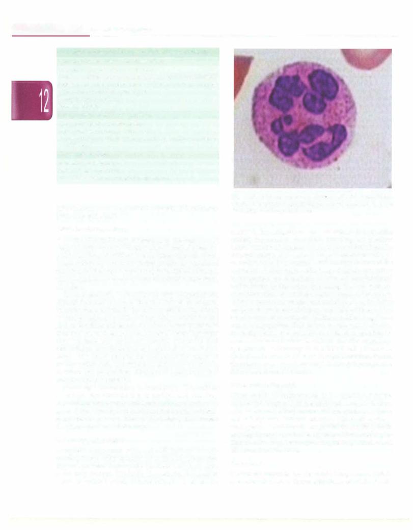
Complete hemogram with red cell indices reveals macrocytic red cells (usually >110 fl) and cytopenias. Hypersegmented neutrophils (nucleus with 6 or more lobes) may be seen (Fig 12.5). The reticulocyte count is useful. If available, serum B12 and folate levels should be
Fig. 12.5: Peripheral smear of a 12-yr-old girl with megaloblastic anemia showinghypersegmented polymorphonuclear cell. Note that the nucleus has more than 5 lobes
assayed. Investigations are performed if a specific underlying cause is suspected. Schilling test involves administration of vitamin B12 (1 mg of unlabeled B12 by intramuscularrouteto saturate hepaticreceptorsand 1 µg of radiolabeled crystalline B12 orally) followed by collection of 24-hr urine collection; adequate absorption is suggested by detection of >7% of administered radioactivity in the urine. Repeating the test with co administration of intrinsic factor helps differentiate between pernicious anemia and malabsorption, the latter being morecommoninchildren. Anychild with morethan one abnormalhematological cell line shouldundergo bone marrow aspiration. This helps to rule out leukemia, myelodysplasia and aplastic anemia. In megaloblastic anemia the bone marrow is cellular and shows nuclear cytoplasmic asynchrony in red blood cell precursors. Granulocyte precursors may also be abnormal. Serum chemistry may reveal elevated lactic dehydrogenase (LDH) and indirect bilirubin.
Differential Diagnosis
Other causes of macrocytosis to be considered in the differential diagnosis of megaloblastic anemia include aplastic anemia, other marrow failure syndromes (pure red cell aplasia, Fanconi anemia, transient erythro blastopenia of childhood), congenital dyserythropoietic anemia,chronicliverdisease,hypothyroidism, coldagglu tinin disease, neoplasms, myelodysplastic syndromes and HIV infection (Fig. 12.3).
Treatment
Treatment depends on the underlying cause. While evaluating for cause, therapeutic doses of folate should
be administered along with vitamin B12. While folate therapy corrects anemia, it does not correct neurological changes induced by cobalamin deficiency and results in the progression of neuropsychiatric symptoms. Folate deficiency due to dietary insufficiency or increased demands is best treated with supplements, while that induced by use of anti-folate medications requires reduc ing or discontinuing the administration of the implicating drug and addition of supplements. Folate is available as 5 mg tablet and overdose is not associated with any adverse effects; a dose of 1-5 mg daily is advocated for 3--4 weeks. Vitamin B12 is administered at a dose of 250-1000 µg by intramuscular route; young children should receive lower doses (250-500 µg) since tremors and extrapyramidal toxicity are reported. Therapyis given daily for 1-2 weeks and then weekly until the hematocrit is normal. Patients with pernicious anemia and malabsorption require monthly supplementation for life. Patients with neuro logical complications should receive 1000 µg daily for 2 weeks, then every 2 weeks for 6 months and monthly for life. The absorption of oral supplements is variable and may be insufficient in patients with malabsorption. Patients with dietary insufficiency should receive nutri tional counseling; if diet cannot be altered due to social and cultural reasons, lifelong vitamin B12 supple mentation is required, either as oral supplements daily or parenteral doses every 3-12 months.
Therapy is associated with improvement in hemato logical parameters, including reticulocytosis, decline in MCV and improvement in platelet and neutrophil counts within a few days of therapy.
Suggested Reading
Chandra J, Jain V, Narayan S, et al. Folate and cobalamin deficiency in megaloblastic anemia in children. Indian Pediatrics 2002;39:453-57 Stabler SP, Allen RH. Vitamin B12 deficiency as a world wide prob
lem. Annu Rev Nutr 2004;24:299-26
Approach to Hemolytic Anemia
The term hemolytic anemia refers to conditions in which the rate of red celldestructionis accelerated and the ability of the bone marrow to respond to anemia is unimpaired. Undermaximalstimulation,the normal marrowis capable of increasing its production rate about six to eight times its basal level. Hemolytic disorders may be divided into inherited and acquired varieties (Table 12.8). This classification has a pathogenetic significance because the nature of hereditary lesions differs from that of acquired. Most intrinsic defects are inherited and the extrinsic are acquired. An exception is paroxysmal nocturnal hemoglobinuria, an acquired disorder characterized by an intrinsic red cell defect.
Cllnlcal Features
In acute hemolysis, symptoms are related to the rate of fall of hemoglobin; patients with rapid hemolysis have
Hematological Disorders -
Table 12.8: Causes of hemolytic anemia
Acquired
Mechanical: Macroangiopathic. (Artificial heart valves, march hemoglobinuria); microangiopathic (disseminatedintravascular coagulation, hemolytic uremic syndrome, thrombotic thrombocytopenic purpura)
Infections, e.g. malaria, kala-azar, Clostridium welchii Antibody mediated: Autoimmune hemolytic anemia (warm and cold types)
Transfusion reactions: Immediate and delayed Hemolytic disease of the newborn
Drugs: Cefotetan, ceftriaxone
Hypersplenism
Cryopathy, e.g. cold agglutinin disease, paroxysmal cold hemoglobinuria
Physical injury, e.g. bums
Chemical injury, e.g. snake bite, lead and arsenic toxicity
Inherited
Hemoglobinopathies, e.g. thalassemia, sickle cell disease Red cell membrane defect, e.g. glucose-6-phosphate dehydro- genase deficiency
Disorders of the cytoskeletal membrane, e.g. hereditary spherocytosis
Unstable hemoglobins
Lipid membrane defects, e.g. abetalipoproteinemia Porphyria
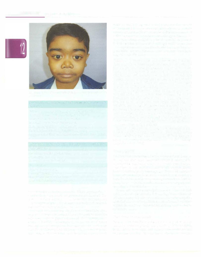
an acute and severe presentation. Anemia is suggested by weakness, pallor and fatigue. Jaundice is a prominent finding of some hemolytic anemias. Red urine suggests hemoglobinuria inintravascular hemolysis. Splenomegaly is seen in autoimmune and several congenital causes of hemolytic anemias. Other useful clues to etiology are the presence of gallstones (spherocytosis), hemolytic or thalassemic facies (Fig 12.6) and leg ulcers (sickle cell anemia); however laboratory tests are still required to confirm the cause.
Laboratory Findings
The reticulocyte count is useful in determining the rate of red cell destruction. The normal reticulocyte count value in the newborn is 3.2±1.4% and in children 1.2±0.7%.
Laboratory test findings in hemolytic anemia can be divided into three groups:
1. Increase in erythrocyte destruction (Table 12.9)
ii.Compensatory increase in the rate of erythropoiesis (Table 12.10) and
iii.Features specific to particular etiologies of hemolytic anemia.
An elevated corrected reticulocyte count may be the only manifestation of mild hemolytic anemia in a well compensated child. Hemoglobin and heme released from red cells following intravascular hemolysis bind to the proteins haptoglobin and hemopexin. These protein complexes are removed from circulation. Hence, haptoglobin and hemopexin levels are low inpatients with
__E_s_s_ _en_t_iat_ _P_ed_ _ia.t.ric_ _s __________________________________
Fig. 12.6: Child with hemolytic anemia, showing hemolytic facies and icterus
Table 12.9: Laboratory signs of accelerated erythrocyte destruction
Fall in blood hemoglobin level at>1.0 g/dl per week Increased serum level of unconjugated bilirubin Increased urinary urobilinogen excretion
Increased serum lactate dehydrogenase Reduced haptoglobin and hemopexin Reduced glycosylated hemoglobin
Decreased erythrocyte life span (labeled with radioisotope s1cr)
Table 12.10: Laboratory signs of accelerated erythropoiesis Peripheral blood
Polychromasia or reticulocytosis Macrocytosis
Increase in nucleated red cells
Bone marrow
Erythroid hyperplasia Iron kinetic studies
Increased plasma iron turnover Increased erythrocyte iron turnover
intravascular hemolytic anemia. When haptoglobin is saturated, free plasma hemoglobin can be detected. The level of indirect bilirubin is an insensitive measurement of hemolysis as it is only elevated when liver function is impaired or when hemolysis is extensive.
A peripheral smear is useful in evaluation of hemolytic anemias. It may show malarial parasites; presence of bite cells may suggest glucose 6 phosphate dehydrogenase enzyme (G6PD) deficiency. Spherocytes are seen in hereditary spherocytosis, but may also be seen post transfusion. Microcytosis withmany fragmented red cells may suggest thalassemia and thrombocytopenia with
schistocytosis (fragmented red cells) can be seen in disseminated intravascular coagulopathy or hemolytic uremic syndrome. The Coombs test is the most important initial test to perform to define the etiology of hemolysis. A positive direct antiglobin (direct Coombs) test means that the erythrocyte is coated with IgG or C3 component of complement, and is positive in most cases of immune hemolytic anemia. However, the test may be falsely negative in 2-5% cases.
Hallmarks of intravascular and extravascular hemolysis.
Followingintravascularhemolysis,hemoglobinisreleased into the plasma (hemoglobinemia). A part of the circu lating free hemoglobin is converted to methemoglobin, which binds with albumin to form methemalbumin that confers a brown color to plasma for several days following the hemolytic event. When the amount of hemoglobin exceeds the haptoglobinbindingcapacity it is excreted in the urine (hemoglobinuria). Some of this hemoglobin is reabsorbed in the proximal renal tubules; the loss of hemeladen tubular cells is seen as hemosiderinuria. Similar to intravascular hemolysis, unconjugated bilirubin, lactate dehydrogenase and reticulocyte count areelevatedin extravascularhemolysisandhaptoglobulin may be decreased. However, in extravascular destruction of red cells, there is nofreehemoglobin or methemoglobin in the plasma; hence, hemoglobinemia, hemoglobinuria and hemosiderinuria are absent.
Specific tests such as hemoglobin electrophoresis, osmotic fragility, enzyme assays for G6PD and pyruvate kinase deficiency and assay for CDSS/59 for paroxysmal nocturnal hemoglobinuria are based on the etiology suspected (Table 12.8).
Management
It is important to maintain fluid balance and renal output during and after acute hemolysis. Shock is managed by appropriate measures. However, blood transfusions, considered useful in acute anemia of other types, should be usedcautiously when managing patients with acquired hemolytic anemias. Even with careful blood matching, transfused blood cells may undergo hemolysis, leading to an increase in the burden on excretory organs and, sometimes, thromboses.
Acute autoimmune hemolytic anemia is treated with corticosteroids (prednisone 1-2 mg/kg/day), which are tapered gradually over several months after ongoing hemolysis has resolved. Patients with chronic hemolysis require thoroughinvestigation for etiology and treatment specific to cause.
Hereditary Spherocytosis
Patients with hereditary spherocytosis may have one of several membrane protein defects. Many of these result in instability of spectrin and ankyrin, the major skeletal membrane proteins. The degree of skeletal membrane
proteindeficiency correlates with the degree of hemolysis. Membrane protein deficiency leads to structural changes including membrane instability, loss of surface area, abnormal membrane permeability and reduced red cell deformability. These defects are accentuated during depletion of metabolites, demonstrated as an increase in osmotic fragility after 24-hr incubation of blood cells at 37°C. Non-deformable erythrocytes are destroyed during passage through spleen.
Laboratoryfindings. Patients with hereditary spherocytosis may have a mild to moderate chronic hemolytic anemia. The red cell distribution width (RDW) is increased due to the presence of spherocytes and increased reticulocytes. The MCV is decreased and MCHC increased due to cellular dehydration.
Prese11tation. The age of presentation ranges from early childhood to adulthood. Patients chiefly present with jaundice of varying intensity. Splenomegaly is found in 75% patients. Gallstones are frequent, particularly in older patients and represent pigment calculi.
Management. Patients require lifelong folic acid supple mentation of 1-5 mg daily to prevent folate deficient due to the high turnover of red cells and accelerated erythro poeisis. While splenectomy does not cure the hemolytic disorder, it may reduce the degree of hemolysis. It is the treatment of choice in patients with severe hemolysis and high transfusion requirement. Splenectomy is delayed or avoided in patients with mild hemolysis. Splenectomy may diminish the risk of traumatic splenic rupture in children with splenomegaly. Splenectomy is usually performed beyond 6 yr of age following immunizations against Haemophilus influenzae type B, Streptococcal pneumoniae and Neisseria meningitidis. Post splenectomy, patients should receive penicillin prophylaxis, to prevent sepsis, usually up to early adulthood.
As with other hemolytic anemias, patients with here ditary spherocytosis are also susceptible to aplastic crisis with human parvovirus B19. This organism selectively invades erythroid progenitor cells and causes transient arrest in red cell production. Patients usually recover in 4-6 weeks.
Abnorma/lties in Red Cell Glycofysis
Glucose is the primary metabolic substrate for erythro cytes. Since mature red cells do not contain mitochondria, glucose is metabolized by anerobic pathways; the two main metabolic pathways are Embden Meyerhof pump (EMP) and the hexosemonophosphateshunt. The Embden Meyerhof pump pathway accounts for 90% of glucose utilization. The inability to maintain adenosine triphos phate impairs cellular functions, including deformability, membrane lipid turnover and membrane permeability, leading to shortened red cell life. The hexose monophos phate shunt is responsible for 10% of glucose metabolism.
Hematological Disorders -
This pathway generates substrates that protect red cells from oxidant injury. A defect in this shunt causes collection of oxidized hemoglobin (Heinz bodies), lipids and membrane proteins in red cells, which result in hemolysis. The reticulocyte count is raised and bone marrow shows erythroid hyperplasia. Demonstration of autohemolysisis ausefulscreening test; diagnosis requires specific enzyme assays.
Glucose-6-phosphate dehydrogenase deficiency
Glucose-6- phosphate dehydrogenase (G6PD) deficiency is the most common red cell enzyme deficiency. It is an X linked recessive disease with full expression in affected males. Many variants are identified based on differences in antioxidant reserve and enzyme levels. After an oxidant exposure, hemoglobin is oxidized to methemoglobin and denatured to form intracellular inclusions also known as Heinz bodies. These Heinz bodies get attached to the red cellmembraneandaggregateintrinsicmembraneproteins such as band 3. Reticuloendothelial cells detect these membrane changes as antigenic sites and ingest a part of the red cell. This partly phagocytosed cell, called 'bite' cell, has a shortened half-life.
The child may present withjaundice in neonatal period. Findings during a hemolytic crisis are pallor, icterus, hemoglobinemia, hemoglobinuria and splenomegaly. Plasma haptoglobin and hemopexin are low. The peripheral blood smear shows fragmented bite cells and polychromasia. Special stains demonstrate Heinz bodies during the initial few days of hemolysis. Diagnosis of G6PD deficiency is suggested by family history, clinical findings, laboratory features and exposure to oxidants prior to the hemolytic event (Table 12.11). Confirmation of the diagnosis requires quantitative enzyme assay or molecular gene analysis.
Management consists of supportive care during the acute crisis (hydration, monitoring and transfusions if needed) along with folic acid supplementation. Coun seling to avoid intake of oxidant drugs (Table 12.11) is imperative.
Pyruvate kinase deficiency This is the most common enzyme defect in the Embden Meyerhof pump and is inherited in an autosomal recessive manner.Homozygotes present with splenomegaly, icterus and hemolytic anemia, but the clinical spectrum is variable. Folic acid supplementation is required to prevent megaloblastic
Table 12.11: Drugs that cause oxidant stress and hemolysis in patients with glucose-6-phosphate dehydrogenase deficiency
Sulfonamides: Sulfamethoxazole Antimalarials: Primaquine, quinine
Analgesics: Aspirin, non-steroidal anti-inflammatory drugs, phenazopyridine (pyridium)
Others: Nitrofurantoin, dapsone, methylene blue, rasburicase, toluidine blue, nalidixic acid, furazolidine, quinidine
--E-s s en t ia__lPe diatrics
complications due to relative folate deficiency. Splenec tomy is considered a therapeutic option in patients with pyruvate kinase deficiency, it does not stop the hemolytic process. The reticulocytecountincreasesdramaticallyafter splenectomy.
Autoimmune Hemolytic Anemia
An autoimmune phenomenon targeting the red cells may occur in isolation, or arise as a complication of an infection (viral hepatitis B, upper respiratory tract viral infections, mononucleosis and cytomegalovirus infection), systemic lupus erythematosus (SLE) or other autoimmune syndromes, immunodeficiency states or malignancies.
Clinical features The disease usually has an acute onset, manifested by weakness, pallor, fatigue and dark urine. Jaundice is a prominent finding and splenomegaly is common. Some cases are chronic. Clinical features may suggest an underlying disease (e.g. SLE or HIV).
Laboratory findings The anemia is normochromic and normocytic and may vary from mild to severe. The reticulocyte count is usually increased. Spherocytes and nucleated red cells may be seen on the peripheral blood smear. Other features suggesting hemolysis include increased levels of lactic dehydrogenase, indirectandtotal bilirubin, aspartate aminotransferase and urinary urobili nogen. Intravascular hemolysis is indicated by hemo globinemia or hemoglobinuria. Serologic studies help define pathophysiology, plan therapeutic strategies and assess prognosis. The direct antiglobulin test is positive in almost all cases. Further evaluation allows distinction into one of three syndromes.
Autoimmune hemolytic anemia due to warm reactive auto antibodies is caused by IgG antibodies against the patient's red blood cells, with specificity for Rh-like antigen. These antibodies have maximal invitro antibody activity at 37°C and do not require complement for activity. The condition can occur in isolation (primary), associated with immune disorders (e.g.SLE,lymphoproliferativedisease,immuno deficiency), or with use of certain drugs (e.g. penicillin, cephalosporins) due to the 'hapten' mechanism (tight binding of drug to the red cell membrane is followed by immune destruction of cells by newly formed or pre existingantibodiestothedrug). Extravasculardestruction of the red cells by reticuloendothelial system occurs, resulting in splenomegaly.
In contrast, patients with cold reactive autoimmune hemolytic anemia have antibodies, primarily of the IgM class, that require complement for their activity, have optimal reactivity in vitro at 4°C, and are specific to the i or I antigen on red cells. Detection of complement alone on red blood cells help make a diagnosis. While the condition is usually seen in adults, children may develop DonathLandsteinerhemolyticanemia,whichis associated with an acute viral syndrome and is mediated by cold
hemolysis. Paroxysmal cold hemoglobinuria is a related condition, usually identical to cold autoimmune hemolytic anemia, except for antigen specificity to P antigen and the evidence of invitro hemolysis. Children may develop cold agglutinins following infections, such as mycoplasma, Epstein-Barr virus (EBV) and cytomegalovirus (CMV), in association with intravascular hemolysis.
IgG andcomplementassociated autoimmune hemolytic anemia may occasionally occur due to warm antibody or, veryrarely,due to drug associatedautoimmunehemolytic anemia.
Management Medical management of any underlying disease is important. Most patients with warm auto immune hemolytic anemia respond to prednisone 1 mg/ kg, given daily for 4 weeks or till hemoglobin is stable. After initial treatment, corticosteroids may be tapered slowlyover4-6months.Whileuseof intravenous immune globulin (!VIG) (1 g/kg/day for 2 days) may induce remission, the response is not sustained. The rate of remissionwith splenectomy may be as high as 50%, parti cularly in warm reactive autoimmune hemolytic anemia. However, this option should be considered carefully in younger patients due to a high risk of infections with encapsulated organisms.Hence, splenectomy is withheld until other treatments have been tried. In severe cases unresponsive to conventional therapy, immunosuppres sive agents such as cyclophosphamide, azathioprine and cyclosporine may be tried alone or in combination with corticosteroids. Danazol is effective in 50-60% of cases of chronic hemolytic anemia. Refractory cases may respond to rituximab (monoclonal antibody to B cell CD20) or to hematopoietic stem cell transplantation.
Patients with cold autoimmune hemolytic anemia and paroxysmalcoldhemoglobinuria are less likely to respond to corticosteroids or intravenous immunoglobulin (IVIG). When associated with infections, these syndromes have an acute,self-limitedcourseandsupportivecare is allthat is necessary. Plasma exchange is effective in severe cold autoimmune (IgM) hemolytic anemia and may be helpful in severe cases because the offending antibody has an intravascular distribution.
Transfusion may be necessary because of the complication of severe anemia but should be monitored closely. In most patients, cross-match compatible blood will not be found and the least incompatible unit should be identified by the blood bank. Transfusions must be conducted carefully, beginning with a test dose.
Prognosis In general, children with warm autoimmune hemolytic anemia are at greater risk for more severe and chronicdiseasewithhighermorbidityandmortalityrates. Hemolysis and positivity of antiglobulin tests may continue for months or years. Patients with cold auto immune hemolytic anemia or paroxysmal cold hemo globinuria have acute self-limited disease.
Suggested Reading
Gupta N, Sharma S, Seth T, et al. Rituximab in steroid refractory autoimmune hemolytic anemia. Indian J Pediatr DOI: 10.1007/sl2098- 0ll-0544-4
Ware RE. Autoimmunehemolytic anemia. In. Orkin SH, Nathan DG, Ginsburg D, et al. (Eds.) (2009), Nathan and Oski's Hematology of In fancy and Childhood, (7th edn). Philadelphia, PA: Saunders Elsevier
Thalassemias
The word thalassemia is a Greek term derived from thalassa, whichmeans 'the sea' (referring to the Mediter ranean sea) and emia, which means 'related to blood'. The disease is more common in populations in the geographic belt from Southeast Asia to Africa. Thalas semias, caused by defects in the globin gene, are themost common monogenic disease. More than 200 mutations are described and the defects are inherited in an auto somal recessive manner. The carrier rates for p thalas semia in north Indians are reported to vary from 3-17% in different ethnic groups.
Pathophysiology
The major hemoglobinin humans, calledHbA, constitutes approximately 90% of total hemoglobin in children beyond one year of age. A minor component, HbA2, accounts for 2-3% of hemoglobin. The main hemoglobin in fetal life is HbF, of which only traces remain after one year. Each of these hemoglobins has two a chains that are associated with two p globin chains in HbA, o chains in HbA2 and y globin chains in HbF.
Thalassemias are inherited disorders of hemoglobin synthesisthatresultfromanalterationintherateofglobin chain production. A decrease in the rate of production of globins (a, p, o, y) impedes hemoglobin synthesis and creates an imbalance with normally produced globin chains. Because two types of chains (a and non-a) pair with each other at a ratio close to 1:1 to form normal hemoglobin, an excess of the normally produced type is present and accumulates in the cell as an unstable product, leading to early the destruction of the red cell. The type of thalassemia usually carries the name of the chain or chains that is not produced. The reduction may vary from a slight decrease to a complete absence. When p chains are produced at a lower rate, the thalassemia is termed P+, whereas p0 thalassemia indicates a complete absenceofproductionof Pchainsfrom theinvolvedallele.
Presentation
Thalassemia should be considered in the differential diagnosis of any child with hypochromic, microcytic anemia that does not respond to iron supplementation. Children with p thalassemia major usually demonstrate nosymptomsuntil about 3-6 months ofage, when pchains are needed to pair with a chains to form HbA, since y chains production is turned off. However, in some cases, the condition may not be recognized till 3-5 yr of age due to a delay in cessation of HbF production.
Hematological Disorders -
Severe pallor and hepatosplenomegaly are almost always present. Icterus is usually absent, but mild to moderate jaundice may occur due to liver dysfunction from iron overload and chronic hepatitis. Symptoms of severe anemia such as intolerance to exercise, irritability, heart murmur or even signs of frank heart failure may be present. Bony abnormalities, such as frontal bossing, prominent facial bones and dental malocclusion are usually present (Fig12.6). Ineffective erythropoiesis leads to a hypermetabolicstate associated with fever and failure to thrive. Hyperuricemia may be encountered.
Spectrum of Disease
p thalassemia trait. Patients have mild anemia, abnormal red blood cell indices and abnormal hemoglobin HPLC results with elevated levels of HbA2, HbF or both. The peripheral bloodfilmexamination usuallyrevealsmarked hypochromia, microcytosis and presence of target cells. Anisocytosis,usuallyprominentinirondeficiencyanemia, is not seen.
Thalassemia intermedia. This condition may occur due to a compound heterozygous states, resulting in anemia of intermediate severity, which usually does not require regular blood transfusions. This is primarily a clinical diagnosis and requires monitoring of the child over time to see the clinical spectrum of disease.
Thalassemia major. This condition is characterized by transfusion-dependent anemia, splenomegaly, bone deformities, growth retardation and hemolytic facies in untreated or inadequately treated individuals. Organomegaly is marked in patients receiving irregular or inadequate transfusion support. Examination of the peripheral blood smear shows severe hypochromia, microcytosis, marked anisocytosis, fragmented red blood cells, polychromasia, nucleated red cells and occasionally, immature leukocytes.
Associated variants. p thalassemia may be associated with p chain structural variants. The most significant condition in this group of thalassemic syndromes is the HbE/P thalassemias. Patients with HbE/P thalassemia may present with severe symptoms identical to that of patientswith p thalassemia major, or with milder course similar to that of patients with thalassemia intermedia or minor. The variation in severity can be explained because of the difference in p globin chain production, i.e. P+ or po, the co-inheritance of a thalassemia gene, level of HbF production and the presence of other modifying genes.
Laboratory Studies
Complete blood count and peripheral blood film examinationareusually sufficienttosuspectthe diagnosis. In thalassemias major and intermedia, the hemoglobin

__Essentia l _Pe_diatrics_------------------------- |
--------- |
_ |
|
level ranges from 2-8 g/dl, MCV and MCH are signif icantly low, reticulocyte count is elevated to 5-8% and leukocytosis is usually present. A shift to the left reflects thehemolyticprocess.The platelet count is usuallynormal unless the spleen is markedly enlarged, causing hypersplenism. Peripheral blood film examination reveals marked hypochromasia and rnicrocytosis, polychromato philic cells, nucleated red blood cells, basophilic stippling and occasional immature leukocytes (Figs 12.7 and 12.8). High performance liquid chromatography (HPLC) for hemoglobin must be sent prior to the first blood transfusion. The test confirms the diagnosis of thalas semia. Absence of HbA and elevation of HbF suggest thalassernia major; the level of HbA2 is not important for diagnosis. The presence of elevated HbA2 alone suggests the diagnosis of thalassemia trait.
Complications and Management
Genetic counseling is needed for the couple and their family to prevent the birth of other children with thalassemia major and prenatal testing can be used to detect thalassemia major in the fetus. Carriers are relatively easy to identify and screen. Prenatal diagnosis and genetic counseling programs have led to a dramatic reduction in the frequency of births of children with thalassemia major in many countries.
The introduction of hematopoietic stem cell transplan tation offers the possibility of cure in severe forms of thalassemia. However, this option is available only to a relatively small number of patients. Treatment of non transplanted children consists of regular blood trans fusions and iron-chelating agents; if both are pursued vigorously, these children survive into adulthood. Major challenges in chronic care of these children are ensuring safety of blood products and meeting the costs of life-long iron-chelating agents.
Patients with thalassemia major require medical supervision to monitor for complications and transfusion therapy. Blood transfusion should be initiated at an early age when the child is asymptomatic and attempts should be made to keep pretransfusion hemoglobin 9-10 g/dl (to promote growth and prevent deformity). Chelation therapyto deal with theaccumulated ironoverloadis vital topreventiron overload and organ dysfunction. A normal diet is recommended, with supplements of folic acid and small doses of vitamins C and E. Iron supplements should not be given. Drinking tea with meals has been shown to decrease absorption of iron in the gut.
Iron overload. Iron overload is the major causes of morbidity and organ toxicity. The excessive load of iron is due to increased gastrointestinal iron absorption as well asrepeatedtransfusions. Hence,avoidanceof transfusions alone will not eliminate the iron overload problem. Patients with signs of iron overload demonstrate signs of endocrinopathy caused by iron deposits, including
,
B
Figs 12.7A and B: Peripheral smears from a transfusion dependent patient with beta thalassemia major showing marked anisopoikilo cytosis, microcytosis, hypochromia, polychromatophilia, nucleated red blood cells and few fragmented erythrocytes. Jenner-Giemsa x 1000
Fig. 12.8: Peripheral smear from an asymptomatic patient with hemoglobin E disease, showing microcytosis, hypochromia, target cells and nucleated red blood cells. Jenner-Giemsa x 1000
diabetes,hypothyroidism, hypoparathyroidism, decreased growth and lack of sexual maturation.
The simplest method for monitoring of iron status is by measurement of serum ferritin. However, the test may underestimate liver and cardiac iron. A liver biopsy or liver MRI and echocardiography may be useful in accurately assessing the iron status. A highly accurate and noninvasive tool to assess the heart iron status is the cardiac T2 magnetic resonance.
Chelation therapy. The introduction of chelating agents capable of removing excess iron from the body has dramatically increasedlife expectancy. Thecost,however, has resulted in poor compliance and inadequate dosing of iron chelators inmany Indian patients. The optimal time to initiate chelation therapy is dictated by the amount of accumulated iron. This usually occurs after 1-2 yr of transfusions when ferritin level is about 1000-1500 µg/1. The standard till now has been deferoxamine which must be administered parenterally because of its short half-life. Prolonged subcutaneous infusion is the most effective route. A total dose of 40-60 mg/kg/day is infused over 8-12 hours during the night for 5-6 days a week by a mechanical pump. Patients should be warned about orange discoloration of urine due to the excretion of iron deferoxamine complex (ferrioxamine). Higher doses of deferoxamine (6-10 g) may be administered intra venously, as inpatient when serious iron overload such as cardiac failure occurs. Severe toxicity may develop if chelation therapy is started prematurely. Eye exami nations, hearingtestsandrenalfunctiontestsare required to monitor the effects of deferoxarnine therapy.
Deferiprone is an oral chelating agent which is less effective than deferoxamine in preventing organ damage. It is administered at a dose of 75 mg/day. Since the agent maycausearthritis,neutropeniaandeven agranulocytosis, its administration requires careful monitoring both to preventserious complications and to assess the adequacy of chelation.
Deferasirox is another oral chelating agent that has shown efficacy similar to parenteral agent deferoxamine in maintaining or reducing liver iron. The molecule is a tridentate ligand that binds iron with a high affinity, forming a 2:1 complex that is excreted in bile and elimi nated primarily via the feces. This chelator is highly selec tive for iron and chelates both intracellular and extra cellular deposits excess in the liver, heart and reticulo endothelial system. The recommended starting dose is 30 mg/kg/day.It may cause skin rash, nausea, vomiting and renal and hepatic toxicity; hence, the serum creatinine, liver function tests and urine for proteinuria need to be monitored.
Hematopoietic stem cell transplantation Hematopoie tic stem cell transplantation is the only known curative treatmentforthalassemia.Pooroutcomeafterhematopoietic
Hematological Disorders -
stem cell transplantation correlates with the presence of hepatomegaly, portal fibrosis and inadequate chelation prior to transplant. The event-free survival rate for patients who have all three features is 59%, compared to 90% for those who do not have these features.
Reactions. After multiple transfusions, many patients develop reactions; these may be minimized by using leukocyte filtersduringtransfusion or byusingleukocyte depletedpackedredcells.Administrationofacetaminophen and diphenhydramine hydrochloride before each transfusion minimizes febrile or allergic reactions. Rarely alloimmunization to red blood cell antigens can occur.
Infections. The major complications of blood transfusions are those related to transmission of infections such as hepatitis B and C and HIV. Hepatitis B vaccination and regular assessment of the hepatitis and HIV status are required.
Lactoferrin, a prominent component of the granules of polymorphonuclear leukocytes, is bacteristatic for many pathogens. The very high transferrin saturation attained in patients with iron overload compromise the bacterio static properties of this protein. Infection with Yersenia enterocolitica can occur in patients with iron overload and presents with fever and diarrhea. Treatment with trimethoprim-sulfamethoxazole and gentamicin is required. Other important infections which may occur are mucormycosis (Rhizopus oryzae) and Listeria
monocytogenes.
Hypersplenism. Thespleenactsas astore for nontoxic iron, protecting the body from extra iron. Hence, early removal of the spleen maybe harmful. Splenectomyis justifiedonly in hypersplenism, which is associated with excessive destruction of erythrocytes that increases the need for frequent blood transfusions, resulting in further iron accumulation. Patients who require more than 200250 ml/kg of packed red blood cells per year to maintain hemoglobin maybenefitfromthisprocedure.This israrely required in children receiving adequate transfusion therapy. Presplenectomy immunizationsandprophylactic antibiotics have significantly decreased infections in splenectomized children. The procedure is usually delayed until the child is aged 7 yr or older.
Bone disease. The classic "hair on end" appearance of the skull, results from widening of the diploic spaces. The maxillamayovergrow,resultinginmaxillaryoverbiteand prominence of the upper incisors. These changes contribute to the classic hemolytic or 'chipmunk' facies observed in patients withthalassemiamajor. Osteoporosis and osteopenia may result in fractures. Such children may need treatment with calcium, vitamin D and bisphosphonates to improve bone density.
Extramedullary hematopoiesis. These occur in patients with severe anemia, e.g. thalassemias intermedia, who are not receiving transfusion therapy. The process may cause
__E_s_s_e_n_ tiia P_e_d_ ia _tr-ics_________________________________
neuropathy or paralysis from compression of the spine or peripheralnerves.Compression fracturesandparavertebral expansion of extramedullary masses, which behave clinicallylike tumors, aremore frequent duringthesecond decade of life.
Psychosocial complications. As these children are surviving into adulthood newer problems related to employment marriage and having families, as well as the stress of chronic illness will need to be addressed.
Management of other Tha/assemic States
Patients with thalassemia intermedia require monitoring to assess the need for transfusion, as persistently low hemoglobin may retard growth. Hydroxyurea at a dose of 15-20 mg/kg/day may be used to increase HbF pro duction and reduce the need for transfusion support. This therapyis most effective in children with XLM1 mutation.
Patients with thalassemia trait do not require medical followup after the initial diagnosis. Iron therapy should not be used unless a definite deficiency is confirmed. Genetic counseling is indicated to create awareness and prevent thalassemia major in subsequent offspring.
SuggestedReadngi
Nadkarni A, Gorakshakar AC, Krishnarnoorthy R, et al. Molecular pathogenesis and clinical variability of beta thalassemia syndromes among Indians: Am J Hematol 2001; 68:75-80
Samaik SA. Thalassemia and related hemoglobinopathies. Indian J Pediatr 2005;72:319-24
SickleCellAnema i
Sickle cell anemia is an autosomal recessive disease that results from the substitution of valine for glutamic acid at position 6 of the beta-globin gene.Patients who are homo zygous for the HbS gene have sickle cell disease. Patients who are heterozygous for the HbS gene have sickle trait. The gene frequency for sickle cell anemia in India is 4.3%, but the disease is reported chiefly from Orissa, Maha rashtra, Madhya Pradesh and Jharkand. Deoxygenation of the heme moiety of sickle hemoglobin leads to hydro phobic interactions between adjacent sickle hemoglobin (HbS) molecules that aggregate into larger polymers. Sicklered bloodcells are less deformable and obstruct the microcirculation, resulting in tissue hypoxia, which further promotes sickling. Thesered blood cellsarerapidly hemolyzed and have a life span of only 10-20 days.
Clinical Features
Patients with sickle cell anemia can present with serious and varied manifestations.
Pain is the most common presentation of vaso-occlusive crisis.Presentationwithpainsuggestsacutechest syndrome if pleuritic in nature and arthritis or osteomyelitis if joint or bone are involved. Painful crises tend to recur, precipitated by triggers suchas dehydrationor fever. Shortness of breath or dyspnea suggests an acute chest syndrome, while
unilateralweakness,aphasia, paresthesias, visualsymptoms may suggest stroke or infarct. Sudden increase in pallor, syncope or sudden pain or fullness in the left side of the abdomen mass may indicate a splenic sequestration crisis.
The usual presentations in a young child are icterus due to elevated unconjugated bilirubin, pallor and mild splenomegaly.The diseasemaymanifestasa febrile illness since these children areprone topneumococcal, Salmonella and other bacterial infections. Tachypnea suggests pneumonia, congestive heart failure, or acute chest syndrome, while hypoxia is common with acute chest syndrome. Children with aplasticcrisis may present with congestive heart failure (CHF) due to severe anemia. Hypotension and tachycardia are signs of septic shock or sequestrationcrisis. Growthretardationandgallstonesare common in children with sickle cell anemia.
Types of Crisis
Vasa-occlusive crisis. A vaso-occlusive crisis occurs when the microcirculation is obstructed by sickled red blood cells resulting in ischemic injury. The major complaint is pain, usually affecting bones such as femur, tibia and lower vertebrae. Alternatively, vaso-occlusion may presentas dactylitis,handandfootsyndrome (painful and swollen hands and/or feet), or an acute abdomen. The spleen may undergo auto-infarction and is often not palpable beyond 6 yr of age. Involvement of the kidney results in papillary necrosis leading to inability to concentrate urine (isosthenuria). Other presentations include acute chest syndrome, retinal hemorrhages, priapism, avascular necrosis of the femoral head and cerebrovascular accidents.
Acute chest syndrome. This is a type of vaso-occulsive crisis that affects the lung and presents with chest pain, cough, tachypnea, dyspnea, hypoxemia, fever or a new pul monary infiltrate. This requires urgentadmission; oxygen support, antibiotics (also should cover for mycoplasma and chlamydia), intravenous fluids, bronchodilators and use of steroids may be of benefit.
Sequestration crisis. This is due to sickled cells that block splenicoutflow,leading to thepoolingofperipheralblood in the engorged spleen resulting in splenic sequestration.
Aplastic crisis. Aplastic crises can occur when the bone marrow stops producing red blood cells. This is most commonly seen in patients with infection or folate deficiency. This is usually self-limited and may follow viral infections of which parvovirus B19 is the most commonly implicated. Usually only supportive care and occasionally packed red blood cell transfusions are required.
Infections
Affected children have increased susceptibility to encapsulated organisms (e.g. Haemophilus influenzae,