Добавил:
Опубликованный материал нарушает ваши авторские права? Сообщите нам.
Вуз: Предмет: Файл:

Ghai Essential Pediatrics8th

.pdf
Скачиваний:
6
Добавлен:
03.11.2022
Размер:
70.46 Mб
Скачать

Infections and Infestations -


Table 10.16: Therapy of helminthiasis


Infestation

Drug

Oral dose

Comments

Roundworm

Albendazole1

400 mg once

Drug of choice


Mebendazole

100 mg q 12 hr for 3 days or 500 mg once

Drug of choice


Ivermectin2

150 to 200 µg/kg once

Drug of choice


Nitazoxanide

Age 1-3 yr: 100 mg q 12 hr for 3 days

Option


Age 4-11 yr: 200 mg q 12 hr for 3 days


Age >11 yr: 500 mg q 12 hr for 3 days


Hookworm

Albendazole1

400 mg once

Drug of choice


Mebendazole

100 mg 12 hr for 3 days or 500 mg once

Drug of choice


Pyrantel pamoate3

11 mg/kg (max 1 g) daily for 3 days

Drug of choice

Pinworm4

Albendazole1

400 mg once; repeat in 2 weeks

Drug of choice


Mebendazole

100 mg once; repeat in 2 weeks

Drug of choice


Pyrantel pamoate3

11 mg/kg base (max 1 g)once; repeat in 2 weeks

Drug of choice

Trichuriasis,

Albendazole1

400 mg once daily for 3 days

Drug of choice

whipworm

Mebendazole

100 mg q 12 hr for 3 days

Alternative


Ivermectin2

200 µg/kg once daily for 3 days

Alternative

Filariasis

Diethylcarbamazine

2 mg/kg q 8 hr for 12 days

Drug of choice


Doxycycline

100-200 mg once daily for 6-8 weeks

Adjunctive; kills symbiotic


bacteria within worms


Albendazole1

400 mg once

Adjunctive; reduces


microfilaria


Antihistamines


Adjunctive; reduce allergic


or steroids


reactions to disintegrating


microfilariae

Strongyloidiasis5

Ivermectin2

200 µg/kg once daily for 2 days

Drug of choice


Albendazole1

400 mg once daily for 7 days

Alternative

Adult tapeworm

Praziquantel6

5-10 mg/kg once

Drug of choice


Niclosamide7

50 mg/kg once

Alternative

Dwarf tapeworm

Praziquantel6

25 mg/kg once

Drug of choice


Niclosamide7

5-15 kg: 1 g on day 1; then 500 mg/day for 6 days

Alternative


>15 kg: 1.5 g on day 1; then 1 g/days for 6 day


Cysticercosis

Albendazole1

7.5 mg/kg (max 400 mg) q 12 hr for 8-30 day;

Drug of choice


Praziquantel6

33.3 mg/kg q 8 hr on day 1; then 16.7 mg/kg

Drug of choice


Anticonvulsants;

q 8 hr for 29 days

Adjunctive role in


corticosteroids;


neurocysticercosis


surgery


Hydatid cyst

Albendazole1

7.5 mg/kg (max 400 mg) q 12 hr for 1-6 mo

Drug of choice

Visceral larva

Albendazole1

400 mg q 12 hr for 5 days (up to 20 days)

Drug of choice

migrans

Mebendazole

100-200 mg q 12 hr for 5 days (up to 20 days)

Drug of choice


Corticosteroids


Adjunctive role in severe


disease, eye involvement

Cutaneous larva

Albendazole1

400 mg daily for 3 days

Drug of choice

migrans

Ivermectin2

200 µg/kg once daily for 1-2 days

Drug of choice

Tropical pulmonary

Diethylcarbamazine

2 mg/kg q 8 hr for 12-21 days

Drug of choice

eosinophilia


1Taken with food; fatty meal increases bioavailability

2 Taken on an empty stomach with water; safety not established for children <15 kg 3 Suspension can be mixed with milk or fruit juice

4Entire household should be treated

5 Severely affected and irnrnunocompromised individuals may require longer treatment course and/or combination of the two therapies 6Taken with liquids during a meal

7Must be thoroughly chewed or crushed and swallowed with a small amount of water

Treatment Eradication of worms is achieved with albendazole, mebendazole or pyrantel pamoate (Table 10.16) Anemia is treated with oral iron therapy. Severe anemia may require a packed cell transfusion.

Filariasis

Lymphatic filariasis is caused by Wuchereria bancrofti, Brugia malayi or Brugia timori. These thread-like parasites reside in the lymphatic system of the host. Most heavily

--Essent-ialPed- ia.trics..__________________________________

infested areas in India are the States of Andhra Pradesh, Tamil Nadu, Kerala, Orissa, Bihar and Eastern Uttar Pradesh. In India, most of the cases (98%) are accounted for by bancroftian filariasis. Brugian filariasis is mostly found in Kerala.

Epidemiology W. bancrofti is mainly transmitted in India through Culex quinquefasciatus and has no animal reservoirs. Brugia malayi is transmitted through Mansonia annulifera and M. uniformis mosquitoes; reservoirs have been observed in cats, dogs and monkeys.

The infected mosquito bites a person and deposits the larvae in the skin. These may remain in the skin or cross this barrier to enter the lymphatics. In humans, larvae develop into adult male or female worms over a period of 4-6 months. Adult worms reside in afferent lymphatics. Adult female worms produce microfilariae that circulate in the bloodstream. The life cycle of the parasite is completed when a mosquito ingests microfilariae during a blood meal. Mosquito serves as the intermediate host in whom the microfilariae develop intoinfectivelarvalstage.

Clinical features Following the inoculation of infective larvae into man, a time lag of 8-16 months may occur before the clinical symptoms appear. Alternatively, microfilaremia may remain asymptomatic.

Acute clinical manifestations are characterized by recurrent attacks of fever, associated with inflammation of the lymph nodes (lymphadenitis) and lymph vessels (lymphangitis). Episodes can recur as frequently as 10 times per yr and usually subside spontaneously over 7 to 10 days. Lymph nodes are enlarged. Both upper and lower limbs are involved in bancroftian and brugian infections. Genital lymphatic involvement occurs exclusively in bancroftian infections.

Chronic stage. Lymphadenitis may progress to lymphatic obstruction resulting in changes associated with elephan­ tiasis. The chronic signs usually do not develop before the age of 25 yr. In bancroftian filariasis, major chronic signs consist of hydrocele, chyluria, lymphedema and elephantiasis. In brugian filariasis, leg below the knee and arm below the elbow are characteristically involved.

Management Microfilariae can be detected in blood, urine (inchyluria),hydrocelefluid, ortissues. Examination of a thick blood film is still the best diagnostic tool. Adult worm may be detected in biopsy of lymph nodes. Lymphoscintigraphy may demonstrate lymphatic abnor­ malities even in asymptomatic patients. Diethylcarba­ mazine is the drug ofchoice for lymphatic filariasis and is active against both adult worms and microfilariae. Repeated courses may be required for complete parasitic cure (Table 10.16). Ivermectin is effective against micro­ filariae in a single oral dose of 400 µg/kg of body weight. A combination of ivermectin and albendazole is also effective in clearing microfilariae.

Tropical Pulmonary Eosinophilia

It occurs in persons having filarial infection. The most common age group is 20-30 yr. The syndrome is thought to representanallergicandinflammatoryresponseelicited by rapid clearance of microfilariae from bloodstream by immune mechanisms. There is paroxysmal nocturnal cough. dyspnea, fever, wheeze, loss of weight and easy fatigability. There may be lymphadenopathy and hepato­ splenomegaly. Chest radiographs may be normal but generally show increased bronchovascular markings, discrete opacities or diffuse miliary mottling. The diag­ nosis is suggested by residence in filarial endemic area, compatible clinical presentation, eosinophilia (>2000/ mm3), elevated IgE levels (>1000 IU/rnl) and high titers ofantimicrofilarial antibodies in theabsenceof microfilaria in bloodstream. Microfilariae may be demonstrable in the lungs and lymph nodes. Patients respond to diethyl­ carbamazine with improvement in symptoms within 3 to 7 days.

Visceral Larva Migrans

Visceral larva migrans is caused by nematodes that are normallyparasitic forotherspecies,namely,Toxocara canis (dog roundworm) or Toxocara cati (cat roundworm). Following ingestion of Toxocara eggs,thelarvae hatchand invadetheintestinal mucosa to be carried by bloodstream to different organs. There is intense host inflammatory response.

Clinical manifestations include fever, cough, wheezing hepatomegaly, pulmonary, infiltration endophthalmitis and neurological disturbances. Patients may have low grade fever with recurrent respiratory tract infections. Markedeosinophilia is present. The diagnosis is confirmed by ELISA for toxocaral antibodies.

Albendazoleandmebendazoleareeffectivedrugs (Table 10.16). Alternative drugs include diethylcarbamazine and thiabendazole (25 mg/kg twice daily for 1-3 weeks).

Taenia solium and saginata

These worms are also known as the pork tapeworm (T. solium) and beef tapeworm (T. saginata), reflecting the principal intermediate hosts for each of them. Man is the only definitive host for both the parasites. Pork tapeworm consists of a scolex with suckers and hooks by means of which it attaches to the intestinal wall. Hooks are absent in T. saginata. Usually, only one adult worm is found in the intestine which may live for up to 25 yr.

Clinical features Most infections with adult worms are asymptomatic. Some children may develop nonspecific complaints like nausea, pain in abdomen, and diarrhoea. Carriers have an increasedrisk ofdevelopingcysticercosis

by repeated autoinfection.

Cysticercosis Infection with the intermediate stage (larvae) of T. solium results in cysticercosis. Neuro-

cysticercosis is the most common parasitic infection of the CNS and may account for as high as 20-50% cases of seizures. Following ingestion, eggs develop into larvae in the small intestine. Larvae migrate across the intestinal wall and are carried to the target organs by bloodstream. The common target organs for cysticerci are brain, muscle and subcutaneous tissue. The larvae mature into cysticerci in about two months time. The size of cysticerci varies from 2 mm to 2 cm. Clinical manifestations depend on the location, number and size of cysts in the brain and host inflammatory response. Viable cysts generally do not elicit a strong response. On the other hand, degenerating cysts provoke a vigorous host response. Most cysts remain viable for 5-10 yr. Neurocysticercosis may manifest as partial or generalized seizure, raised intracranial tension, focal neurological deficits, or disturbances uncon­ sciousness or behavior.

Management The diagnosis of taeniasis is established by the demonstration of eggs or proglottids in the stools. patients may pass motile segments of worms through anus. Diagnosis of neurocysticercosis is made by CT and MRI of brain. Detection of antibodies by enzyme-linked immunotransferblot (EITB) has more than 90% sensitivity and specificity. CSF eosinophilia is a helpful finding.

Treatment of taeniasis is similar for both infestations. Niclosamide or praziquantel are effective drugs.

There is no consensus on the management of neuro­ cysticercosis. Various options include observation, anti­ convulsant medication, antiparasitic drugs, surgery or a combination of these. Children having seizures and calcified, inactive lesions on CT do not require specific therapy, apart from anticonvulsants. There are two effective anti-cysticercal drugs: albendazole and prazi­ quantel (Table 10.16). During treatment, dying parasities can provoke severe life-threatening inflammatory response, which can be prevented by giving steroids for 2-3 days before and during treatment. The response to albendazole is better than praziquantel. Anti-cysticercal therapy is contraindicated for spinal or ocular disease as drug induced inflammation may produce irreversible organ damage. These lesions, as well as those within the ventricular system, are best managed surgically.

Infections and Infestations -

Echinococcosis

Human echinococcosis is caused by larval stages of E. granulosus or E. multilocularis, and is characterized by production of unilocular or multilocular cystic disease of lung and liver. Clinical manifestations are also known as hydatid disease or hydatidosis.

Clinical feature Symptoms occur due to mass effect of the cysts and are related to the organ in which they occur. Liver cysts present with abdominal pain and a palpable mass. Lung cyst may present with chest pain, hemoptysis and breathlessness. There may be passage of cysts in the urine (hydatiduria) and hematuria following hydatid disease of the kidneys. Rupture or leakage from a hydatid cyst may cause fever, itching, rash, anaphylaxis and

dissemination of infectious scolices.

Management Diagnosis is made by ultrasonography and CT scan. USG can reveal the internal membranes of cyst, floating ectogenic cyst material (hydatid sand) and daughter cysts within the parent cyst. These findings are of value in differentiating hydatid cyst from simple cysts ofliver.Diagnosticaspiration isgenerallycontraindicated because of risk of infection and anaphylaxis. Antibody detection by ELISA is more sensitive but less specific.

When feasible, surgical removal of cyst is the definitive treatment. Recently surgical excision has been replaced by USG or CT-guided percutaneous aspiration instillation of hypertonic saline or another scolicidal agent and aspiration after 15 min. Medical therapy includes albendazole ina dose of15mg/kg/day bid fortwoweeks repeated for 3-12 courses with 15 days drug-free interval in between two courses (Table 10.16). The efficacy rate is 40-60%. The response to medical therapy is monitored by serial ultrasonography. Surgical removal can be contemplated for a large solitary cyst following albend­ azole therapy.

Suggested Reading

Bethony J, Brooker S, Albonico M, et al. Soil-transmitted helrninth

infections: ascariasis, trichuriasis, and hookworm. Lancet 2006; 367:1521-32

KeiserJ,UtzingerJ. Thedrugswe have andthedrugswe needagainst major helrninth infections. Adv Parasitol 2010;73:197-223

Diseases of Gastrointestinal

System and Liver

GASTROINTESTINAL SYSTEM

Most diseases of thegastrointestinal (GI) tract present with a few symptoms, such as vomiting, dysphagia, abdominal pain, distension, diarrhea, constipation, gastrointestinal bleeding and failure to thrive. Additionally, other diseases may present with symptoms attributed to the gastro­ intestinal tract. Appropriate evaluationrequiresan assess­ ment of symptoms and signs, listing differential diagnosis and planning investigations in order of least to most invasive.

Investigations

The chief tools for evaluating gastrointestinal disorders are broadly classified as follows:

Stool examination. pH, reducing substances, microscopy for pus cells, blood, fat, eosinophils or parasites; culture; ELISA for specific pathogens, toxin assay for Clostridium difficile.

Radiological imaging. These provide information on both structural and functional abnormalities, and include plain abdominal radiograph; barium studies (swallow, meal, follow-through,enteroclysis andenema); ultrasonography and guided tissue sampling; computed tomography (CT) plain, angiography, enterogrphay or enteroclysis); magnetic resonance imaging (MRI); plain, angiography, enterography or cholangiopancreatography.

Transit studies. Include video fluoroscopy for swallowing, small bowel and colonic transit.

Nuclearmedicinetestsusingradioisotopes. Meckel scan, 99mTc tagged red blood cell scan for GI bleeding, gastric emptying scan, and urea breath test for Helicobacter pylori.

Endoscopy. Upper GI study, colonoscopy, enteroscopy, double balloon enteroscopy, capsule endoscopy, endo­ scopic retrograde cholangiopancreatography (ERCP).

Manometry. Esophageal, antroduodenal, colonic and biliary; useful in diagnosing motility disorders of

Anshu Srivastava, Barath Jagadisan, SK Yachha

esophagus, small and large intestine and sphincter of Oddi. 24 hr pH metry and impedancemonitoring provide information on gastroesophageal reflux.

Tests for evaluation of ma/absorption. D-xylose assay; estimation of fecal fat, fecal a-1 antitrypsin (protein-losing enteropathy); chymotrypsin or elastase (pancreatic insufficiency); pancreatic stimulation test; lactose or lactulose hydrogen breath test; Schilling test (vitamin B12 malabsorption).

Histopathology. Biopsy of esophagus, stomach, or small or large intestine; special stains and immunohistochemistry enable detection of pathogens or tumor cells.

Serologi;. Antibodies to H. pylori; anti-tissue transgluta­ minase (celiac disease); antinuclear cytoplasmic antibodies (inflammatory bowel disease); antienterocyte antibody (autoimmune enteropathy) and serology for HIV.

Vomiting

Vomitingrefersto acuteexpulsionofgastriccontents through the mouth. Vomiting should be differentiated from regurgitation, especially in infants. Regurgitation is the involuntary and effortless expulsion of small amounts of gastriccontentsthatisnotaccompanied bynausea. Recurrent or persistent vomiting requires thorough evaluation and treatment. Persistent vomiting may be complicated by dehydration, hypokalemic hypochloremic metabolic alkalosis, malnutrition andconstipation. Vigorous vomiting can uncommonly result in esophageal tear (Mallory-Weiss syndrome) or rupture (Boerhaave syndrome).

Vomiting is a common, but often nonspecific, symptom that may be acute, chronic or recurrent (Table 11.1). Short­ lasting vomiting with acute onset is the most common form and is often caused by viral infections. Chronic vomiting may be (i) cyclic, characterized by 5 stereotype episodes occurring at high intensity ( 4 emesis/hr) and infrequently ( 2 episodes/week), with normalcy in between; or (ii) chronic, characterized by frequent episodes

278


Diseases of Gastrointestinal System and Liver

Table 11.1: Causes of vomiting

or photophobia. These patients need immediate

Gastrointestinal

Nongastrointestinal

investigation in a hospital.

Workup for chronic vomiting should include evaluation


Acute


for cause with blood chemistry (blood sugar, electrolytes,


Gastroenteritis

Infections, e.g. urinary tract

serum amylase and liver enzymes); ultrasound abdomen,

Hepatitis

infection, meningitis, ence­

upper gastrointestinal endoscopy and, as indicated by

Appendicitis

phalitis, pertussis

available clues, barium studies (meal and small bowel

Small intestinal obstruction,

Raised intracranial tension

follow-through), gastric emptying scan, CT or MRI brain,

(malrotation, volvulus,

Diabetic ketoacidosis

metabolic testing or urine analysis. It is important to

intussusception)

Defects in fatty acid oxidation

remember that children with cyclic vomiting should be

Cholecystitis

or respiratory chain

evaluated during symptomatic attack before starting

Pancreatitis

Drug or toxin induced

intravenous fluids since test results are typically non­


Chronic


contributory during asymptomatic periods.


Gastroesophageal reflux

Raised intracranial tension

Evaluation of a child with acute vomiting should

include assessment of hydration, electrolytes, creatinine

Gastritis

Chronic sinusitis

Gastric outlet obstruction

Uremia

and plain X-ray abdomen (in suspected surgical causes).

(hypertrophic pyloric

Overfeeding

Promethazine andondansetron are usefulin postoperative

stenosis, peptic ulcer)


vomiting and to abort episodes of cyclical vomiting.

Small bowel obstruction


Ondansetron, given alone or with dexamethasone, is

(duodenal stenosis, annular


preferred for chemotherapy related vomiting. Domperi­

pancreas, superior


done and metoclopramide are useful in patients with

mesenteric artery syndrome)


gastroparesis.Antihistaminicslikediphenhydraminehelp

Food allergy


in motion sickness. Management of the underlying

Achalasia cardia


condition is essential.

Gastroparesis


Some common disorders presenting with vomiting are

Eosinophilic esophagitis


described below:

Recurrent


Cyclic vomiting

Urea cycle defects

Idiopathic Hypertrophlc Pyloric Stenosls

Abdominal migraine

Diabetic ketoacidosis

Hypertrophicpyloricstenosisisthe mostcommonsurgical

Malrotation with volvulus

Addison disease

disorder of thegastrointestinal tract in infants. The pylorus


(>2/week) at low intensity (1-2 emesis/hr). While chronic

is thickened and elongated with narrowing of its lumen

due to hypertrophy of the circularmusclefibersof pylorus.

vomiting is usually caused by a gastrointestinal etiology,

Clinical presentation. The classical presentation is with non­

cyclic vomiting is predominantly due to neurologic,

bilious vomiting that gradually increases infrequency and

metabolic and endocrine causes.

severity to become projectile in nature. The disorder is


Evaluation


4-6 times more common in boys than girls. Most patients

A detailed history and examination often gives clue to the

present with vomiting starting beyond 3 weeks of age;

however, about 20% are symptomatic since birth and

diagnosis. The etiology of vomiting varies according to

presentation is delayed until 5 months of age in others.

age; while infectious causes occur across all ages, most

Constipation is common.Recurrent and persistent vomi­

congenitalanomalies,e.g. atresiaor stenosis andmetabolic

ting causes dehydration, malnutrition and hypochloremic

disorders, present in the neonatal period or infancy. The

alkalosis. As the stomach muscles contract forcibly to

first step is to find out whether the vomitus is bilious or

overcome the obstruction, a vigorous peristaltic wave can

nonbilious. This determines the site of disease. Lesions

be seen to move from left hypochondrium to umbilicus,

beyond the ampulla of Vater cause bilious vomiting and

particularly on examination after feeding. A firm olive­

those proximal to it lead to nonbilious vomiting. Asso­

shaped mass is palpable in the midepigastrium in 75-80%

ciated features may indicate etiology, e.g. vomitus con­

infants, especially after feeds.

taining stale food of previous day (suggests gastric outlet


obstruction), visible peristalsis (obstruction), vomiting in

Evaluation. Ultrasound abdomen is the diagnostic

early morning (intracranial neoplasm or cyclic vomiting

investigation and shows muscle thickness of >4 mm and

syndrome), vertigo (middle ear disorder) and hypotonia

pyloruslengthof>16mm. Theultrasoundis100%sensitive

(mitochondrial disorders). The 'red flag' symptoms and

and nearly 90% specific in diagnosis of hypertrophic

signs in a child with vomiting are the presence of blood

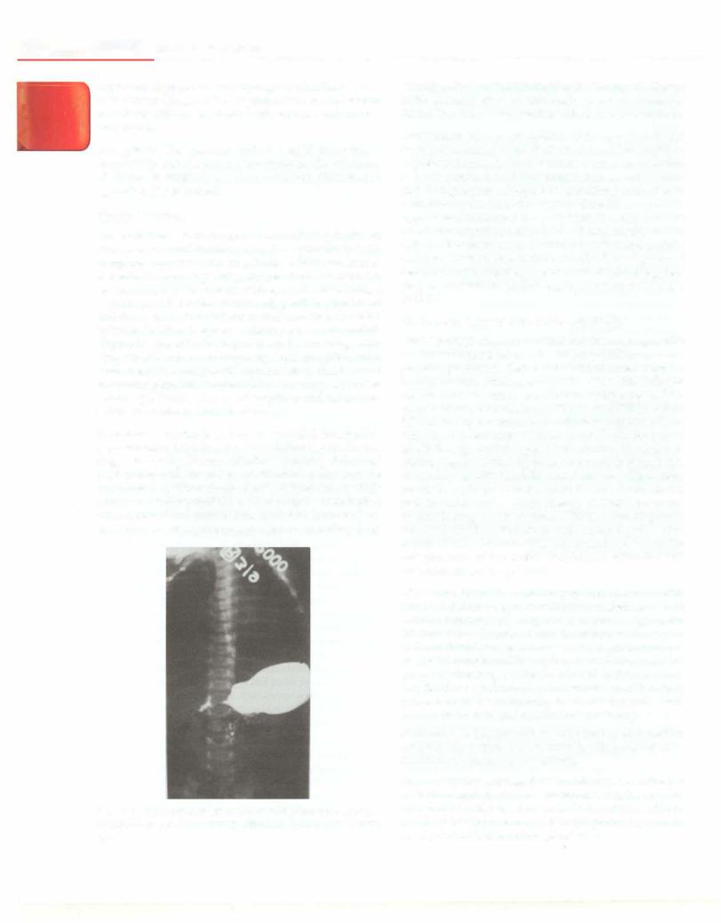
pyloric stenosis. However, in case of doubt, an upper GI

or bile in the vomitus, severe abdominal pain with

barium study can show a consistent elongation of the

abdominal distension or tenderness, projectile vomiting,

pyloric channel (Fig. 11.1) or an upper GI endoscopy is

persistent tachycardia or hypotension, neckstiffness and/

performed.

__e_ss_ e_n_t_i_a lPe diat _ric_s---------------------

-------------


l

111

,.,

Differential diagnosis. Gastroesophageal reflux disease, cow


milk protein allergy, antral or pyloric web are considered


in patients without a palpable pyloric mass and normal


ultrasound.


Management. The treatment includes rapid correction of


dehydration and electrolyte abnormalities. The treatment


of choice is surgical; a pyloromyotomy (Ramstedt's


operation) is performed.


Cyclic Vomiting


This is defined as occurrence of stereotypic episodes of


intense nausea and vomiting as defined previously, with


complete normalcy between episodes and the absence of


a metabolic, neurologic or gastrointestinal disorder. An


episode maylast for an hour to 10days and occurs at least


1 week apart. Episodes are often triggered by physical or


emotional stress. The patient should have had at least 5


episodes in all or 3 episodes during a 6-month period.


Typically the attacks begin in early morning with


symptoms of autonomic surge, e.g. lethargy, pallor, mild


fever, headache, tachycardia, hypertension, diarrhea and


abdominal pain. Most subjects have onset in preschool or


school age. Family history of migraine and/or motion


sickness is noted in 30-40% cases.


Evaluation. A thorough history and physical examination


at presentation helps identify children that require further


diagnostic testing. Presence of bilious vomiting, abdominal


tenderness and/or severe abdominal pain; attacks


precipitated by intercurrent illness, fasting and/or high


protein meal; abnormalities on neurological examination


(altered sensoriurn, papilledema or motor asymmetry), or


development of continuous or chronic vomiting help

Fig. 11.1: Upper gastrointestinal barium study show narrowing and elongation of pyloric channel in idiopathic hypertrophic pyloric stenosis

identify subjects withhigh likelihood of an organicdisease of the gastrointestinal, or neurologic system or a metabolic defect. Symptoms may, overlap with abdominal migraine.

Management. Known precipitants of the episodes should be avoided. Management of an attack includes providing a quietenvironment,administrationof intravenous fluids, use of serotonin 5 HT3 antagonists such as ondansetron (0.3-0.4 mg/kg/dose IV q 4-6 hr upto 20 mg) andsedation with lorazepam (0.05-0.1 mg/kg/dose IV every 6 hr). Agents recommended for prophylaxis against future attacks are cyproheptadine (0.25-0.5 mg/kg/day in two or three divided doses) in children below 5yr and, inolder children, amitriptyline (initiate at 0.25-0.5 mg/kg/day at bedtime; may increase upto maximum of 1.0-1.5 mg/kg/ day) or propranolol (0.25-1 mg/kg/day; up to 10 mg q 8-12 hr).

Gastroesophageal Reflux Disease (GER)

GERis passage of gastric contents into the esophagus with or without regurgitation and vomiting. GER is a normal physiologic process that occurs several times a day in healthy infants, children and adults. When this reflux of gastric contents causes troublesome symptoms and/or complications, it is known as GER disease (GERD). About 50% of healthy 3-4-month-old infants regurgitate at least once per day and most of them outgrow this by 1 yr of age. Followup studies suggest that infants with persistent spilling beyond 3 months of age are at an increased risk of developing GERD in childhood. Children with obesity, repaired esophageal atresia, cystic fibrosis, hiatal hernia, preterm babies and a family history of GERD are at risk of developing severe chronic GERD. Neurologically impaired children like those with cerebral palsy have increased riskof severe GERDdueto multiple factors like low pressure of the lower esophageal sphincter and predominant supine position.

Clinicalfeatures Thecommonsymptomsassociatedwith GERD in children are: (i) recurrent regurgitation with or without vomiting; (ii) weight loss or poor weight gain; (iii) irritability (in infants); (iv) heartburn or chest pain (older children); (v) hematemesis, dysphagia and odyno­ phagia (if complicated by esophagitis or stricture esopha­ gus); (vi) wheezing, stridor, cough and hoarseness; and (vii) Sandifer syndrome, an uncommon manifestation, characterized by spasmodic torsional dystonia with arching of the back and opisthotonic posturing.

Evaluation A detailed history and physical examination are generally sufficient to establish the diagnosis of GER. Useful investigations are as follows.

24 hr ambulatory esophageal pH monitoring is a validated method for quantitativemeasurement of esophageal acid exposure. It is also used to evaluate the efficacy of anti­ secretory therapy and to correlate symptoms (e.g. cough, chest pain) with acid reflux episodes.

Diseases of Gastrointestinal System and Liver

Combined 24 hr multiple intraluminal impedance and pH monitoring detects acid, weakly acid and nonacid reflux episodes. It is thus superior to pH monitoring alone which detects only acid reflux episodes. Its main utility is to evaluate the temporal relationship between symptoms and GER episodes.

Upper GI endoscopy may show erosions or mucosal breaks in the distalesophagealmucosa,the mostreliableevidence of reflux esophagitis. Mucosal erythema and pallor are highlysubjective and nonspecific findings. Complications likestrictureesophagus (Fig. 11.2) and Barrett's esophagus can be picked up at endoscopy. Histological features like elongated rete pegs, basal cell layer hyperplasia and dilated intercellular spaces, alone or in combination, are suggestive of reflux esophagitis.

Endoscopic biopsy is important to evaluate other causes of esophagitis and to diagnose Barrett's esophagus. While not useful for the diagnosis of GERD, barium contrast radiography helps rule out anatomic abnormalities of the upper gastrointestinal tract that may cause symptoms similar to those of GERD. This investigation should be done in all infants with vomiting.

Tests ofesophageal motility are not required for diagnosis of GERD.

Nuclear scintigraphy has a role in the diagnosis of pulmonary aspiration in patients with chronic and refractory respiratory symptoms due to GERD.

Empiric trial of acid suppression. Using proton pump inhibitors for up to 4 weeks is justified in older children or adolescents with typical symptoms suggesting GERD (heart burn or chest pain). There is no evidence to support its use as a diagnostic test in infants and young children where symptoms of GERD are less specific.

Fig. 11.2: Upper gastrointestinal endoscopy showing ulceration with stricture formation in reflux esophagitis

Management Treatment ofGERD depends on patient's age and nature and severity of symptoms and includes lifestyle changes, pharmacologic therapy and surgery.

Lifestyle changes Parentaleducation, guidanceand support are essential and usually sufficient to manage healthy, thriving infants with symptoms due to physiologic GER. Infants should be placed in left lateral position with the head end elevated by 30° in the postprandial period to reduce the frequency of reflux. Cow milk protein allergy is sometimes a cause of unexplained crying and vomiting in infants. Therefore, formula-fed infants with recurrent vomiting may benefit from a 2--4 weeks trial of an exten­ sively hydrolyzed protein formula. Adding thickening agents such as rice cereal (one tablespoon, i.e. -10 g in 60 ml milk) to formula does not decrease the time with pH <4 (reflux index) measured by esophageal pH studies, but it does decrease the frequency of overt regurgitation. Infants with inadequate weight gain because of losses by regurgitation may benefit from increasing the energy density of formula. Careful followup with charting of caloric intake and weight gain is essential.

For children andadolescents with GERD, measures that are useful include: dietary modification (to avoid caffeine, chocolate and spicy foods), weight loss if obese, sleeping in the left lateral position with elevation of the head-end of the bed, avoidance of alcoholand cessation of smoking.

Pharmacologic therapies. Medications usedforGERDinclude agentstobufferorsuppressgastricacidsecretion.Histamine- 2 receptor antagonists like ranitidine decrease acid secretion by inhibiting receptors on gastric parietal cells. They are superior to placebo but less effective than proton pump inhibitors (PPI) for relief ofsymptoms and healing of eso­ phagealmucosa. Development of tachyphylaxis is a major problemwiththese agents. They are chieflyusedininfants with GERD in whom PPI are still not approved for use.

Proton Pump Inhibitors (PPis) inhibit acid secretion by blocking Na+-K+-ATPase, the final common pathway of parietal cell acid secretion. PPis are the agent of choice for GERD. PPI maintain intragastric pH :?:4 for long periods and inhibit meal-induced acid secretion. PPis currently approved for use in children are omeprazole (0.7-3.3 mg/ kg/day, max 80 mg), lansoprazole (0.6-1.6 mg/kg/day; weight <30 kg: 15 mg, >30 kg: 30 mg; max 60 mg) and esomeprazole (<20 kg: 5-10 mg, >20 kg: 10-20 mg OD). Children with typical symptoms of chronic heartburn should be treated with lifestyle changes and 2--4 weeks trial of PPL In patients with endoscopically diagnosed refluxesophagitis orestablished nonerosiverefluxdisease, PPis for 3 months constitute initial therapy. Headache, diarrhea, constipation and nausea may be seen in 10-14% of children on PPls. In addition, PPis may increase rates of community-acquired pneumonia and gastroenteritis.

Antacid therapy is not recommended for most patients with GERD. Currently, there is insufficient evidence to

E

s


_____________

_s_s_ _en_t_ia_l_P__deiat _ri_c

_______________

_


justify the routine use of prokinetic or other agents such as cisapride, metoclopramide, domperidone, bethanechol, erythromycin or baclofen for GERD.

Surgical therapy. Fundoplication decreases reflux by increasing the baseline lower esophageal sphincter (LES) pressure, decreasing the number of episodes of transient lower esophageal sphincter relaxation and the nadir pressure during swallow-induced relaxation, increasing the length of intra-abdominal esophagus, accentuating the angle of His and reducing a hiatal hernia if present. Antireflux surgery may be of benefit in selected children with chronic-relapsing GERD. Children with underlying disorders predisposing to the most severe GERD are also at the highest risk for operative morbidity and post­ operative failure.

Suggested Reading

Li BU, Lefevre F, Chelimsky GG, et al; North American Society for PediatricGastroenterology,Hepatology and Nutrition. Consensus state­ ment on the diagnosis and management of cyclic vomiting syndrome. J Pediatr Gastroenterol Nutr 2008;47:79-93

Pediatric Gastroesophageal Reflux ClinicalPracticeGuidelines:Joint

Recommendations of the North American Society for Pediatric Gastro­ enterology, Hepatology and Nutrition (NASPGHAN) and theEuropean Society for Pediatric Gastroenterology, Hepatology and Nutrition (ESPGHAN). J. Pediatr Gastroenterol Nutr 2009;49:498-547

Dysphaga i

Dysphagia refers to a sensation of food being hindered in its passage from the mouth to the stomach, i.e. difficulty in swallowing. Odynophagia is painful swallowing and globus is the sensation of a lump in the throat. Dysphagia can be divided into two distinct groups:

Oropharyngeal or transfer dysphagia. Presence of drooling, choking, coughing and nasal regurgitation suggests oropharyngeal dysphagia. Disorders involving chewing, oral transfer or pharyngeal phase of swallowingcause this. The main causes are cerebral palsy, bulbar poliomyelitis, muscular dystrophy, brainstem tumors and neuropathy.

Esophageal dysphagia. This occurs due to conditions involving esophageal phase ofswallowing, i.e. coordinated peristalsisofesophagealbody with simultaneousrelaxation of LES. Etiology of esophageal dysphagia can be broadly divided into two groups: motor causes (e.g. achalasia cardiaanddiffuse esophageal spasm) andstructural causes (e.g. strictures, foreign body, Schatzki's ring, esophageal web,eosinophilicesophagitis andextrinsiccompressionby aberrant vessel or mediastinal mass).

Differential Diagnosis and Evaluation

The important causes of dysphagia and their evaluation are shown in Table 11.2 as well as discussed below.

Congenital esophageal stenosis. This may be of three types: web or diaphragm, fibromuscular stenosis and stenosis due to cartilaginous tracheobronchial remnants. Symptoms

of vomiting or chest infection due to aspiration typically develop around 6 months of age when weaning is started. The cartilaginous type occurs in distal esophagus and should be differentiated from achalasia and peptic stricture. Children with this type of congenital stenosis have high risk of perforation on endoscopic dilatation and should be treated surgically.

Foreign bodies in esophagus. Infants and toddlers are at maximum risk due to the habit of oral exploration. Sharp foreign bodies and batteries can cause damage by perforation secondaryto pressure or chemicalnecrosis and can present with dysphagia, mediastinitis and/or upper gastrointestinal bleeding. The vast majority of foreign bodies pass unimpeded through the GI tract. The most frequent sites of impaction are at the cricopharynx, mid esophagus at tracheal bifurcation and just above the LES.

The childpresentsacutelywith choking and dysphagia. Sometimes the ingestion is unwitnessed and the foreign body is completely asymptomatic initially, presenting days to weekslaterwith complications like abscess, fistula or bleed. Guidelines for management recommend that no foreign body should be left in esophagus for more than 24 hr. Endoscopic removal under sedation or anesthesia is the standard of therapy.

Achalasia cardia. This is a motor disorder of the esophagus characterized by loss of esophageal peristalsis, increased LES pressure and absent or incomplete relaxation of LES with swallow.

Childrenpresent with dysphagia, vomiting, weight loss, respiratory symptoms and slow eating whereas toddlers present with coughing and feeding aversion with failure to thrive. The onset is gradual and the average age at diagnosis in children is around 8-9 yr. The barium swallow shows esophageal dilatation with beak-like narrowing at the LES (Fig. 11.3). Manometry is the most sensitive and specific tool and shows absent peristalsis in esophagus; incomplete/ absent LES relaxation and raised intraesophageal pressure. Endoscopy is useful to exclude other etiologies, of dysphagia. The esophagus is dilated and the scope passes through the gastroesophageal junction with some gentle pressure.

Endoscopic pneumatic balloon dilatation or Heller's cardiomyotomy with antireflux procedure are the two main therapeutic modalities. Botulinum toxin injection has been tried but due to short duration of action (a few months) and limited experience, it cannot be recommended as a first line of treatment in children.

Eosinophilic esophagitis. This entity has attracted attention in the recent past. This is a clinicopathological syndrome characterized by symptoms of food impaction, dysphagia and regurgitation, presence of 15 eosinophils per high power field in esophageal biopsies from upper, mid and lower esophagus and exclusion of other disorders with similar presentation, such as GERD. Diagnosis is based

Diseases of Gastrointestinal System and Liver

Table 11.2: Evaluation and management of esophageal lesions that cause dysphagia

EtiologiJ

Investigation

Finding

Treatment

Corrosive (acid or alkali) stricture

Barium swallow and meal;

Narrowing in one or

Endoscopic dilatation (Balloon


upper GI endoscopy

multiple, short or long,

or Savary Gilliard dilators)


segments of esophagus;


may show contracted


stomach or pyloric stenosis

Stricture after repair of tracheo­

Barium swallow

Narrowing of a short

Endoscopic dilatation

esophageal fistula


segment of esophagus


Congenital stricture

Barium swallow; CT chest

Stricture in middle or

Endoscopic dilatation or surgery


lower esophagus; carti­


laginous tissue in


stricture


Postsclerotherapy stricture

Barium swallow; upper

Narrowing in lower end

Endoscopic dilatation


GI endoscopy

of esophagus


Peptic stricture

Barium swallow;

Narrowing in lower

Endoscopic dilatation; proton pump


endoscopy; 24 hr pH

esophagus; may

inhibitor after dilatations


sh1dy

show hiatus hernia or


erosions


Foreign body

Plain X-ray; upper GI

Type of foreign body

Endoscopic retrieval


endoscopy

and site of impaction


Achalasia cardia

Barium swallow;

Beak-like narrowing in

Pneumatic dilatation; Heller's


esophageal manometry

lower esophagus

cardiomyotomy

Infectious esophagitis (in

Upper GI endoscopy;

White curd-like deposits

Fluconazole (Candida); ganciclovir

immunocomprornised children)

endoscopic biopsy

(Candida); ulcers (cyto- (cytomegalovirus) or acyclovir


megalovirus, herpes)

(herpes)

GI gastrointestinal

Fig. 11.3: Barium swallow showing achalasia cardia

on typical endoscopic features of plaques, furrows or concentric rings (called 'trachealization' of esophagus) and histology. Treatment options include the use of elemental formula, elimination diets, or topical corticosteroids. Resolution of the condition with food elimination diets provides evidence that it is a food-antigen driven process.

Suggested Reading

Furuta GT, Liacouras CA, Collins MH, et al; First International Gas­ trointestinal Eosinophil Research Symposium Subcommittee. Eosino­ philic esophagitis in children and adults: a systematic review and con­ sensus recommendations for diagnosis and treatment. Gastroenterol­ ogy 2007;133:1342-63

Gariepy CE, Mousa H. Clinical management of motility disorders in children. Sernin Pediatr Surg 2009;18:224-8

Constipation

Constipation is defined as a delay or difficulty in defecation, present for 2 or more weeks and sufficient to cause significant distress to the patient. It is increasingly being recognized as a very common problem in children and is associated with both physical and psychological morbidity and a poor quality of life. The normal stool frequency decreasesfrom4 or more per dayduring infancy to once per day at 4 yr of age. A stool frequency of <2/week is considered abnormal for all ages. Consti-pation can be dividedinto two groups: functional or organic. Theorganic causes of constipation are listed in Table 11.3. In a study of 135 Indian children with constipation, 85% had functional constipation and 15% had an organic etiology (most commonly, Hirschsprung disease, cerebral palsy and meningomyelocele).

Approach

A detailed history and physical examination is the most useful tool for making a diagnosis of constipation. The

- E s:s en tial P edi:atrics _______________________________

:;::,: ;;,::,:.,: :.;:; .:;.;;:;;:_

details about pattern of stooling, time of first passage of meconium, presence of blood in stools, diet, stressful life events, drug intake and previous surgeries should be known. A predominantly liquid and low fiber diet (milk based) is common and contributes to constipation. A complete physical and neurological examination is essential. Examination for features of spina bifida (pigmentationortuftofhaironlowerback),powerinlower limbs, perianal sensation, voluntary contraction and tone of anal sphincter and amount and consistency of stool in rectum on per rectal examination are extremely useful for diagnosis. Presenceof 'redflags' like failureto thrive, b ood. in stools, recurrent fever with loose stools (enterocohhs), recurrent vomiting, lump in abdomen, recurrent chest infections and features of hypothyroidism shouldalert the physician to suspect organic etiology.

Functional Constipation

The increase in intake of low residue diet and sedentary lifestyleis responsible for theincreaseinfunctionalconsti­ pationinchildren.Functionalconstipationisd fined?Yt e presence of at least 2 or more of the followmg cntena:

(i)twoorfewerdefecationsinthetoiletperweek;(ii)atleast 1 episode of fecal incontinence per week; (iii) histor of retentive posturing or excessive volitional stool retention; (iv) history of painful or hard bowel movements; (v) pre­ sence of alargefecalmassin therectum; and (vi) history of passageoflargediameterstoolsthatmayobstructthetoilet.

Children with functionalconstipation pass large orhard stools and display stool withholding behavior, charac­ terizedbystiffeningofwholebodyandscreamingininfants, to walking on tiptoes or tightening of buttocks in older children. This is often misunderstood by parents as if the childis trying to defecate. Often an acute illness, chang in diet,coercivetoilettrainingornonavailabilityofcleantoilet leads to nonpassage of stools. The stools become hard and cause pain on passage which leads to association of defecationwithpainandwithholding.Thisfurtherincreases stoolsize and hardness with more pain on defecation and a vicious cycle of constipation is initiated. Children with functionalconstipationoftenhaveabdominalpain(10-70%), anorexia(10-25%),enuresisorurinarytractinfections (30%) and psychological problems (20%).

Table 11.3: Causes of constipation

Intestinal nerve/muscle disorders: Hirschsprung disease, intestinalneuronal dysplasia, pseudo-obstruction, spinalcord abnormalities (tethered cord, myelomeningocele)

Anorectal: Anteriorly placed anus, analstenosis, rectal stricture, pelvic mass (sacral teratoma)

Systemic disease: Hypothyroidism, celiac ?isease,_ d(abetes insipidus, diabetes mellitus, hypercalcemia, cystic f1bros1s, myotonic dystrophy

Developmental: Mental retardation, autism

Drugs: Opiates, anticholinergic agents, phenobarbitone, vincristine, lead

Management Noinvestigationsarerequiredfordiagnosis in the majority of children with functional constipation. However, an X-ray abdomen may be done to document impaction in select situations, e.g. an obese child who is not willing for a per rectal examination. Two main steps in the management are disimpaction and maintenance therapy.

Disimpaction is required in patients who have a rectal impaction, i.e. presence of a large hard mass of feces on per rectal examination. Rectalimpactionisresponsiblefor progressive dilatation of the rectum over time and increased threshold volume for rectal sensation and defecation. This 'clean out' is essential if maintenance therapy is to be effective. The oral route is preferred over rectal as it is less invasive. Total bowel wash is done to clean the entire colon using polyethylene glycol (PEG) in a dose of 1.5 g/kg/dayfor 3-4days at home. Alternatively, PEG electrolyte solution can be given in the dose of 15-40 ml/kg/hrtill therectaloutput is clear and devoid of solid fecal material. In young children, this should be done usinganasogastrictube and in hospitalundersupervision. The child should be fasting for PEG administration. Intravenous fluids may be required in small children during this period to maintain adequate hydration. An alternative to oral administration of PEG is the use of phosphateenemaor sodium dioctylsulfosuccinate enema, 30-60 ml/10 kg body weight to a maximum of 120 ml, once or twice daily for 1-2 days. Repeated rectal enemas should be avoided in children.

The aim of the maintenance phase is to promote regular stooling and prevent reimpaction. Success of this therapy is defined as passage of 1-2 soft stools per day and no soiling. It includes the following components:

i.Behavioral training involves establishing a positive routineof sitting on toiletforpassingstools aftermeals regularly (2-3 times per day for 5-10 min) and docu­ menting all stool passage. Embarrassment or punish­ ment should be avoided.

ii.Dietary changes. A nutritiousdiet with fruit/vegetables and adequate fluids is given. A short trial of milk and milk product free diet may be done in cases suspected to have milk allergy.

iii.Medication. Regular and tailor-made (as per response) laxative use is the key to success and this should be explained to the family. Osmotic laxatives like lactulose (1-3 ml/kg/day) and PEG (0.8-1.0 g/kg/ day) are the first line agents. Stimulant laxatives like senna or bisacodyl are to be used only intermittently as a rescue therapy to avoid impaction. Prokinetics likecisapride arenot recommended. Ininfants,mineral oil and stimulant laxatives should not be used. Glycerin suppository is preferred over enema for impaction in infants. Premature withdrawal of medications is a very common cause of relapse.

Prognosis Most of thechildrenneed maintenance therapy for 6-24 months. About 50-60% patients achieve success