Добавил:
Опубликованный материал нарушает ваши авторские права? Сообщите нам.
Вуз: Предмет: Файл:

Ghai Essential Pediatrics8th

.pdf
Скачиваний:
6
Добавлен:
03.11.2022
Размер:
70.46 Mб
Скачать

Suggested Reading

Chang AB. Pediatric cough: children are not miniature adults. Lung 2010;188:533--40

De Blasio F, Virchow JC, Polverino M, et al. Cough management: a practical approach. Cough 2011;7:7

Marchant JM, Masters IB, Taylor SM, et al. Evaluation and outcome of young children with chronic cough. Chest 2006;129:1132--41

Empyema Thoracis

Empyema thoracis is defined as collection of pus in the pleural cavity. This is commonly caused as a complication of staphylococcal (rarely S. pneumoniae or gram-negative bacilli) pneumonia or rupture of subdiaphragmaticorliver abscess in the pleura.

Clinicalfeatures include fever, breathingdifficulty,toxic appearance of child. There is decreased movement of respiration with decreased air entry and vocal resonance. The percussion note is dull. Occasionally, it may manifest as a pulsatile swelling over chest, empyema necessitans.

An X-ray film of the chest shows shift in mediastinum withobliteration ofcostophrenicangle andvarying degree ofopacification. Pleural tap shows purulent fluid with pus cells, high protein and low sugar. Gram stain and culture may show causative agent. Empyema should be differen­ tiated from other causes of pleural effusion including tuberculosis and neoplasia.

The treatment consist of administration of antibiotics active against Staphylococcus, e.g. cloxacillin, vancomycin. The pus collected in the pleural cavity is drained by inter­ costal drainagetube. Drainage of fluidunder thoracoscopy

Disorders of Respiratory System -

is preferred if facility exists. If the lung fails to expand, despite intercostals drainage and antibiotics, a CT scan of chest is done.

Suggested Reading

Brims FJ, Lansley SM, Waterer GW, Lee YC. Empyema thoracis: new insights into an old disease. Eur Respir Rev 2010;19:220-8

Pulmonary Manifestations of HIV Infection

Pulmonary diseases are an important cause of morbidity and mortality in HIV infected children. Respiratory tract symptoms are the initial symptoms in more than 50% of these children. These illnesses range from recurrent upper respiratory tract infections to serious bacterial infections to opportunistic infections and exclusive conditions like lymphoid interstitial pneumonitis. In the west, the incidence of Pneumocystis carinii pneumonia has declined significantly due to early diagnosis of HIV infection and institution of cotrimoxazole prophylaxis. Also, the wide use of highly active antiretroviral therapy has improved the quality of life and survival in HIV infected children. Common pulmonary infections in HIV infected children are discussed in Chapter 10.

Suggested Reading

Hull MW, Phillips P, Montaner JS. Changing global epidemiology of pulmonary manifestations of HIV/AIDS. Chest 2008;134:1287-98

Theron S, Andronikou S, George R, du Plessis J, Goussard P, Hayes M, Mapukata A, Gie R. Non-infective pulmonary disease in HIV posi­ tive children. Pediatr Radio! 2009;39:555-64

Disorders of Cardiovascular System

R Krishna Kumar, R Tandon, Manu Raj

Diseases of the cardiovascular system are an important cause of childhoodmorbidityandmortality. The majority of heart diseases presenting in early childhood are congenital, occurring due to structural defects during development. Despite substantialdecline in the incidence of rheumatic fever, rheumatic heart disease continues to be prevalent in India. Systemic hypertension is increasingly recognizedinchildhood andmay predispose to cardiovascular morbidity in adulthood. A variety of othercardiovascularconditionsmaypresent inchildhood.

The management of children with cardiovascular diseasesrequiresanintegrated approachwithinputsfrom various specialties. There have been substantial advances in the field of pediatric cardiology. Many congenital heart defects that were considered universally fatal can be corrected and affected children can expect to survive into adulthood. These developments include an improved understanding of the pathophysiology of disease, advances in diagnostic capability and successful surgical and medical management of various heart diseases. However, the access to pediatric cardiology services in developing nations is limited. The development of comprehensive pediatric heart programs across regions is essential to improve the management of children with cardiovascular diseases.

CONGESTIVE CARDIAC FAILURE

Congestive cardiac failure is the inability of the heart to maintain an output, at rest or during stress, necessary for the metabolic needs of the body (systolic failure) and inability to receive blood into the ventricular cavities at lowpressure duringdiastole (diastolic failure). Thus, due to systolic failure it is unable to propel blood into the aorta and in diastolic failure it receives inadequate amount of blood. Diastolic heart failure is recognized by clinical features of heart failure with evidence of increased filling pressures with preserved systolic function and in many instances, cardiac output. An

increase in left sided pressures results in dyspnea from pulmonary congestion. An increase in right sided pressures results in hepatomegaly and edema. Besides hypertrophied ventricles, diastolic failure occurs in restrictive heart disease and constrictive pericarditis. While mitral and tricuspid valve stenoses result in elevatedatrial pressure,theyarenot, in the strictest sense diastolic heart failure.

Etiopathogenesis

The common causes of diastolic failure are indicated in Table 15.1. The causes of congestive failure can be classified according to age (Table 15.2). Rheumatic fever and rheumatic heart disease is typically encountered beyond 5yr age; its prevalence appears to be declining in selected urban populations.

Heart failure from congenital heart disease typically happens within the first 1-2 yr of life. Patients with left to right shunts tend to develop CCF around six to eight weeksoflife. Unlikeleft torightshunts, congenitalleakage of the mitral or the tricuspid valve can result in heart failure at an early age. Congenital tricuspid regurgitation (TR) manifests early because the elevated pulmonary

Table 15.1: Heart failure due to diastolic dysfunction

Mitral or tricuspid valve stenosis* Constrictive pericarditis Restrictive cardiomyopathy

Acute ventricular volume overload (acute aortic or mitral valve regurgitation)

Myocardial ischemia#

Marked ventricular hypertrophy (hypertrophic cardio­ myopathy, storage disorders, severe hypertension, severe aortic or pulmonary valve stenosis)

Dilated cardiomyopathy#

*Results in elevated atrial pressures with normal ventricular diastolic pressures (filling pressures)

#Often have combined systolic and diastolic dysfunction

396

Table 15.2: Causes of congestive cardiac failure Infants

Congenital heart disease

Myocarditis and primary myocardial disease Tachyarhythmias, bradyarhythmias Kawasaki disease with coronary occlusion

Pulmonary hypertension (persistent pulmonary hypertension of the newborn; primary pulmonary hypertension; hypoxia, e.g. upper airway obstruction)

Miscellaneous causes Anemia Hypoglycemia Infections Hypocalcemia

Neonatal asphyxia (myocardial dysfunction, pulmonary hypertension)

Children

Rheumatic fever, rheumatic heart disease

Congenital heart disease complicated by anemia, infection or endocarditis

Systemic hypertension

Myocarditis, primary myocardial disease Pulmonary hypertension (primary, secondary)

artery pressures increases its severity. If the TR is not severe, it may improve with time as pulmonary vascular resistance declines.

The age of occurrence of heart failure may point towards the underlying cause (Table 15.3). Heart failure at an unexpectedly early age for a patient thought to have a simple shunt lesion should prompt the search for an associated condition such as coarctation.

Arrhythmias are an important cause of congestive cardiac failure in infancy. Three-quarters of infants with paroxysmal supraventricular tachycardia are below 4 months old. Heart rates above 180/min tend to preci­ pitate heart failure. There is usually no failure in the first 24 hr. If the tachycardia persists for 36 hr, about 20% will develop heart failure and almost 50% will do so in 48 hr. There is a tendency for recurrences of tachycardia if the

Disorders of Cardiovascular System -

onset is after 4 months of age. Any long-standing tachyarrhythmia can be associated with ventricular dysfunction that may mimic cardiomyopathy. Typical examples include ectopic atrial tachycardia and perma­ nent junctionalre-entranttachycardia.Severe bradycardia, typically from complete heart block, can also result in heart failure.

With a normalheart, hemoglobin levels5 g/dl can result in heart failure. In a heart compromised by disease, failure may be precipitated even with hemoglobin levels of 7-8 g/dl. Younger infants are more susceptible to develop failure with anemia.

Clinical Features

The recognition of cardiac failure in older children is based on the same features as in adults.

Symptoms. Slow weight gain is related to two factors. The infant takes small feeds because of easy fatigability and there is an excessive loss of calories from increased work of breathing. Uncommonly, there may be an unusual gain in weight due to collection of water, manifesting as facial puffiness or rarely as edema on the feet. The difficulty in feeding may manifest itself as 'poor feeder', a complaint that the baby does not take more than one to two ounces of milk at a time or that he is hungry within a few minutes after taking a small feed. Shortness of breath or fatigue from feeding results in the baby accepting only small amounts of milk at a time. A few minutes rest relieves the baby and since hunger persists, the result is an irritable infant crying all the time. Often a mother may state that the baby breathes too fast while feeding or that the baby is more comfortable and breathesbetter when held against the shoulder-which is the equivalent of orthopnea in older children. Not infrequently, the baby is brought with persistent hoarse crying, wheezing,excessive perspiration and less commonly, because of facial puffiness (Table 15.4).

Signs. Left sided failure is indicated by tachypnea and tachycardia. Persistent cough, especially on lying down, hoarse cry and wheezing are other evidences of left sided failure; basal rales in the chest are usually not audible.


Table 15.3: Time of onset of congestive failure

Age

Lesion

Birth-1 week

Duct dependent systemic circulation (hypoplastic left heart syndrome, critical aortic stenosis, severe


coarctation, arch interruption); total anomalous pulmonary venous return (obstructed), congenital


mitral and tricuspid valve regurgitation, neonatal Ebstein anomaly

1-4 weeks

Patent ductus arteriosus (PDA) in preterms, ventricular septal defect (VSD) with coarctation, persistent


truncus arteriosus, transposition with large VSD or PDA, severe coarctation; critical aortic stenosis,


congenital mitral or tricuspid regurgitation, single ventricle physiology with unrestrictive pulmonary


blood flow

1-2 months

Transposition with VSD or PDA, endocardial cushion defects, VSD, PDA, severe coarctation; total


anomalous pulmonary venous return, anomalous left coronary artery from pulmonary artery, single


ventricle physiology with unrestrictive pulmonary flow

2-6 months

VSD, PDA, endocardial cushion defect; anomalous left coronary artery from the pulmonary artery,


coarctation, single ventricle physiology with unrestrictive pulmonary blood flow


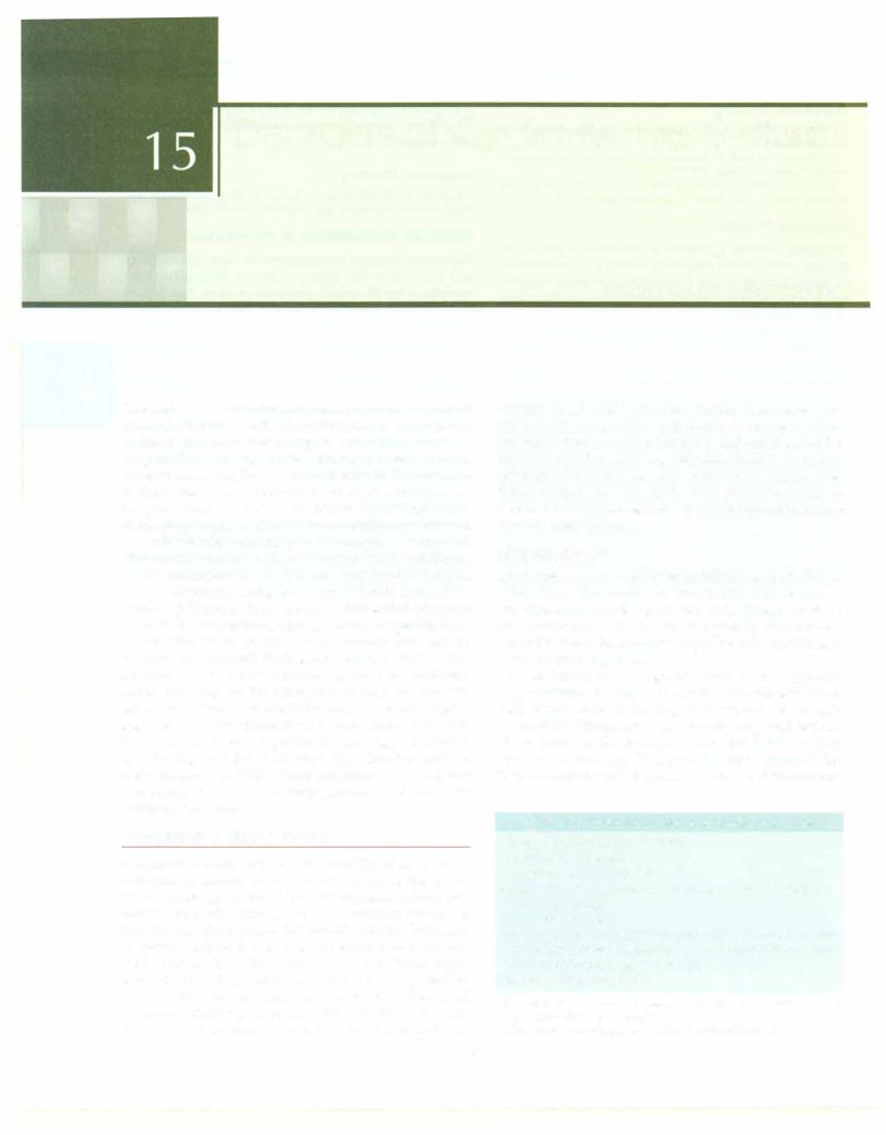
Essen tiaiPed ia trics


_______________________


__

_

_

_ _

_

_ _

_

_

_ _ _

_ _

_ __________


J, Penph:r resistance J

J, Venous return


Poor weight gain


Table 15.4: Symptoms of cardiac failure


Difficulty in feeding


i Cardiac

outputj

J, LVEDP---i

1


Breathesshouldertoo fast; breathes better when held against the


Persistent cough and wheezing


_!


-


i


Irritability, excessive perspiration and restlessness

Improved tissue 02

J, Pulmonary congestion


Pedal edema


l


-l


Right-sidedfailure is indicatedbyhepatomegalyandfacial


l Fatigue,


l DyspneaJ


puffiness. Examination of the neck veins in small babies


Better work capacity


is not helpful. Firstly, it is difficult to evaluate the short


neck with baby fat and secondly, hemodynamic studies

Fig. 15.1: By reducing the systemicvascularresistance and decreasing


show thatrightatrial mean pressures stays normal in more

the venous tone vasodilators provide better work capacity. LVEDP


than one-half of infants with congestive failure. Edema

left ventricular end-diastolic pressure


on thefeetoccurslate. Common to bothleft and right sided


failure is the presence of cardiac enlargement, third sound

50%, improves impaired oxygenation secondary to


gallop and poor peripheral pulses with or without


cyanosis (Table 15.5).


pulmonary congestion.


If the infant or thechild isrestless or dyspneic, sedatives


l


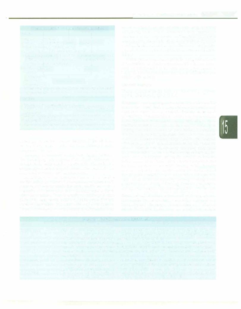
Table 15.5: Signs of congestive cardiac failure

are used. Morphine sulfate in doses of 0.05 mg/kg SC


10

Left-sided


Failure of either side

Right-sided failure

provides effective sedation. A benzodiazepine such as


failure


Cardiac enlargement

Hepatomegaly

midazolamisusefulforsedationinselectedcircumstances.


Tachypnea


Sedatives reduce anxiety and lower the catecholamine


Tachycardia

Gallop rhythm (S3)

Facial edema

secretion, thusreducing physical activity, respiratory and


Cough


Peripheral cyanosis

Jugular venous

heart rate. Requirement of oxygen for body tissues goes


Wheezing


Small volume pulse

engorgement

down, and this reduces the cardiac workload.


Rales in chest

Lack of weight gain

Pedal edema

Fever, anemia or infection also increase the work of the


heart. In infants and smaller children the presence of


superadded pulmonary infection is difficult to recognize.


Treatment


Antibiotics are therefore, sometimes administered


Management of heart failure is a four-pronged approach

empirically. In older children antibiotics are used, only if


evidence of infection is present.


for correction of inadequate cardiac output. The four­

Anemia

imposes stress on the heart because of the


prongs are: (i) reducing cardiac work, (ii) augmenting


myocardial contractility, (iii) improving cardiac perfor­

decreased oxygen carrying capacity of blood. Anemia


mance, (iv) correcting the underlying cause. Identification

results in tachycardia and in a hyperkinetic circulatory


of the cause is important since it has dir ct bearing on survival.

state.Correctionofanemiawill result indecreasedcardiac


If a newborn has heart failure due to duct dependent

work. If transfusion is indicated packed red cells can be


administered. Typicallypacked cell volumes of 10-20 ml/


systemic circulation (critical coarctation, aortic stenosis,

kg are required to correct severe anemia; a single dose of


interrupted aortic arch), administration of prostaglandin

frusemide IV is often given prior to the transfusion. Less


to open the closing duct improves survival.

commonconditionscausingstress tothe heart arerepeated


Reducing Cardiac Work

(Fig. 15. l)


pulmonary emboli, thyrotoxicosis

and obesity.


The work of the heart is reduced by restricting patient

Vasodilators counteract the compensatory mechanisms in


activities, sedatives, treatment of fever, anemia, obesity, and by

heart failure and improve cardiac output (Fig. 15.1).


vasodilators. Mechanical ventilation helps when heart failure

Arteriolarandvenousvasoconstriction is mediatedthrough


is severe by eliminating the work of breathing.

catecholamines. Arteriolar constriction maintains blood


Neonates with heart failure are nursed in an incubator.

pressure by increasing the systemic vascular resistance,


They are handled minimally° . The baby is kept propped

which increases the work of heart (Fig. 15.2). Veno­


up at an incline of about 30 . The pooling of edema fluid

constriction results in decreased venous capacitance and


in the dependent areas reduces the collection of fluid in

increased venous return, increasing thefillingpressures of


lungs, thus reducing° the work of breathing. At a

the ventricles to increase the cardiac output. Since


temperature of 36-37 C, the overall circulatory and

compensatory mechanisms are inappropriately excessive,


metabolic needsare minimal, thus reducing work of heart.

vasodilators, by reducing the arteriolar and venous


Humidified oxygen to maintain a concentration of 40 to

vasoconstriction, reducetheworkofheart.Nitratesareused

aspreferentialvenodilators andhydralazineasan arteriolar dilator.

ACE inhibitors (captopril, enalapril) are effective for treating heart failure in infants and children. These agents are effective vasodilators, suppress renin-angiotensin­ aldosterone system, reducing vasoconstriction and salt and water retention. By suppressing catecholamines, they prevent arrhythmias and other adverse effects on the myocardium. The major side effect of ACE inhibitors is cough, which can be troublesome. Persistent cough may necessitate the use of angiotensin receptor blockers, such as losartan. Initially it is necessary to monitor the renal function: urinalysis, blood levels of creatinine and electro­ lytes once a week for six to eight weeks. These medications may cause first-dose hypotension; the first dose should be one-quarter of the calculated dose. The patient should ideally remain recumbent for the first 6 hr to prevent an unusual fall in blood pressure.

Although beta-blockers might precipitate CCF, they improve symptoms and survival especially in patients withdilatedcardiomyopathy, whocontinuetohavetachy­ cardia. Useful agents include metoprolol and carvedilol. The latter is preferred since it has properties of beta­ blockerswithperipheral vasodilation; treatment is started at low dose and increased depending on tolerability (0.08 to 0.4 mg/kg/day, maximum 1.0 mg/kg/day). Calcium channel blockers have adverse effects in heart failure and should be avoided unless indicated for systemic hypertension.

Intheacutecaresetting, sodium nitroprussideisused as a vasodilator, since it acts on the venous and arterial sys­ tems. Phosphodiesteraseinhibitorssuchasmilrinonehave become popular especially in postoperative period. These agents have powerful vasodilatory and inotropic effects. Specific indications for use of vasodilators include acute mitral or aortic regurgitation, ventricular dysfunction resultingfrommyocarditis,anomalouscoronaryarteryfrom pulmonary artery and in the early post operative setting.

Augmenting Myocardial Contractility

Augmenting myocardial contractility by inotropic agents like digitalis improves cardiac output. In infants and children,onlydigoxin is used. It hasa rapidonset of action and is eliminated quickly. It is available for oral and parenteral administration. Oral digoxin is available as 0.25 mg tablets and as digoxin elixir (1 ml = 0.05 mg) (Table 15.6). Parenteraldigoxin (0.5 mg/2 ml) isavailable; its dose is 70% of the oral dose.

Infants tolerate digitalis well. In a hospitalized patient full digitalization should be sought to maximize benefit. Children are digitalized within a 24 hr period; 1h of the calculated digitalizing dose is given initially, followed by % in 6-8 hr and the final % after another 6-8 hr. The maintenancedose isusuallyone-quarter of thedigitalizing dose (Table 15.6). Before the third daily dose, an electrocardiogram is done to rule out digitalis toxicity.

Disorders of Cardiovascular System


Congestive cardiac failure


.J.. CO --+ Vasoconstriction


t


Arteriolar


Venous


i


i


r


+


[1'SVR


J i Venous tone]


i Afterload


i Venous return


I

,_____,


Increased work of heart


Fig. 15.2: Low cardiac output (CO) results in vasoconstriction,


increasing systemicvascularresistance (SVR) andvenous tone leading


to increase in the work of heart


Table 15.6: Dosage of digoxin and diuretics


Digitalizing


Maintenance


dose, mg/kg


(fraction of

I


digitalizing dose)

Digoxin


Premature, neonates

0.04

1/4


1 month to one year

0.08


l/3tol/4

1 to 3yr

0.06


l/3tol/4

Above 3yr

0.04

1/3


Diuretics


Frusemide

1-3mg/kg per day orally or


1 mg/kg per dose IV


Spironolactone

1 mg/kg orally every 12 hr


Toxicity can be controlled by omitting thenextone or two doses. The PR interval is a useful indicator; if it exceeds the initial intervalby 50%, digitalis toxicity is present. The upper lirnit of normal PR interval in infants is O.14 second.

Digitalisisusedwithcautioninthefollowingsituations:

(i)prematureneonates; (ii) heartfailure duetomyocarditis; and (iii) very cyanotic patients. In other situations, it is better to use half the calculated digitalizing and the main­ tenance dose initially. Myocardial damage, gross cardio­ megaly, hypoxia, acidosis, hepatic, renal and pulmonary insufficiencyincreasethesensitivity ofthemyocardiumto digitalis. Digoxin is beneficial for symptom relief and is advised inpatientswith mild,moderatelysevereorsevere congestivefailure, with or withoutsinus rhythm. Digoxin can becombinedwithACE inhibitorsforsynergistic effect. By increasing cardiac output, digoxin lowers systemic impedance indirectly, unloading the ventricles.

lnotropic Agents

These agents belong to two groups: (i) catecholamine inotropes,like dopamine, dobutamine and adrenaline and (ii) phosphodiesterase inhibitors like amrinone and milrinone. These agents combine inotropic effects with peripheral vasodilation. If blood pressure is low, dopa-

__E_s_s_ _en_t_ia_P_ed_iat _rics _________________________________ _

mine should be used, as an intravenousinfusion. At a dose of less than 5 microgram/kg per minute, dopamine causes peripheral vasodilation and increases myocardial contractility. Renal blood flow improves, resulting in natriuresis; higher doses result in peripheral vaso­ constriction. The dose of dobutamine is 2.5 to 15 micro­ gram/kg/min; the dose should be increased gradually until the desired response is achieved. In patients with dilated cardiomyopathy, dobutamine is used as 24 hr infusion once ortwice a week and retains its effectiveness for varying lengths of time.

Improving Cardiac Performance

by Reducing Venous Return (Preload)

Diuretics reduce the blood volume, decrease venous return and ventricular filling. This tends to reduce the heart size. The larger the heart, the more the wall tension and the poorer is its performance. With reduction in heart size and volume, the myocardial function and the cardiac output improve. Diuretics reduce the total body sodium thereby, reducing blood pressure and peripheral vascular resistance. This helps in increasing the cardiac output and reducing the work of the heart.

Diuretics are the first line of management in congestive failure. The action of oral frusemide starts within 20 min. Frusemide should be used in combination with a potassium sparing diuretic (triamterene, spironolactone, amiloride) instead of using potassium supplements. The combination prevent potassium and magnesium loss and reduces the risk of arrhythmias. Frusemide activates the renin angiotensin aldosterone axis, which is responsible for vasoconstriction and sodium and water retention. When frusemide is combined with ACE inhibitors, the combination suppresses the axis and is therefore synergistic.

The other method of altering the body fluid volume is by restricting the sodium intake. Sodium restriction is difficultto implement in infants and youngchildren. Low sodiumdietsshouldbeusedonly if theheartfailurecannot be controlled with digitalis, diuretics and ACE inhibitors. However, it is prudent to advise such patients to avoid salt rich foods such as chips andpickles.Since heart failure increases calorie requirements, adequate intakes is advised.

Correcting the Underlying Cause

Non-invasive tests (especially echocardiography) allow identificationof the cause in most children with suspected heart disease. Many of these are managed by curative or palliative operations. A diagnosis of idiopathic dilated cardiomyopathy requires exclusion of conditions that are known to cause ventricular dysfunction. The conditions that might be missed are sustained tachyarrhythmias, coarctation of aorta and obstructive aortitis, anomalous origin of the left coronary artery from pulmonary artery and hypocalcemia. It is important to look for subtle

evidence of sustained tachyarrhthmias. Anomalous origin of the left coronary artery is treated surgically.

The presence of CCF in a child with rheumatic heart disease does not necessarily mean presence of active carditis. In any patient of rheumatic heart disease, if active carditis has been excluded and an adequate trial has been given to medical management, operative treatment should be considered. Uncommon causes of CCF in infants and children include upper respiratory obstruction, hypoglycemia, neonatal asphyxia and hypocalcemia.

Prognosis

The mortality of CCF in children is high and prognosis depends on the underlying cause.

CONGENITAL HEART DISEASE

Congenitalheart disease (CHD) encompassesa broad and diverse range of conditions that manifest from prenatal period to late adulthood. In common usage, CHD refers to structural heart defects that are present at birth. History, physical examination, chest X-ray, ECG and echocardio­ graphy help in identifying the presence of CHD, except perhaps in the early newborn period where the diagnosis can be challenging. Palliative or corrective surgery is feasible for most patients with CHD, provided if under­ taken in a timely fashion. It is also possible to identify and determine the severity of specific lesions through echocardiography.

A substantial proportion of patients with CHD have significant problems involving other organ systems, or specific chromosomal and single gene disorders. Pedia­ tricians need to identify associated conditions, since they mighthavesignificant bearing on outcomes. It is important to recognize that, in spite of significant recent advances, longterm concerns after palliation and corrective surgery are significant and many children need lifelong followup.

Epidemiology and Etiology

CHD accounts for nearlyone-third of all majorcongenital anomalies.The prevalence of CHD in infancy is estimated at 6--8 per 1000 live births; 25% are life threatening and require early intervention. A proportion of patients with CHD have an identifiable genetic basis (Table 15.7). Table 15.8 shows the association of CHD with acquired disorders and teratogens.

Physiology of Congenital Heart Disease

Pressure, Flow and Resistances

The pressures and resistances in the pulmonary and systemic circulations are indicated in Table 15.9. The pulmonary and systemic flows are equal if there are no abnormal communications between the two sides.

According to Poiseuille's equation, modified for application to blood flow through vessels.

Pressure = Flow x Resistance

Disorders of Cardiovascular System -

Table 15.7: Common genetic conditions linked to occurrence of congenital heart disease

Syndrome

Genetic mutation; inheritance

Cardiac lesions

Other features

CATCH 22

Microdeletion in 22q;

Interrupted aortic arch,TOF,

Cleft palate,hypocalcemia,


autosomal dominant (AD)

VSD,persistent truncus

thymic hypoplasia,nasal


arteriosus,double outlet

regurgitation,gastroesophageal


right ventricle

reflux,learning disability

Williams Beuren

Microdeletion in elastin

Supravalvar aortic stenosis,

Elfin facies,mental retardation,


(7qll.23) ; AD

pulmonary stenosis,

hypersocial personality,short


hypertension

stature,hypercalcemia

Down

Trisomy 21; Robertsonian

AV canal defect,perimemb­

Characteristic fades,


translocation or mosaicism

ranous VSD,TOF

clinodactyly,mental retardation;


hypotonia

Turner

45XO or 46/45XO; mosaic

Bicuspid aortic valve,

Shortstature,gonadaldysgenesis


coarctation

lymphedema,

Noonan

PTPNll; AD

Pulmonic stenosis,hypertro­

Short stature,dysmorphic fades,


phic cardiomyopathy,ASD

webbed neck,developmental


delay,cryptorchidism

VATER association

Sporadic

VSD,TOF

Vertebral,renal and limb defects,


anal atresia,tracheoesophageal


fistula

Holt Oram

TBXS; AD

Ostium secundum ASD; VSD

Radial ray anomalies

CHARGE association

CHD7; often de nova

Branch pulmonary artery

Coloboma,growth failure,


stenosis,TOF,VSD

choanal atresia,genital


hypoplasia,ear anomalies

Alagille

JAG1; most cases are de nova

Pulmonary stenosis,TOF

Dysmorphic fades,cholestatic


jaundice,butterfly vertebrae,


renal anomalies

AD autosomal dominant; AV atrioventricular; ASD atrial septa! defect; TOF tetralogy of Fallot; VSD ventricular septa! defect

Table 15.8: Prenatal exposure that increase risk of congenital heart disease

Gestational diabetes (transposition,atrioventricular septal defects,hypoplastic left heart,cardiomyopathy,PDA)

Febrile illness in first trimester (increased risk)

Rubella (PDA,peripheral pulmonary stenosis,VSD)

Lupus (complete heart block)

Phenylketonuria (VSD,TOF,PDA,single ventricle)

Vitamin deficiency (increased risk of heart disease)

Teratogens, (first trimester) e.g. anticonvulsants,NSAIDs, cotrimoxazole,thalidomide,retinoic acid

Exposure to organic solvents,herbicides,pesticides,ionizing radiation

NSAIDs nonsteroidal anti-inflammatory drugs; PDA patent ductus arteriosus; TOF tetralogy of FaUot; VSD ventricular septa! defect

The pressure is measured in mm of mercury, flow in liters/ min and resistance in dynes/sec/cm5 or units (80 dynes/ sec/cm5 = 1 unit). Although this equation is not strictly accurate when applied to flow of blood in pulmonary and systemic circuits, it does serve a useful purpose in understanding the hemodynamics. Thus:

Systemic pressure =

systemic flow x peripheral vascular resistance

Pulmonary arterial pressure =

pulmonary flow x pulmonary vascular resistance

It is thus obvious that the pressure in a vessel is dependent on theflow through the vessel and the resistance, offered by the vessel to the flow of blood. It is possible to increase the pressure in a vessel either by increasing the flow or by increasing the resistance. Increase in flow through the pulmonary artery means a left to right shunt, as occurs in atrial or ventricular septal defect or patent ductus arteriosus. Generally, this increase in flow is not associated with significant increase in pressure as the resistance Jails or remains the same. At the same time the distensibility characteristics of the pulmonary artery are suchthatit canaccommodate almostthreetimesthenormal flow without an increase inpressure. Hence, largelefttoright shunts can take place without an increase in pressure.

Increase in pulmonary vascular resistance means obstructive disease in the pulmonary circuit. The pulmonary vessels develop medialhypertrophyand later intimal changes are added, to further obstruct the flow of blood through the pulmonary circulation. Afteracertainstage it is an irrever­ sible process. The increase in resistance to flow in the pulmonary circuit is associated with reduction inflow. The increase in pressure in the pulmonary artery associated with normal resistance is called hyperkinetic pulmonary arterial hypertension whereas when the pressure is increa­ sed due to increase in pulmonary vascular resistance, it is called obstructivepulmonary arterial hypertension. Clinically both situations are seen and can be separated from each other on the bedside.

-----------------------------Essential Pediatrics

Table 15.9: Systolic and diastolic pressures and resistance in the pulmonary and systemic circuits

Chamber(Vessel

Pressure (mm Hg)

Superior vena cava

0-6

Right atrium

0-6

Right ventricle

25/0---6

Pulmonary artery

25/10

Resistance, dynes/sec/cm5


Pulmonary vascular

80-240

Fetal Circulation (Fig. 15.3)

The heart assumes its normal four-chambered shape by the end of six weeks of intrauterine life. From then on only minor changes occur and consist mainly in the growth of the heart as a whole with increasing age of the fetus. For the exchange of gases the fetus is dependent on placental circulation, whereas the neonate is dependent on the lungs. Immediately following birth, with the first inspira­ tion, the lungs expand with air and the gas exchange functionis transferred from the placentato thelungs. This

1 10 necessitates circulatory adjustments following birth to transform the fetal circulation to the postnatal circulation.

Blood oxygenated in the placenta is returned by way of umbilicalveins,whichenter the fetus at the umbilicus and join the portal vein (Fig. 15.3). The ductus venosus provides a low resistance bypass between the portal vein and the inferior vena cava. Most of the umbilical venous blood shunts through the ductus venosus to the inferior

Ductus arteriosus

Fig. 15.3: Fetal Circulation: Details of the circulation are provided in the text. Saturations of blood(%) in various chambers and vessels are indicated in black font and pressures(mm Hg) are indicated in red font

Chamber(Vessel

Pressure (mm Hg)

Pulmonary vein

6-10

Left atrium

6-10

Left ventricle

80-120/5-10

Aorta

80-120/60-85

Systemic vascular

800-1600

vena cava. Only a small proportion mixes with the portal venous blood and passes through the liver. Blood from inferior vena cava comprising that from hepatic veins, umbilical veins and that from lower extremities and kidneys enters the right atrium. On reaching the right atrium the blood stream is divided into two by the inferior margin of septum secundum-the crista dividens. About one-third of the inferior vena cava blood enters the left atrium, through the foramen ovale, the rest two-thirds mixes with the venous return from the superior vena cava to enter the right ventricle.

The blood reaching the left atrium from the right atrium mixes with small amount of blood reaching the left atrium through the pulmonary veins and passes to the left ventricle. The left ventricle pumps out the blood into the ascending aorta for distribution to the coronaries, head and upper extremities. The superior vena cava stream, comprising blood returning from the head and arms, passes almost directly to the right ventricle. Only minor quantities (1 to 3%) reaches the left atrium. The right ventricle pumps out blood into the pulmonary trunk. A small amount of this blood enters the pulmonary circulation, the rest passes through the ductus arteriosus into the descending aorta to mix with the small amount of blood reaching the descending aorta from the aortic arch (derived from the left ventricle).

The main differences between the fetal and postnatal circulation are: (i) presence of placental circulation, which provides gas exchange for the fetus; (ii) absence of gas exchange in the collapsed lungs; this results in very little flowof blood tothe lungs andthus littlepulmonary venous

returnto leftatrium; (iii)presenceofductusvenosus,joining the portalvein with the inferior vena cava, providing alow resistance bypass for umbilical venous blood to reach the inferiorvenacava; (iv)widelyopenforamenovaleto enable oxygenatedblood (through umbilicalveins)to reach the left atrium and ventricle for distribution to the coronaries and thebrain;andlastly(v) wideopenductusarteriosustoallow right ventricular bloodto reach the descending aorta, since lungs are non-functioning.

Circulatory Adjustments at

Birth-Transitional Circulation

Circulatory adjustments continue to occur for a variable period following birth. This change is brought about because of a shift from placental dependence for gas

Disorders of Cardiovascular System

exchange in the fetus to pulmonary gas exchange in the neonate. Loss of placental circulation and clamping of the umbilical cord, after birth, results in a sudden increase in systemic vascular resistance with the exclusion of the low resistance placental circulation. This tends to increase the aortic blood pressure and the left ventricular systolic pressure.Theleft ventricular diastolic pressure also tendsto riseandincreases theleftatrialpressure.Thelossofplacental circulation results in a sudden reduction of flow through the ductus venosus that closes off. Flow through the ductus venosus disappears by the 7th day of postnatallife. The loss ofplacentalflow results in adecreaseinthe volumeofblood returning to the right atrium. The right atrial pressure decreases. The left atrial pressure becomes higher than the right atrial pressure and the septum primum, which acts as a valve of the fossa ovalis, approximates with the septum secundum to closeoff the foramen ovale. Functional closure of theforamenovaleoccurs relatively quickly. Overaperiod of months to years, the septum primum and septum secundum become firmly adherent resulting in anatomical closure of the foramen ovale.

Sudden expansion of lungs with the first few breaths causes a fall in pulmonary vascular resistance and an increased flow into the pulmonary trunk and arteries. The pulmonary artery pressure falls due to lowering of pulmonary vascular resistance. The pressure relations between the aorta and pulmonary trunk are reversed so that the flow through the ductus is reversed. Instead of blood flowing from the pulmonary artery to aorta, the direction of flow through the ductus, is from the aorta to pulmonary trunk. The increased saturation following birth causes the ductus arteriosus to constrict and close. Some

functional patency and flow canbe demonstratedthrough the ductus arteriosus for a few days afterbirth. The ductus arteriosus closes anatomically within 10 to 21 days.

This results in the establishment of the postnatal circulation. Over the next several weeks, the pulmonary vascular resistance continues to decline. There is fall in the pulmonary artery and right ventricular pressures.The adult relationship of pressures and resistances in the pulmonary andsystemic circulations is establishedby the end of approximately two to three weeks (Fig. 15.4).

Hemodynamic Classification of

Congenital Heart Disease

CHD has beenbroadly classified as cyanotic and acyanotic heart disease (Table 15.10). While broad classifications work for most situations, there are patients who cannot be classified into common physiologic categories. Additionally there are often specific issues such as valve regurgitation that determine the clinical manifestations. The following physiological concepts are important to understand common congenital malformations:

i.Pre-tricuspid versus post-tricuspid shunts

11.The VSD-PS physiology

u1. Single ventricle physiology

1v. Duct dependent lesions

vi. Unfavorable streaming and parallel circulation

Pre-tricuspid versus post-tricuspid shunts Acyanotic heart disease with left to right shunts is traditionally classified as pre-tricuspidandpost-tricuspidshunts.There areimportantdifferencesinphysiologythat impactclinical manifestations and natural history. Left to right shunts at

IVC JSVC


At birth


PVLPV

SVC

Adult


PVLPV


10


10

IVC J 0-6


6-10


RA

LA


RA


LA


60/10


60/10


25/0-6


120/0-10


RV


LV


RV


LV


PA


60/40


60/40


Ao


PAI


25/10


120/75


DA


Ao


PVR = 250 U


PVR = 1 - 2 U

Fig. 15.4: Pressure and resistance in the right and left-sided chambers and vessels at birth compared to adults. Ao aorta; DA ductus arteriosus; IVC inferior vena cava; LA left atrium; LV left ventricle; PA pulmonary artery; PV pulmonary vein; PVR peripheral vascular resistance; RA right atrium; RV right ventricle; SVC superior vena cava


s


___________


E s s ent ia1P ediatric

_______________

_


__

________

_______

_______


Table 15.10: Broad physiologic categories of congenital heart disease

Acyanotic heart disease: Left to right shunts

Pre-tricuspid: Partial anomalous pulmonary venous drainage, atrial septa! defect

Ventricular: Ventricular septa! defects (VSD)

Great artery: Aorto-pulmonary window, patent ductus; ruptured sinus of Valsalva

Both preand post-tricuspid: Atrioventricular septa! defect, left ventricle to right atrial communications

Acyanotic heart disease: Obstructive lesions

Inflow: Cor-triatriatum, obstructive lesions of the mitral valve Right ventricle: Infundibular stenosis, pulmonary valve stenosis, branch pulmonary artery stenosis

Left ventricle: Subaortic membrane, valvar aortic stenosis, supravalvar aortic stenosis, coarctation of aorta

Miscellaneous: Coronary artery abnormalities, congenital mitral and tricuspid valve regurgitation

Cyanotic heart disease

Reduced pulmonary blood flow

Intact interventricular septum: Pulmonary atresia with intact ventricular septum, critical pulmonic stenosis with right to left shunt at atrial level, Ebstein anomaly; isolated right ventricular hypoplasia

Unrestrictive ventricular communication: All conditions listed under VSD with pulmonic stenosis

Increased pulmonary blood flow

Pre-tricuspid: Total anomalous pulmonary venous communication, common atrium

Post-tricuspid: All single ventricle physiology lesions without pulmonicstenosis,persistenttruncus arteriosus, transposition of great vessels

Pulmonary hypertension

Pulmonary vascular obstructive disease (Eisenmenger physiology)

Miscellaneous

Pulmonary arteriovenous malformation, anomalous drainage of systemic veins to LA

or proximal to the level of the atria are known as pre­ tricuspid shunts. They include atrial septal defects and partialanomalous pulmonary venousconnection. The left to right shunt and the consequent excessive pulmonary blood flow is dictated by relative stiffness of the two ventricles. Since the right ventricle is relatively stiff (non­ compliant) at birth and during early infancy the shunt is small. Over the years the right ventricle progressively enlarges to accommodate the excessivepulmonary blood flow. Thepulmonaryvasculaturealsobecomes capacious to gradually accommodate the excessive blood flow. This explains why atrial septal defects (ASD) seldom manifest with symptoms of pulmonary over-circulation during infancy and childhood. The clinical signs are also easily explained by the physiology of pre-tricuspid shunts. The

diastolic flow murmur of ASD is across the much larger tricuspid valve and is therefore relatively subtle or even inaudible. The excessive blood in the right ventricle is ejected into the pulmonary artery resulting in an ejection systolic murmur. The second heart sound splits widely and is fixed because of the prolonged right ventricular ejection time and prolonged "hang-out" interval resulting from increased capacitance of the pulmonary circulation. Pulmonaryarterialhypertension (PAH)istypically absent or, at most, mild. The presence ofmoderate or severe PAH in ASD is uncommon but worrisome and may suggest the onset of irreversible changes in the pulmonary vasculature.

Post-tricuspidshunts are different in that there is direct transmission of pressure from the systemic to the pulmonary circuit at the ventricular level (VSD) or great arteries (PDA and aorto-pulmonary window). The shunted blood passes through the lungs and finally leads to a diastolic volume overload of the left ventricle. The hemodynamic consequences are dictated by the size of the defect. For patients with large post-tricuspid shunts, symptoms begin in early infancy, typically after some regression of elevated pulmonary vascular resistance in the newborn period together with progressive development of the pulmonary vascular tree.

The excessive pulmonary blood flow returns to left atrium and flows through the mitral valve resulting in apical diastolic flow murmur that is a consistent marker oflargepost-tricuspidshunts.The leftatriumand ventricle are dilated as a result of this extra volume. Elevated pulmonary artery pressure is an inevitable consequence of large post-tricuspid shunts, and is labeled hyperkinetic PAH. This needs to be distinguished from elevated pulmonary vascular resistance that results from long­ standing exposure to increased pulmonary blood flow.

VSD-PS physiology (Fa/lot physiology) This situation is characterized by a large communication at the ventri­ cular level together with varying degrees of obstruction to pulmonary blood flow. Typically, this is in the form of subvalvar (infundibular), valvar, annular (small annulus) andoccasionallysupra-valvarstenosis. The freecommuni­ cation between the two ventricles results in equalization of pressures. Severity of PS dictates the volume of blood flowingthroughpulmonaryarteriesandthereforeamount of oxygenated blood returning via pulmonary veins. Severe PS results inrighttoleftshuntacross the VSD with varying degrees of hypoxia and, consequently, cyanosis. Cyanosis is directly proportionate to the severity of PS. Because the rightventricleis readily decompressedby the large VSD, heart failure is unusual. The best example of VSD-PS physiology is tetralogy of Fallot (TOF). In its least severe form, TOF is often not associated with cyanosis (pink TOF). Here PS is significant enough to result in a large pressure gradient across the right ventricular outflow tract (RVOT), but not severe enough to result in a