

Ghai Essential Pediatrics8th
.pdfDisorders of Kidney and Urinary Tract -
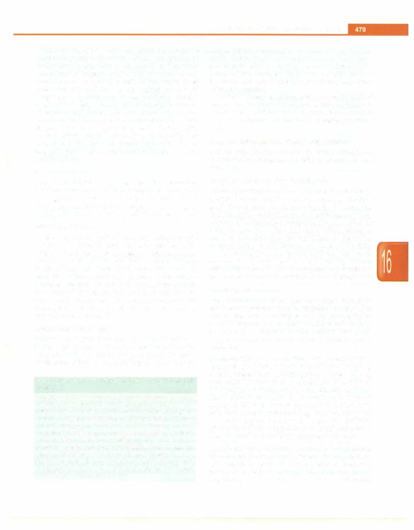
Figs 16.8A and B: (A) Poststreptococcal GN. Moderately severe proliferation and exudative changes with infiltration of neutrophils. Few open capillary lumina are seen; CB) lmmunofluorescence examination showing extensive fine granular deposition of lgG along the capillary wall and in mesangium with a starry sky appearance
elevated in cases ofstreptococcal skin infection. The titers decreaseto lowlevelswithin4-6 weeks. Thelevel of serum C3 is low in 90% patients but normalizes by 8-12 weeks. Persistent low C3 levels indicate other forms of GN.
Management
Patients with mild oliguria and normal blood pressure can be managedathome. Closeattentiontobloodpressure and dietary intake is essential. Once acute GN has occurred, treatment with penicillin has no effect on the course of the disease, but may be given if active pharyngitis or pyoderma is present. The principles of management of patients with severe oliguria and acute kidney injury are discussed later.
Diet. The intake of sodium, potassium and fluids should be restricted until blood levels of urea reduce and urine output increases. Overhydration is a dangerous compli cation as it may increase hypertension and precipitate left ventricular failure. Patients with azoternia requireaccurate measurement of urine output and daily weight, and restriction of fluid intake to an amount equal to insensible losses and 24 hr urine output.
Diuretics. Patients showing modest edema are treated with oral frusemide at a dose of 1-3 mg/kg; the edema disappears withthereturn of renal function. Therapy with IV frusernide (2-4 mg/kg) is necessary in subjects with pulmonary edema.
Hypertension. Mild hypertension may be controlled by restriction of salt and water intake. Effective anti hypertensive agents include arnlodepine, nifedipine or diuretics. Beta-blockers and angiotensin converting enzyme inhibitors carry risk of hyperkalemia. Patients with hypertensive emergencies need prompt treatment with IV nitroprusside or labetalol.
Left ventricularfailure. Hypertension should be controlled and IV frusemide given to induce diuresis, leading to improvement in heart failure. If diuresis is not noted, dialysis is initiated. Respiratory support with positiveend expiratory pressure may be needed.
Prolonged oliguria. Treatment, as outlined above, should be continued and levels of blood urea and electrolytes monitored. Dialysis is required in children with severe renal failure and prolonged oligoanuria, fluid overload and life threatening electrolyte disturbances. Occurrenceof secondary infections should be avoided.
Outcome and Prognosis
Acute poststreptococcal GN has an excellent prognosis in childhood.The symptomsbegin toresolvein thefirst week with loss of edema and fall in blood pressure. Gross hematuria and significant proteinuria disappear within 2-weeks, although microscopic hematuria and slight proteinuria may persist for several months. Hypertension subsides within 2-3 weeks, but rarely may persist for several weeks. Patients with acute GN of nonstreptococcal etiology have variable and unpredictable outcome. These cases need closefollowup over several years with periodic urinalyses and measurements of blood pressure.
Renal biopsy. A biopsy is rarely indicated in those suspected to have poststreptococcal GN except when renal function is severely impaired beyond 7-10 days or serum C3 remains depressed beyond 6-8 weeks. Patients with unresolving acute GN (persistent oliguria or azoternia past 7-10days,hypertension or gross hematuria past 2-3 weeks) or those with features of a systemic illness (e.g. systemic lupus) require a kidney biopsy (Table 16.6).

|
|
n i l |
i |
i |
|
______________________ |
|
Ess_e__t a_Ped__atr cs |
|||||
|
__ |
|
|
|
__________ |
|
|
|
|
|
|
||
Crescen tic Glomerulonephritis
Rapidly progressive GN (RPGN) is defined as an acute nephritic illness accompanied by rapid loss of renal function over days to weeks. The histopathological correlate is the presence of crescents (crescentic GN) involving 50% or more glomeruli (Fig. 16.9) suggesting severe glomerular injury. The chief forms of RPGN are:
(i) immune complex crescentic GN (irnmunofluorescence showing immunoglobulinand C3 deposits; normal orlow C3), (ii) pauci-immune crescentic GN (related to small vessel vasculitis; positive antineutrophil cytoplasmic antibodies; scant immune deposits) and (iii) anti glomerular basement membrane GN (with anti-GBM antibodies; linear IgG deposits). The severity of clinical and histological features often correlates. Patients with circumferential crescents involving more than 80% glomeruli show advanced renal failure; those with noncircumferential crescents in fewer glomeruli have an indolent course. Renal biopsy should be performed in all patients with severe nephritic features, which do not resolve within 1-2 weeks.
The outcome is related to histological severity and prompt institution of therapy. Without appropriate treat ment, patients are at risk for progressive renal failure. Satisfactory results have been obtained with initial administration ofIVand oralcorticosteroidsandIVcyclo phosphamide, followed by maintenance immunosup pression.Plasmapheresisisrecommendedinpatientswith pauci-immunecrescenticGNand Goodpasture syndrome.
Nephritisin Henoch-Schonlein Pur pura
Henoch-Schonlein purpura (HSP) is the most common vasculitis in children (Fig. 16.10). Mild renal involvement indicated by microscopic hematuria and mild proteinuria is common. Serum IgA levels may be elevated. Renal
Fig. 16.9: Large cellular crescent with compression of glomerular tuft (Masson trichome X 200)
Fig. 16.10: Henoch-Schonlein purpura in a 6-yr-old girl admitted with severe abdominal pain. Note purpuric rash over the lower limbs
biopsy shows mesangial proliferation with mesangial deposition of IgA. Most patients recover without any specific treatment. However, longterm observation is necessary to detect insidious renal damage. Rarely a patientmaypresentwithnephriticornephroticsyndrome, hypertension, azotemia and crescentic GN. Therapy with a combination of oral/IV corticosteroids and cyclophos phamide initially, followed by maintenance steroids and azathioprine is recommended. Longterm outcome depends on the severity of renal manifestations.
l m munoglobulin A Nephropathy
Predominant deposition of IgA in the glomeruli, chiefly in the mesangium and occasionally in capillary walls is characteristic.Theusualclinicalmanifestation is recurrent episodes of gross hematuria following upper respiratory infections; each episode lasts for 2-5 days. In between these episodes, microscopic hematuria and mild protei nuria may persist. An acute nephritic or nephrotic syndrome is rarely the initial manifestation. Renal histo logy shows mesangial proliferation of varying severity. Patients with hematuria and non-nephrotic proteinuria are treated using angiotensin converting enzyme inhi bitors. Therapywithcorticosteroids and alkylating agents is indicated in patients with nephrotic range proteinuria or deranged renal function.
Lupus Nephritis
Variable clinical and renal histological patterns are observed in patients with systemic lupus erythematosus. Asymptomatic proteinuria and/or hematuria, acute nephritic syndrome and nephrotic syndrome are most common. Rarelyrenal involvement may bemanifested as rapidly progressive GN. Renal biopsy may show almost normal glomeruli, focal or diffuse proliferative GN or membranous nephropathy. Immunofluorescence studies show mesangial and capillary wall deposits of IgG and

C3 and usually Clq and IgA. Antinuclear and double stranded DNA autoantibodies are present in most cases with lupus nephritis; C3 levels are reduced.
Remissions and relapses and progressive renal damage are characteristic. Infections and end stage renal disease are the chief cause of mortality. Judicious use of cortico steroids, cytotoxic agents (cyclophosphamide, myco phenolate mofetilandazathioprine), calcineurin inhibitors (cyclosporine, tacrolimus) and monoclonal antibodies and prompt treatment of infections has improved outcomes.
Suggested Reading
Brogan P, Bagga A Leukocytoclastic vasculitis. In: Cassidy JT, Petty RE, Laxer RM, Lindsay CB, eds. Textbook of Pediatric Rheumatology, 6 th ed. Philadelphia, Saunders Elsevier 2011;483-97
Brogan P, Eleftheriou D, Dillon M. Small vessel vasculitis. Pediatr Nephrol 2010:25;1025-35
Gulati A, Bagga A. Management of lupus nephritis. Indian J Rheumatol 2012; ?(Suppl 1):69-79
Hogg RJ. Idiopathic immunoglobulin A nephropathy in children and adolescents. Pediatr Nephrol 2010;25:823-9
NEPHROTIC SYNDROME
Nephrotic syndrome is characterized by massive pro teinuria, hypoalbuminemia and edema; hyperlipidemia is often associated. Some patients show hematuria and hypertension. Heavy proteinuria (more than 1 g/m2 per day) is the underlying abnormality, leading to hypoalbuminemia (serum albumin below 2.5 g/dl). The resultantfall inplasmaoncoticpressureleadsto interstitial edema and hypovolemia. This stimulates the renin angiotensin-aldosterone axis and antidiuretic hormone secretion that enhances sodium and water retention. The pathogenesis of edema may however be different in patients with significant glomerular lesions, who show primary sodium retention and expanded intravascular volume. Hypoalbuminemiaalsoinduceshepaticsynthesis of -lipoproteins resulting in hypercholesterolemia.
More than 90% of childhood nephrotic syndrome is primary (or idiopathic).Othercausessuchas amyloidosis, vasculitis, systemic lupus erythematosus, postinfectious GN andhepatitis Bnephropathyare infrequent.Nephrotic syndrome in children can be divided into two groups based on renal histological characteristics: (i) minimal change nephrotic syndrome (MCNS); and (ii) nephrotic syndrome with significant lesions (Table 16.7).
Steroid sensitive nephrotic syndrome (which is usually MCNS) has a satisfactory longterm outcome. In contrast, the steroid resistant form (usually associated with significant glomerular lesions)hasless satisfactory course and a significant proportion progress to chronic renal failure.
STEROID SENSITIVE NEPHROTIC SYNDROME
MCNS accounts for 80% cases of nephrotic syndrome in children. Renal biopsy does not show significant
Disorders of Kidney and Urinary Tract -
Table 16.7: Features of idiopathic nephrotic syndrome
Features |
Minimal lesion |
Significant lesions |
Age at onset |
2-6 yr |
Older children |
Sex incidence |
Higher in boys |
Equal |
Hematuria |
Rare |
Usual |
Blood |
Normal |
Normal or increased |
pressure |
|
|
GFR |
Normal |
Normal or decreased |
Renal biopsy |
Normal glomeruli; |
Changes of varying |
|
mild mesangial |
severity;C3, |
|
proliferation; |
imrnunoglobulin |
|
often IgM deposits |
deposits |
SerumC3 |
Normal |
Low in MPGN |
Selectivity of |
High |
Low |
proteinuria |
|
|
Response to |
Remission in >95% |
Unsatisfactory |
steroids |
|
|
Prognosis |
Good; relapses stop |
Variable progression of |
|
by second decade |
renal damage |
MPGN membranoproliferative glomerulonephritis
abnormalities on light microscopy (Fig. 16.llA). Electron microscopy shows nonspecific obliteration of epithelial foot processes. Immunofluorescence studies do not demonstrate deposition of immune reactants except occasional mesangial IgM. On the other hand, patients with focal segmental glomerulosclerosis (FSGS) show evidence of sclerosis involving a segment of the glome rular tuft (Fig. 16.llB). The pathogenesis of MCNS is obscure. There is evidence to suggest perturbation of cell mediated immunity, which through yet undefined mechanisms alters the permselectivity of the glomerular filter, resulting in massive proteinuria. A proportion of patients have a primary abnormality of the epithelial foot processes (podocytes).
Clinical Features
The onset is insidious with edema first noticed around the eyes and subsequently on legs. It is soft and pits easily on pressure. Gradually edema becomes generalized, with ascites, hydrothorax and hydrocele (Fig. 16.12). With increasing edema, urine output may fall. The blood pressure is usually normal; sustained elevation suggests the possibility of significant glomerular lesions. The bloated appearance and relative well-being of thechildis misleading and after the loss of edema, severe muscle wasting isrevealed. Infections may be present at the onset and during relapses.
Laboratory Findings
Urineexaminationshowsheavy (3-4+) proteinuria. Gross hematuria or persistent microscopic hematuria suggests the likelihood of significant glomerular lesions; hyaline and granular casts are present. Serum albumin is low and values below 1 g/dl are often obtained. Hypercholestero lemia mayimpartamilky appearance to theplasma. Blood

-.__EssentialPediatrics_________________________________
_______________
Figs 16.11 A and B: (A) Renal histology in a 4-yr-old boy with steroid dependent nephrotic syndrome. There is normal morphology of glomerular capillary loops, mesangial matrix and cells suggestive of minimal change disease; (B) histological features in a 6-yr-old girl with
steroid resistant nephrotic syndrome secondary to focal segmental glomerulosclerosis. Note the hilar sclerosis involving large areas of the glomerulus and adhesions to the Bowman's capsule
Fig. 16.12: An 8-yr-old boy with steroid dependent nephrotic syndrome. Anasarca is seen affecting upper limbs (including dorsa of hands), trunk and ascites. Note the cushingoid features and striae on lower abdominal wall and upper legs
urea and creatinine values are within the normal range except when there is hypovolemia and fall in renal perfusion.
Blood levels of IgG are low and those of IgM elevated; C3 level is normal. The severity of glomerular damage is reflected in the passage of proteins of large molecular weight, chiefly globulin. Protein selectivity is the ratio of clearance of high molecular weight (e.g. IgG) to low molecular weight proteins (e.g. transferrin, albumin). A low ratio indicates highly selective proteinuria, as in MCNS. However, this information does not offer diagnostic help.
Evaluations considered at onset of nephrotic syndrome include: (i) urinalysis for proteinuria, red cells, casts;
(ii)blood levels of urea, creatinine, albumin, cholesterol;
(iii)complete blood counts and (iv) tuberculin test. Depen ding on clinical and laboratory findings, the following additional tests may be required: (i) C3 and antistrepto lysin O (gross or persistent microscopic hematuria); (ii) chest X-ray (positivetuberculin test; history of contact with tuberculosis); (iii) hepatitis B surface antigen (recent jaundice, raised levels of transaminases); (iv) antinuclear antibodies (suspected systemic lupuserythematosus); and
(v)urine culture (suspected urinary tract infection). A renal biopsy is not required to confirm the diagnosis of MCNS prior to starting treatment. A biopsy is recom mended in children with atypical features at the onset (age below 12 months, gross or persistent microscopic hema turia, low blood C3, hypertension or impaired renal function). Patients who continue to show nephrotic range proteinuria despite appropriate steroid therapy require a biopsy to determine the underlying disorder.
Management of Initial Episode
The child should receive a high protein diet. Salt is restric ted to the amount in usual cooking with no extra salt given. Any associated infection is treated. The presence of tuberculosis should be looked for. Diuretics are adminis tered only if edema is significant. Frusemide (1-4 mg/kg/ day in 2 divided doses) alone or with an aldosterone antagonist, spironolactone (2-3 mg/kg/day in 2 divided doses)is adequate. Diuretics shouldbe used cautiously and overzealous fluid loss avoided. Therapy with cortico steroids results in abolition of proteinuria (remission) usually by 10-14 days, diuresis and loss of edema.
The first episode of nephrotic syndrome should be treated adequately, both in terms of dose and duration of corticosteroids, since this is considered an important determinant of longterm course. Only prednisolone and prednisone are of proven benefit in the treatment of proteinuria. Either of these agents is given at a dose of 2 mg/kg per day (maximum 60 mg) in single or divided doses for 6 weeks, followed by 1.5 mg/kg (maximum 40 mg) as a single morning dose on alternate days for the next 6 weeks. Therapy with corticosteroids is then stopped. While some experts propose that therapy with corticosteroids should not be stopped abruptly and tapered over the next 8-12 weeks, the benefits of pro longed therapy need to be balanced by the risk of steroid adverse effects.
Parent Education
The parents should be explained about the disease and the usual outcome and their cooperation ensured. They are taught how to examine urine for protein, which should be done periodically to detect a relapse early. During the periods of remission, no dietary restrictions are imposed.
Subsequent Course
A small proportion of patients have only a single episode of the illness, while the majority shows relapses. Some patients have three or less relapses in a year (infrequent relapsers), while others have four or more relapses (frequent relapsers) (Table 16.8). About 15% remain in remission while on prednisolone therapy and relapse whenever the dose is reduced or within 2 weeks of its discontinuation (steroid dependent). About 10-15% patients either donotrespond tothe initial treatment with prednisolone, or do so transiently and later cease to respond (steroid resistant).
Management of Relapse
Relapses are often triggered by minor infections. Symptomatic therapy of infectious illness might result in remission of low grade (1-2+) proteinuria. However, persistence of 3-4+ proteinuria requires treatment for
Table 16.8: Important definitions to clarify course of nephrotic syndrome
Remission: Urine albumin nil or trace (or proteinuria <4 mg/ m2/hr) for 3 consecutive early morning specimens
Relapse: Urine albumin 3+ or 4+ (or proteinuria >40 mg/m2/ hr) for 3 consecutive early morning specimens, having been in remission previously
Frequent relapses: Two or more relapses in initial six months or four or more relapses in any twelve months
Steroid dependence: Two consecutiverelapses when on alternate day steroids or within 14 days of its discontinuation Steroid resistance: Absence of remission despite therapy with daily prednisolone at a dose of 2 mg/kg per day for 4 weeks and alternate day for next 4 weeks.
Disorders of Kidney and Urinary Tract
relapse. Prednisolone is given at a dose of 2 mg/kg/day until protein is negative/trace for three consecutive days, and then on alternate days at a dose of 1.5 mg/kg for 4 weeks. Thus, treatment for a relapse usually lasts for 5-6 weeks and there is no evidence that its prolongation affects the outcome.
The first 2-3 relapses aretreatedin themannerdescribed above. Once the pattern of relapses is known, therapy is individualized.Patientswith infrequent relapses continue to receive treatment for individual relapses as outlined above.
Frequent Relapses and Steroid Dependence
Patients with frequent relapses or steroid dependence require prolonged treatment in order to maintain disease remission.
Longterm Alternate Day Predniso/one
Following treatmentof arelapse, the dose of prednisolone is tapered to maintain the patient in remission; usually a small dose is given on alternate days for 9-18 months. This strategyiseffectiveinmaintainingremissionin many patients. Since infections precipitate relapses, adminis tering the same small dose daily for 5-7 days starting at onset of infections may prevent relapses. However, relapses, while on this therapy, are treated with daily prednisolone at 2 mg/kg/dayuntilremission, afterwhich alternate day therapy is resumed at 1.5 mg/kg. Patients with repeated relapses, while on longterm therapy, should be considered for treatment with a steroid sparing agent.
Steroid Sparing Agents
The additional use of an alternative agent should be consideredinpatientswith: (i)prednisolonethreshold (for maintaining remission) higher than 0.5-0.7 mg/kg on alternate days, or (ii) features of corticosteroid toxicity (growth failure, hypertension and cataract). The agents used, usually in successive order, are listed below and in Table 16.9.
Levamisole. This immunomodulator iseffectivein reducing relapses in aproportionof patientswithfrequentrelapsing or steroid dependent nephrotic syndrome. After inducing remission, levamisole is administered at a dose of 2-2.5 mg/kg on alternate days. Alternate day predniso lone is given in decreasing doses, until a dose of 0.3-0.5 mg/kg is reached, for 3-6 months; it is occasionally possible to discontinue steroids altogether. Treatment with levamisole is given for 1-2 yr or longer. The chief side effect is leukopenia, which should be monitored every 2 months; others include flu like symptoms and rash.
Cyclophosphamide. Treatment with alkylating agents and alternate day prednisolone is effective in many patients with frequent relapsing or steroid dependent nephrotic syndrome. A 12-week course of treatment may induce long-lasting remission in 30-40%cases. Side effects include

___E_s_s_ en_t_ia_i_P_ed_iat r_ics_________________________________ _
|
Table 16.9: Therapy for steroid sensitive nephrotic syndrome |
||
Agent |
Dose |
Duration |
Adverse effects |
Prednisolone |
Maintain on 0.3-0.7 mg/kg on |
9-18 mo |
Cushingoid body habitus, hypertension, short |
|
alternate days |
|
stature, cataract, hirsutism |
Levamisole |
2-2.5 mg/kg on alternate days |
1-2 yr |
Leukopenia, rash, flu-like symptoms |
Cyclophos- |
2-2.5 mg/kg/day |
12 weeks |
Leukopenia; alopecia; gonadal toxicity; nail |
phamide* |
|
|
discoloration (hemorrhagic cystitis; nausea and |
|
|
|
vomiting are more common with IV administration) |
Mycophenolate |
600-1000 mg/m2/day or |
1-3 yr |
Gastrointestinal discomfort, diarrhea; leukopenia |
mofetil |
20-25 mg/kg/day |
|
|
Cyclosporine (CyA)* |
CyA: 4-5 mg/kg/day |
12-36 mo |
Acute and chronic nephrotoxicity, elevated |
or Tacrolimus (Tac)* |
Tac: 0.1-0.2 mg/kg/day |
|
transaminases (both agents); hirsutism, gum |
|
|
|
hyperplasia, hypertension or hyperlipidemia |
|
|
|
(CsA > Tac); hyperglycemia, neurotoxicity with |
|
|
|
headache and seizures (Tac > CsA) |
Rituximab* |
375 mg/m2 IV once a week |
2-3 doses |
Infusion reactions (fever, rash, bronchospasm); |
hypogammaglobulinemia, neutropenia
• Preferred earlier if relapses are life threatening (associated with peritonitis, other serious infections or thrombosis) or in presence of significant steroid toxicity
leukopenia, nausea and vomiting; a high fluid intake is ensuredto prevent hemorrhagiccystitis. Alkylating agents are associated with a risk of gonadal toxicity and malignancies, although at the doses and duration used these risks are minimal. Another alkylating agent, chlorambucil has significant additional toxicitiesanda low margin of safety, and is not recommended.
Mycophenolate mofetil. Prolonged treatment withthis agent is useful in reducing relapse rates and corticosteroid sparing. The lack of renal, hemodynarnic and metabolic toxicity makes it an alternative to calcineurin inhibitors. Chief side effects include gastrointestinal discomfort, diarrhea and leukopenia. The dose of the medication is 600-1000 mg/m2/day or 20-25 mg/kg/day in two divided doses for 12-36 months. Tapering doses of prednisolone are given for 6-12 months.
Cyclosporine and tacrolimus. Therapy with either of these agents is indicated in patients that fail to benefit with levamisole, cyclophosphamide and/or mycophenolate mofetil. Treatment may be associated with significant adverse effects. Cyclosporine A (4-5 mg/kg/day) or tacrolimus (0.1-0.2mg/kg/day) are administered, in two divided doses, for 12-24 months aiming for respective troughlevels of80-120 ng/ml and3-7 ng/ml. Both agents have strong steroid sparing potential, with steroid discontinuation achieved in most patients over 6-9 months.
Adverse effects are common and include acute and chronic nephrotoxicity. A renal biopsy is done after 2-3 yr of continuous therapy. Patients receiving cyclosporine have cosmetic side effects (hirsutism, gum hyperplasia), hypertension and hypercholesterolemia. Treatment with tacrolimus is associated with risk of hyperglycemia, elevated transaminases, diarrhea, tremors, headache and seizures.
Rituximab. This monoclonal anti-CD20 antibody has been used with success in patients with steroid dependent nephrotic syndrome, withremission lasting6-18 months. This agent appears to be useful in patients who fail to respond or show toxicity with other therapies.
Complications in Nephrotic Syndrome
The patient should be maintained in remission, as far as possible. Relapses should be promptly treated so that the childdoesnot develop morethanminimaledema.Several complicationsthat areassociatedwithmassiveedema and ascites.
Edema
Edema is controlled with salt restriction and oral hydro chlorothiazide or frusemide for a few days. Salt must not be totally stopped and the usual amounts usedin cooking should be allowed. For massive edema, higher doses of frusemide along with spironolactone are needed. Infusion of albumin may be necessary in intractable cases where serum albumin levels are extremely low causing poor renal perfusion and oliguria.
Infections
Nephrotic syndrome and steroid therapy render children susceptible to infections. Infection with S. pneumoniae, gram-negative organisms and varicella are common. Children present with serious infections, e.g. peritonitis, cellulitis, pneumonia and meningitis. Peritonitis may manifest with low grade fever, diarrhea and abdominal discomfort. Patients with varicella should receive oral acyclovirfor 7days; severe illnessrequiresadministration of IV acyclovir. Immunization with pneumococcal and varicella vaccinesis advisedoncethepatientis off steroids for 4 weeks.

Thrombotic Complications
Patients with nephrotic syndrome are at risk for throm bosis involving renal, pulmonary and cerebral veins. Aggressive use of diuretics, venepuncture of deep veins and hypovolemia increase the risk of this complication. Treatment with low molecular weight heparin followed by oral anticoagulants is recommended.
Hypovolemia and Acute Renal Failure
Hypovolemia may occur during a severe disease relapse or following administration of diuretics, particularly in children with poor oral intake, diarrhea and vomiting. Features include abdominalpain, lethargy, dizziness and leg cramps, tachycardia, hypotension, delayed capillary refill, low volume pulses and clammy distal extremities. Elevatedratioofbloodureatocreatinine,highhematocrit, urine sodium <20 mEq/1, fractional excretion of sodium 0.2-0.4%andurinarypotassium index [urineK+/(urineK+ + urine Na+)] >0.6 suggest the presence of hypovolemia. Therapy with diuretics should be discontinued. Patients require admission and rapid infusion of normal saline (10-20 ml/kg) over20-30min.Those who do not respond to two boluses of saline should receive infusion of 5% albumin (10-15 ml/kg) or 20% albumin (0.5-1 g/kg).
Steroid Toxicity
Repeated andprolongedcourses of steroids often result in significant toxicity, characterized by cushingoid features, short stature, hypertension, osteoporosis and subcapsular cataract. Timely use of steroid sparing agents (levamisole, alkylating agents, cyclosporin) are recommended.
Longterm Outcome
Children with MCNSusuallyhave an excellentprognosis. The frequency of relapses decreases with time and a majority of patients outgrow the condition by adulthood. Itis unfortunately notpossibleto predictwhen a particular patient will stop getting relapses. The mortality rate of 1-4% is associated with infections and hypovolemia that should be preventable.
STEROID RESISTANT NEPHROTIC SYNDROME
The management of patients with steroid resistant nephrotic syndrome is difficult, with patients showing a variable response to immunosuppression, adverse effects of prolonged therapy and risk of progressive renal damage. Steroid resistance is diagnosed if there is lack of remission despite treatment with prednisolone, at a dose of 2 mg/kg/day (60 mg/m2/day) for 4 weeks. Care is taken to exclude systemic infections (e.g. peritonitis, cellulitis, respiratory tract infections), which might result in persistent proteinuria.
Baseline assessment of renal function, blood levels of albumin and cholesterol, and estimation of proteinuria (24 hr quantitation) guides evaluation. Patients should be
Disorders of Kidney and Urinary Tract
evaluated for secondary causes. Children with steroid resis ce shouldundergorenal biopsybefore instituting specific treatment. While patients with minimal change disease show satisfactory response to therapy, the presence of FSGS with chronic tubulointerstitial changes 1s associated with less satisfactory outcomes. Before start of immunosuppressive therapy, patients with steroid resist t nephrotic syndrome should undergo testing for hepatitis B surface antigen, anti-HCV IgG and HIV.
About 10-20% patients with familial and sporadic steroid resistant nephrotic syndrome have homozygous or compound heterozygous mutations in genes encoding podocyte proteins, including podocin (NPHS2), nephrin (NPHSl) and Wilm's tumor (WT1) genes. These patients are unresponsive to immunosuppressive medications, progresses rapidly to end stage renal disease and unlike nongenetic FSGS (which recurs after transplantation in 30%), does not recur. Where facilities exist, mutational analysis should be offered to patients with (i) congenital nephrotic syndrome (onset below 3 months of age), (ii) family history of SRNS, (iii) sporadic initial steroid resistance that does not respond to therapy with cyclophosphamideor calcineurin inhibitors, and (iv) girls with steroid resistant FSGS.
Management
Patients with steroid resistant nephrotic syndrome secondary to minimal change disease, FSGS or mesangio proliferative GN are treated similarly. The chief factor predicting renal outcome is the response of proteinuria to therapy, rather than the renal histology. The aim of therapy in patients is thus to induce and maintain remission of proteinuria, while avoiding medication related adverse effects. Most regimens use a combination of an immuno suppressive agent with prednisolone (given on alternate days) and an angiotensin converting enzyme inhibitor (Table 16.10). The best resultsareobtained with regimens combining calcineurin inhibitors (cyclosporine or tacrol imus) and tapering doses of corticosteroids. The aim of treatment is the achievement of complete remission, but occurrenceofpartialremission is alsosatisfactory.Patients who respondtotreatmentdo so within 3-6 months; those that fail therapy with one regimen may show response to different agents.
Adjunctive therapy with angiotensin converting enzyme inhibitors (e.g. enalapril 0.3-0.6 mg/kg/day, ramipril 6 mg/m2/day) is associated with decrease in proteinuria and control of hypertension. Adverse effects include dry cough, hyperkalemia and decline in renal function. Angiotensin receptor blockers (e.g. losartan, valsartan) may be used in case of persistent dry cough with ACE inhibitors, or as add-on therapy for better antiproteinuric effect. Therapy with HMG coenzyme-A reductase inhibitors is useful in lowering blood levels of cholesterol in subjects with persistent hyperchole sterolemia.

|
n l |
i |
i |
|
|
|
________ |
|
|
|
|
s |
|
|
|
||
___ |
Esse_tia Pe_datr_c_________________________ |
|
_ |
|||||
|
|
|||||||
|
|
Table 16.10: Agents for management of steroid resistant nephrotic syndrome |
|
|
||||
Agent |
Dose |
|
Duration |
Efficacy |
Adverse effects |
|
|
|
Calcineurin inhibitors |
|
|
|
|
|
|
|
|
Cyclosporine (CsA) |
4-5 mg/kg/day |
12-36 months |
50-80% Acute and chronic nephrotox.icity (both agents); |
|
||||
Tacrolimus (Tac) |
0.1-0.2 mg/kg/day |
12-36 months |
70-85% |
hirsutism and gum hyperplasia (CsA > Tac); |
|
|||
|
|
|
|
|
|
hypertension, high cholesterol (CsA > Tac); |
|
|
|
|
|
|
|
|
hyperglycemia (Tac); elevated transaminases, |
|
|
|
|
|
|
|
|
neurotox.icity, headache and seizures (Tac > CsA) |
|
|
Cyclophosphamide |
|
|
|
|
|
|
|
|
Intravenous |
500-750 mg/m2 |
6 pulses |
40-50% Leukopenia; alopecia; nausea and vomiting; |
|
||||
Oral |
2-2.5 mg/kg/day |
12 weeks |
20-25% |
gonadal toxicity; hemorrhagic cystitis |
|
|||
High dose corticosteroids with cyclophosphamide |
|
|
|
|
||||
Methylprednisolone |
20-30 mg/kg IV |
'Pulses' on |
30-50% Hypertension, hypokalemia, hyperglycemia, steroid |
|
||||
|
|
|
|
alternate |
|
psychosis, systemic infections |
|
|
|
|
|
|
days x 6; once |
|
|
|
|
|
|
|
|
weekly x 8; |
|
|
|
|
|
|
|
|
fortnightly X 4 |
|
|
|
|
Prednisolone |
Tapering doses* |
18 mo |
|
|
|
|
||
Cyclophosphamide |
2-2.5 mg/kg/day•• |
12 weeks |
Side effects of cyclophosphamide therapy and prolonged |
|
||||
|
|
|
|
|
steroid therapy |
|
|
|
*Prednisolone 1.5 mg/kg on alternate days x 4 weeks; 1.25 mg/kg x 4 weeks; 1 mg/kg x 4 mo; 0.5-0.75 mg/kg x 12-18 mo **Cyclophosphamide is administered during 3-15 weeks
Hypertension must be controlled and infections mana ged appropriately. Edemaisminimized withjudicioususe of diuretics. The use of intravenous albumin should be limited to cases with (i) symptomatic hypovolemia, (ii) symptomatic edema or (iii) marked ascites that is causing respiratory compromise. In cases with hypo volemia, 10-20 ml/kg of 4.5-5% albumin should be infused. Severe symptomatic edema or ascites may be treated with0.75-1 g/kg of 20% albumin, infused over2 hr, to expand the circulating volumefollowed by frusemide 1 mg/kg. Close monitoring is essential to avoid fluid overload and pulmonary edema. Albumin infusion augments diuresis when co-administered with frusemide in severely hypoalbuminemic patients with refractory edema.
Congenital NephroticSyndrome
Congenital nephrotic syndrome present in the first 3 months of life with anasarca, hypoalbuminemia and oliguria. The etiology of congenital nephrotic syndrome is heterogeneous. The 'Finnish' form of the disease is inherited in an autosomal recessive manner, with mutations in the gene encoding nephrin (NPHSl). The characteristic renal histology with microcystic dilation of proximal tubules is seen after a few months of life, although ultrastructural abnormalities of the glomerular basement membrane are present at birth. Elevated levels of alpha-fetoprotein (AFP) in maternal serum and amniotic fluid enable antenatal screening. The clinical course is complicated by failure to thrive, recurrent
infections, hypothyroidism and progression to renal failure by 2-3 yr.
Patients with Denys Drash syndrome show mutations in the WTl gene, congenital nephrotic syndrome, male pseudohermaphroditism and high risk of bilateral Wilms' tumor. Renal histology is characterized by diffuse mesangial sclerosis and there is progressive renal failure.
Other causes of congenital nephrotic syndrome include infections (congenital syphilis, cytomegalovirus disease, toxoplasmosis) and mutations in PLCEl or NPHS2 genes; rarely renal histology may be normal (minimal change nephrotic syndrome) or show focal segmental glomerulo sclerosis. Therapy of patients with congenital nephrotic syndrome is supportive with appropriate nutrition, control of edema, thyroxin supplements and reduction of proteinuria through ACE inhibitors and/or indomethacin.
Suggeste d Reading
D'Agati VD, Kaskel Fl, Falk RJ. Focal segmental glomerulosclero sis. N Engl J Med 2011;365:2398-411.
Greenbaum LA, Benndorf R, Smoyer WE. Childhood nephortic syn drome: Current and future therapies. Nat Rev Nephrol 2012;8:445-58 Gulati A, Bagga A, Gulati S, on behalf of the Indian Society of Pediatric Nephrology. Guidelines for management of children with
steroid resistant nephrotic syndrome. Indian Pediatr 2009;46:35-47 HodsonEM, Willis NS, CraigJC.Corticosteroidtherapyfornephrotic
syndrome in children. Cochrane Database Syst Rev 2007; (4):CD001533 Indian Pediatric Nephrology Group. Indian Academy of Pediatrics.
Management of steroid sensitive nephrotic syndrome. Revised guide lines. Indian Pediatr 2008;45:203-14
Sinha A, Bagga A. Nephrotic syndrome. Indian J Pediatr 2012;79: 1045-55
CHRONIC GLOMERULONEPHRITIS
Chronic GN is not a single disease entity, but comprises advanced stages of several forms of GN. In most cases, the glomerular disease is primary and not part of a systemic disorder. However, chronic GN may occur in systemic lupus erythematosus, microscopic polyarteritis, familial nephropathies and nephropathies due to drugs and toxins. Variable glomerular deposition of immunoglobulin, complement and fibrin is found on irnrnunofluorescence studies. Renal biopsy examination in early stages shows several patterns, while later the histologic changes are nonspecific. Most glomeruli are sclerosed with corres ponding tubular, interstitial and vascular changes. Poststreptococcal GN seldom leads to chronic GN.
Clinical Features
Thepatientmaybe asymptomatic and the disease detected on routine urine examination. Others may show failure to thrive, persistent anemia, moderate to severe hyper tension, edema, nocturia, microscopic or gross hematuria, bone pains and deformities.
Differential Diagnosis
It might be difficult to distinguish chronic from acute GN. The presence of anemia, growth retardation, evidence of long-standing hypertension (hypertensive retinopathy, left ventricular hypertrophy) and radiological skeletal changesindicateimpaired renalfunction of long duration. Examination of the renal biopsy is valuable in confirming the diagnosis.
Urinalysis shows proteinuria,hematuria, whitecellsand casts. Urine specificgravity is fixed and low (around1010). Blood urea and creatinine levels are raised and the glomerular filtration rate less than 30 ml/min/1.73 m2. Ultrasonographyshowssmallkidneyswithregularoutline.
Management
There is no specific treatment for chronic GN. Treatment with steroids and immunosuppressive drugs does not offer any benefit. The blood pressure should be controlled and infections treated. If renal function is compromised, the treatment is that of advanced chronic kidney disease.
INTERSTITIAL NEPHRITIS
This is focal or diffuse inflammatory reaction of renal interstitiurn with secondary involvement of tubules and rarely, glomeruli. Acute interstitial nephritis is usually due to infections or drugs (e.g. ampicillin, cephalosporins). Common causes of chronic interstitial nephritis include urinary tract obstruction and vesicoureteric reflux. Interstitial nephritis may be a feature of a systemic disorder (e.g. systemic lupus, vasculitis, associated with uveitis); autoantibodies to tubular basement membrane are found in some cases. In many instances, no cause is determined.
Disorders of Kidney and Urinary Tract
The clinical features are nonspecific and include abdominal pain, anorexia, pallor, headache and edema. Hypertension is absent. The presence of progressive renal insufficiency associated with satisfactory urine output, and urinaryabnormalitiessuchas hyposthenuria andmild proteinuria suggest the diagnosis. Leukocytes and eosinophils are frequently seen in the urine, the latter a feature of drug-associated disease.
A renal biopsyestablishesthe diagnosis and helps assess severity. Drug-related interstitial nephritis is treated with stoppage of the offending drug; treatment with corticos teroids is beneficial. Systemic illness, if any, should be appropriately managed. The treatment of chronic interstitial nephritis is symptomatic.
Suggested Reading
Ulinski T, Sellier-Leclerc AL, Tudorache E, et al. Acute tubulointerstitial nephritis. Pediatr Nephrol 2012;27:1051-7
URINARY TRACT INFECTIONS
Urinarytractinfection(UTI)is acommonmedicalproblem in children, affecting 3-10% girls and 1-3% boys. They are an important cause of morbidity and might result in renal damage, often in association with vesicoureteric reflux (VUR). Beyond infancy, the incidence of UTI is higher in girls. During infancy, UTI are equally common in boys and girls because the route of infection is often hematogenous and boys have a higher incidence of urinary tract anomalies.
Microbiology
In most cases, UTI are caused by E. coli that forms the predominant periurethral flora, and uncommonly by
Klebsie/la, Enterobacterand Staphylococci epidermidis. Proteus and Pseudomonas infections occur following obstruction or instrumentation, while Candida infection occurs in immunocompromised children or after prolonged antimicrobial therapy.
Predisposing Factors
Recurrent UTI are observed in 30-50% children, usually within 3 months of the first episode. Predisposing factors for recurrent UTI includefemale sex, age below 6months, obstructiveuropathy, severevesicoureteric reflux (VUR), habitual postponement of voiding (voiding dysfunction), constipation and repeated catheterization, e.g. for neurogenic bladder. Children with malnutrition and those receiving immunosuppressive therapy are also susceptible.
Clinical Features
Theclinicalfeaturesdepend upon the age and the severity of UTI. Neonates show features of sepsis with fever, vomiting, diarrhea, jaundice, poor weight gain and lethargy. The older infant has unexplainedfever,frequent micturition and occasionally convulsions. Gross hematuria

__E_s_s_e_n_t i a_l_P_e_d_ i_a t_r_ic_s_________________________________
is uncommon. The presence of crying or straining during voiding, dribbling, weak or abnormal urine stream and palpable bladder suggest urinary obstruction.
It is difficult to distinguish between infection localized to the bladder (cystitis) and upper tracts (pyelonephritis). The distinction is not necessary since radionuclide studies show that most UTI in children below 5 yr of age involve the upper tracts. Hence,all children should be managed as ifthey have pyelonephritis. Patientswithhighfever(>39°C), systemic toxicity, persistent vomiting, dehydration, renal angle tenderness or raised creatinine are considered as having complicated UTI. Patients with low grade fever, dysuria,frequency andurgency and absenceof symptoms of complicated UTI are considered to have simple UTI. This distinction is important for purposes of therapy.
Important features on evaluation include history of straining at micturition, incontinence or poor urinary stream, voiding postponement and surgery for meningo myelocele or anorectal malformation. Finding of palpable kidney(s), distended bladder, tight phimosis or vulval synechiae and neurological deficit in lower limbs suggest a predisposing cause.
Diagnosis
The diagnosis of UTI is based on growth of significant number of organisms of a single species in the urine. Significantbacteriuria is definedas a colonycountof >105/ ml of a single species in a clean catch sample. Urine is obtained by suprapubic bladder aspiration or urethral catheterization in children below 2 yr. Any colonies on suprapubic aspiration and >50,000/ml on urethral catheterizationareconsidered significant. The occurrence ofsignificantbacteriuriain absence ofsymptoms istermed asymptomatic bacteriuria.
The presence of >10 leukocytes per mm3 in fresh uncentrifuged sample, or >5 leukocytes per high power fieldin centrifugedsample is usefulfor screening. Dipstick examination,combining leukocyte esterase and nitrite,has moderate sensitivity and specificity for detecting UTI.
Treatment
Once UTI is suspected,a urine specimen is sent for culture andtreatmentstarted.Infants below3monthsof ageand children with complicated UTI should initially receive parenteral antibiotics. The initialchoice of antibiotics is empiric and is modified once culture result is available. While a third generationcephalosporinispreferred,therapywithasingle dailydoseofaminoglycosideisalsosafeandeffective(Table 16.11). Once oral intake improves and symptoms abate, usually after 48-72 hr, therapy is switched to an oral antibiotic. The duration of treatment for complicated UTI shouldbe10-14days.Olderinfantsandpatientswithsimple UTI should receive treatment with an oral antibiotic for 7-10 days. Adolescents with cystitis may receive shorter durationofantibiotics,lasting 72hr.Patientswithasympto matic bacteriuria do not require treatment.
Table 16.11: Antimicrobials for treatment of UTI
Medication |
Dose (mg/kg/day) |
Parenteral |
|
Ceftriaxone |
75-100, in 1-2 divided doses IV |
Cefotaxime |
100-150, in 2-3 divided doses IV |
Amikacin |
10-15, single dose IV or IM |
Gentamicin |
5-6, single dose IV or IM |
Coamoxiclav |
30-35 of amoxicillin, in 2 divided doses IV |
Oral |
|
Cefixime |
8-10, in 2 divided doses |
Coamoxiclav |
30-35 of amoxicillin, in 2 divided doses |
Ciprofloxacin |
10-20, in 2 divided doses |
Ofloxacin |
15-20, in 2 divided doses |
Cephalexin |
50-70, in 2-3 divided doses |
All children with UTI are encouraged to take enough fluids and empty the bladder frequently to prevent stasis of urine. Routinealkalizationofthe urine is not necessary. With appropriate therapy, fever and systemic toxicity reduceand urineculture is sterile within 24-36 hr. Failure to obtain such a result suggests either lack of bacterial sensitivity to the medication or presence of an underlying anomaly of the urinary tract. A repeat urine culture is not required during or following treatment,unless symptoms fail to resolve despite 72 hr of therapy; symptoms recur, suggesting recurrent UTI, or contamination of the initial urine culture is suspected.
Imaging Studies
Following treatment of thefirst episode of UTI, plans are made for evaluation of theurinarytract. Theaimof imaging studies is to identify urologic anomalies that predispose to pyelonephritis, such as obstruction or vesicoureteric reflux, and detect evidence of renal scarring. Renal ultrasonography is useful in detecting hydronephrosis or anomalies of the urinary bladder and may be performed evenduringtherapyforUTI.Micturatingcystourethrogram is necessary for the diagnosis and grading of VUR (Fig. 16.13) and defines urethral and bladder anatomy. This procedure may be performed 2-4 weeks after treatment of theUTI. DMSA scintigraphy detects cortical scars, which are regions of decreased uptake with loss of renal contours or presence of cortical thinning with decreased volume (Fig. 16.SA). In order to differentiate between scars and reversible changes of pyelonephritis, this procedure is done 3-4 months after therapy for UTI.
These investigations should be performed judiciously, such that sufficient evaluation is done but at minimum risks of complications, such as radiation exposure and iatrogenic infections. The recommendations of the Indian Society of Pediatric Nephrology on evaluation following the first UTI are summarized in Table 16.12. All infants (<1 yr) require evaluation using ultrasonography, MCU and DMSA scan, since they are at the highest risk of UTI