Добавил:
Опубликованный материал нарушает ваши авторские права? Сообщите нам.
Вуз: Предмет: Файл:

Ghai Essential Pediatrics8th

.pdf
Скачиваний:
6
Добавлен:
03.11.2022
Размер:
70.46 Mб
Скачать
Metabolic screen

Diseases of Gastrointestinal System and Liver

at 1 yr and 70-80% in the longterm. Nearly 50% patients will have a relapse after successful therapy. In nearly one­ third patients, constipation persists even after puberty.

Other Etiologies of Constipation

A subgroup of children with constipation who fail to respond to medical management despite compliance or have red flags will need evaluation for organic disorders.

The main modalities of investigation are as follows. Rectal biopsy A full thickness rectal biopsy or suction

biopsy with mucosa and submucosa is required to rule out Hirschsprung disease, neuronal intestinal dysplasia

and hypoganglionosis.

Anorectal manometry In normal persons the internal anal sphincter shows relaxation on distension of rectum with a balloon (or stools). This is known as rectoanal inhibitory reflex and its presence excludes the diagnosis of Hirschsprung disease.

It is usefulto lookfor hypothyroidism, cystic fibrosis, hypercalcemia, celiac disease and lead

poisoning.

MRI of lumbosacral spine It is useful for diagnosis of

spina bifida occulta or tethered cord.

Colonic transit study Assessment of total and segmental colonictransittime is done either by radio-opaque markers or by scintigraphy. Based on transit studies, various patterns of colonic motility have been defined: normal colonictransit,slow transitconstipation(prolongedtransit throughout the colon) and outlet obstruction (delayed transit through anorectum).

Hirschsprung Disease

Hirschsprung disease is the commonest congenital gut motility disorder with an incidence of 1 in 5000 and is characterizedbylackofganglioniccells inthe submucosal and myenteric plexuses of the distal intestine. The distal rectumisaganglionicandtheaganglionosisextendsproxi­ mally in a variable length of colon. The absence of enteric neurons leads to tonic contraction of the aganglionic segment and functional obstruction. This multigenic disordercanbefamilialorsporadic.Downsyndromeisone of the most common associated malformations. Hirschsprungdiseaseisclassifiedbythelengthofinvolved intestine, with -75% cases involving only the colon distal to splenic flexure (classic form or short segment disease), -20% involvingcolon proximalto splenicflexure and -5% cases involving entire colon and small bowel as well.

Affected infants present shortly after birth with constipation and signs of distal obstruction. About 60-90% children with Hirschsprung disease fail to pass meconium in first 48 hr of life. Occasionally, the disease is missed and the child presents later with chronic constipation, failuretothrive and episodesofenterocolitis (loose stools with blood and mucus). Presence of empty

rectum on per rectal examination with a gush of liquid stools on withdrawal of finger suggests the diagnosis of Hirschsprung disease.

In neonatal period, plain X-ray abdomen reveals bowel distension with multiple air-fluid levels and paucity of air in pelvis. A carefully performed barium enema without prior colonic preparation and slow injection of contrast clearly delineatesthe narrow aganglionicbowel, transition zone and proximal dilated colon in Hirschsprung disease (Fig. 11.4). In contrast, in functional constipation the rectum is grossly dilated, with a rectum to sigmoid ratio of >1 and absence of transition zone. However, a normal study does not exclude Hirschsprung disease. Absence of rectoanal inhibitory reflex on anorectal manometery suggests Hirschsprung disease. Rectal biopsy is the gold standard in diagnosing Hirschsprung disease, with full thickness biopsy being ideal. Documentation of the absenceof ganglioniccells inthemyenteric andsubmucosal plexi is essential for the diagnosis. Hypertrophied nerves with increase in acetyl cholinesterase activity in the parasympathetic nerve fibers are seen in the aganglionic segment. The main differences between functional constipation and Hirschsprung disease are shown in Table 11.4.

Management depends on timing of diagnosis and is essentially surgical; the role of medical management is restrictedto stabilizingthe generalcondition and treating episodes of enterocolitis. Definitive surgical treatment involves resection of the aganglionic bowel, pull through and anastomosis of normallyinnervated ganglionic bowel close to the anal margin. Effort is made to preserve the anal canal and sphincter mechanism, thus preserving

Fig. 11.4: Barium enema showing Hirschsprung disease. Black arrow shows the dilated proximal segment and white arrow shows the transition zone


n

l


______


E_s_s_ e t_ai_P_ed_iat_rics ___________________________


_

1-rn


Table 11.4: Differences between functional constipation and Hirschsprung disease


Feature


Functional constipation

Hirschsprung disease


Passage of meconium


Normal

Delayed


Onset of symptoms


Beyond 1 yr of age

Within infancy


Encopresis


Yes

No


Stool withholding behavior

Yes

No


Episodes of enterocolitis

No

Yes


Growth failure


No

Yes


Abdomen


Not distended; may have palpable fecoliths

Distended


Per rectal examination

Soft to hard stools present

Empty rectum with gush of stools on


removing the finger


Barium enema


Rectum larger than sigmoid; ratio >1

Rectosigmoid ratio <1; transition zone seen


Anorectal manometry


Rectoanal inhibitory reflex present

Rectoanal inhibitory reflex absent


Rectal biopsy


Ganglions present

Ganglions absent


continence. In patients with delayed presentation, a colostomy in the ganglionic bowel is performed initially to relieve the obstruction and allow the dilated hypertro­ phied proximal bowel to return to normal. Subsequently, definitive surgery is performed. Now less invasive, laparoscopic and single staged surgeries are performed, in comparison to previous 2-3 staged procedure. In the longterm, majority show improvement but nearly two­ thirds of patients have some form of constipation or continence problem.

In subjects with other organic cause of constipation, treatmentis targeted towards the etiology,e.g. gluten free diet for celiac disease, surgery for tethered cord and hormone supplementation for hypothyroidism.

Suggested Reading

Benninga MA, Voskuijl WP, Tininiau JAJM. Childhood constipa­ tion: is there new light in the tunnel? J Pediatr Gastroenterol Nutr 2004;39:448-64

Clinical practice guideline: Evaluation and treatment of constipa­ tion in children: Summary of updated recommendations of North American Society for Pediatric Gastroenterology, Hepatology and Nutrition. J Pediatr Gastroenterol Nutr 2006;43:405--07

Haricharan RN, Georgeson KE. Hirschsprung disease. Semin Pediatr Surg 2008;17:266-75

Khanna V, Poddar U, Yachha SK. Etiology and clinical spectrum of constipation in Indian children. Indian Pediatr 2010;47:1025-30

Abdomnai lPani

Abdominal pain is a common manifestation of multiple pathologies which vary from benign to life-threatening conditions. The pain may be acute or chronic in nature.

To be able to arrive at a diagnosis, careful history and examinationand appropriateinvestigationsare necessary.

An understanding of pain perception in the abdomen and location of pain provides valuable information.

The gut is innervated by the enteric nervous system

which is involved in regulating secretion, motility and in sensory perception of visceral pain. The enteric nervous system is also influenced by the central nervous system.

Stretching of the visceral peritoneum overlying or inflammation results in pain sensation. The pain from the

stomach and proximal intestine is sensed in the epigas­ trium; from the midgut to theperiumbilicalarea; and from the transverse colon to the suprapubic area. The inflammation in the parietal peritoneum causes pain in the overlying abdominal wall. Thus, the pain of appendi­ citis is referred to the periumbilical area when the inflammation is restricted to the visceral peritoneum, but is perceived in the right iliac fossa when the inflammatory fluid comes in contact with the parietal peritoneum. Pain arising from retroperitoneal structures is referred to the back as it is sensed by the somatic nerves in the posterior abdominal wall. Referred pain is common in abdominal pathologies; a subdiaphragmatic collection on the right side may manifest as right shoulder painand ureteric pain is referred to the corresponding side as testicular pain. Radiation of pancreatic pain to the back and ureteric pain from loin to groin are also known.

Informationobtainedfrom the historyandexamination that aid in the diagnosis include the age of the patient, duration, type and frequency of pain, any nocturnal episodes, association with eating or defecation, vomiting, bloodinstools,diarrhea, constipationor obstipation, fever, joint pain, dysuria, hematuria, weight loss, jaundice, abdominal distension, fever and history of drug intake. Other systemic findings are also important as abdominal pain may be a manifestation of metabolic conditions like diabetic ketoacidosis and acute intermittent porphyria. Lower lobe pneumonia has been reported to account for

2.5-5% of abdominal pain.

Physicians must distinguish abdominal pain due to emergent diagnoses like appendicitis or intussusception from benignconditionslikegastroenteritisorconstipation. Examinationshouldbe meticulous including examination of genitalia as torsion of testes or incarcerated hernia can be easily overlooked. The accuracy of abdominal exami­ nation may be improved by distracting a child with toys or engaging in conversation. Ininfantsand young children the manifestation of pain may initially be as incessant cry.

Differential diagnosis should be considered in terms of age as many diagnoses are seen more commonly in children of certain age groups as shown in Table 11.5.

Diseases of Gastrointestinal System and Liver

Table 11.5: Common causes of abdominal pain

lntussusception

Infants and young children (<2 yr of age): Colic, acute

This is acommoncause of intestinal obstruction in children

between 3 months and 6 yr. Intussusception refers to the

gastroenteritis, intussusception, malrotation of gut with

volvulus, incarceratedhernia, trauma, necrotisingenterocolitis

telescoping of a proximal segment of intestine (intussus­

Preschool children (2-5 yr of age): Acute gastroenteritis, urinary

ceptum)intoadistalsegment (intussuscipiens).Thismaybe

ileocolic, colocolic or ileoileal.Most cases occur in infants

tract infections, constipation, intussusception, acute appendi­

during the weaning period following introduction a new

citis, malrotation of gut with volvulus, intestinal perforation

food, vaccination or upper respiratory tract infection. An

with peritonitis, choledochal cyst, lower lobe pneumonia,

areaofenlargedsubmucosal Peyer'spatch probablyactsas

incarcerated hernia, torsion testis, acute pancreatitis, diabetic

ketoacidosis, Henoch-Schonlein purpura, Meckel diverti­

a lead point. Beyond two years of age, the possibility of a

culum, trauma

submucosal lead point like lipoma and polyp that needs

Older children and adolescents: Acute gastroenteritis, gastritis,

surgical resection shouldbe considered as failure to resect

them will lead to recurrence. Inflammatory conditions like

acute appendicitis, Crohn disease, constipation, urinary tract

Henoch-Schonlein purpura also result in intussusception.

infections,dysmenorrhea,pelvicinflammatory disease,ectopic

Asaresult,thereisvenouscongestion,boweledemaleading

pregnancy, Mittelschmerz, renal calculi, acute pancreatitis,

cholecystitis, pneumonia, trauma, early phase of acute viral

toarterialobstruction,bowelischernia,necrosis,perforation

hepatitis, testicular or ovarian torsion, intestinal obstruction,

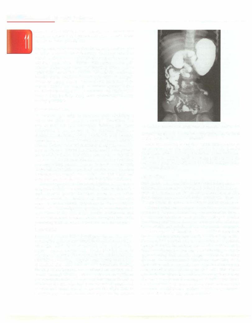
and shock. The classic triad of abdominal pain, red currant

perforation or peritonitis

jellystools(bloodandmucus)andpalpablemassisseenonly


in a small percentage of children. X-ray abdomen shows

The management of these acute conditions includes

paucity of air in right lower quadrant. Ultrasound is the

initial stabilization of the patient followed by specific

investigation of choice thatconfirmsthe diagnosis ('dough

management which may or may not be surgical. Some of

nut'sign)andprovidesinformationaboutpresenceofamass

these acute conditions are described briefly:

as lead point.Vascularityof bowel is bestassessed on color

Acute Appendicitis

Doppler. Barium enema shows a characteristic 'claw' sign

iftheintussusceptioninvolvescolon. Earlyreductioneither


Acute appendicitis is the commonest pediatric surgical

with saline (under ultrasound guidance), barium contrast

emergency and is more common in older children. The

(both diagnostic and therapeutic) or with air insufflation is

condition is considered as occurring due to obstruction of

advisable.Reductionwithairissaferwithlowerrecurrence

the appendiceal lumen by either fecolith or lymphoid

rates. Failure of radiological reduction or suspected

tissue, e.g. following viral infection. The obstruction,

intestinal gangrene may necessitate surgery and resection.

distentionandinfectionintheappendixcausesprogressive

Gallstones (Cholelithiasis)

inflammation and, subsequently, perforation. The patient


presents with fever and anorexia followed by pain in the

Gallstones are of three main types: cholesterol stones with

periumbilical area. Vomiting follows the periumbilical

>50% cholesterol, pigment (black or brown) stones and

pain, unlike in gastroenteritis. As the inflammatory fluid

mixed types. Pigment stones are common in children with

spreads, the pain is then felt in the right iliac fossa

hemolytic anemia. High-riskgroupsfor gallstones include

(McBurney point) towards which the child characteris­

children with hemolytic anemia, obesity, ileal resection or

ticallypointswithafinger.A retrocecalinflamedappendix

disease,intakeofdrugslikeceftriaxone,progressivefamilial

maybe difficult to diagnose and maymanifest asspasm at

intrahepaticcholestasistypeIII andtotalparenteralnutrition.

the hip. The diagnosis is most often based on clinical

Overall, hemolytic anemia and other predisposing

suspicion after historyandexamination. Palpation reveals

conditions account for 20-30% and 30--40% of gallstones,

localized tendernessandisbest elicited if there is rebound

respectively, while 30--40% cases remain idiopathic.

tenderness.

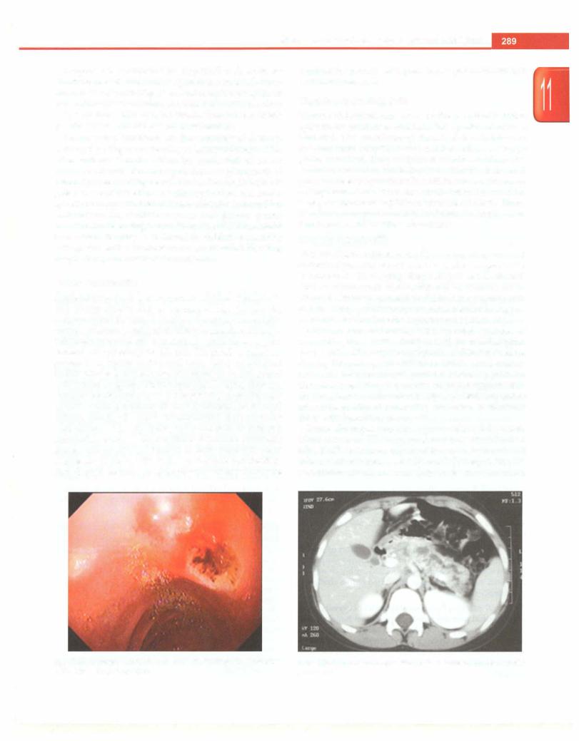
Clinical presentation. Typical presentation is with acute or

Hemogram shows polymorphonuclear leukocytosis.

recurrent episodes of right upper quadrant or epigastric

Urine microscopy should bedone to rule out urinary tract

pain which may radiate to the right shoulder. Icterus and

infection.Abdominal ultrasounddetectsa dilated (>6 mm)

pain radiating to the back is suggestive of a stone in

tubular, aperistaltic structure which is not compressible

common bile duct or ampullacausing pancreatitis. Fever

and is surrounded by fluid. Ultrasound has a sensitivity

does not usually occur; however, if present, it suggests

of 85-90% and specificity of 95-100% for diagnosing

presence of cholecystitis or cholangitis.

appendicitis. Computed tomography may be done


occasionally if the diagnosis is in doubt. In up to a third of

Diagnosis. Serum bilirubin and alkaline phosphatase are

patients, the appendix rupturesbefore surgery. Intravenous

elevated if the stone is in the common bile duct. Raised

fluids and antibiotics for gram-negative and anaerobic

amylase suggests pancreatitis. Ultrasonography is the

coverage should be given in all cases. Early surgery is

investigation of choice for diagnosis of gallstones. MRCP

necessary to prevent complications like perforation,

or ERCP have a better accuracy than ultrasonography in

appendiceal abscess and sepsis.

diagnosing common bile duct stones. These children

___E_s_s_e_n_t_ia_1_P_e_d_i_.atr_ic_s_________________________________

should be investigated with hemoglobin, reticulocyte count, peripheral blood picture and other investigations to look for hemolytic disease.

Management. Symptomaticcholelithiasis istreatedby open or laparoscopic cholecystectomy. Common bile duct calculicanbe removed by ERCP or at surgeryby common bile duct exploration. Children with asymptomatic gall­ stones without underlying predisposing factors can be safely followed up. Patients withsicklecelldisease should be subjected to prophylactic cholecystectomy even if gall­ stones are asymptomatic. Children with other hemolytic anemias should be screened by ultrasonography if they are being considered for splenectomy and cholecystec­ tomy should be done along with splenectomy in those having gallstones.

Choledochal Cyst

Choledochal cyst refers to abnormal cystic dilatation of biliary tree either as a single or multiple dilatations. This may or may not have associated intrahepatic cystic dilatations. It can present in the neonatal period as cholestasis, mimicking biliary atresia (5% of all neonatal cholestasis), or in the older child with recurrent episodes of pain, obstructive jaundice or mass in right upper quad­ rant. Acute or recurrent pancreatitis may be the presen­ tation of choledochal cyst, either due to stone impaction at lower end of common bile duct, or due to anomalous pancreatobiliary junction known to be associated with choledochal cyst. Biliary peritonitis secondary to bile duct perforation can complicate a choledochal cyst. Untreated cases may go on to develop secondary biliary cirrhosis.

Ultrasonography is the investigation of choice to diagnose choledochal cyst. MRCP is done to define the anatomy of the pancreaticobiliary ductal system before surgical excision. Definitive treatment is with cyst resection and hepaticojejunostomy in the majority as there is ariskof malignancy (cholangiocarcinoma) in the epithelium of the cyst if left in situ. Antibiotics and supportivetherapyarerequiredbeforesurgeryfor a child presenting with obstructive jaundice and cholangitis.

Ma/rotation

Rotational abnormalities developing during the matu­ ration of the gut cause recurrent obstruction, occurring as either the Ladd's band or volvulus of the gut over the narrow mesenteric pedicle. About 80-90% cases of volvulus occurwithin the first yearoflife.Abdominal pain with bilious vomiting suggests small bowel obstruction; abdominal distension may not be a prominent finding. Findings of malrotation are confirmed on barium meal follow through (BMFT), which shows duodenojejunal junction on the right of the spine (rather than to the left of midline at the level of pylorus), an abnormally positioned caecum and small bowel loops on the right side of abdomen(Fig. 11.5).Ifvolvulusis alsopresent,thecontrast

Fig. 11.5: Bariummeal follow through showing malrotation ofintestine

abruptly tapers into a corkscrew appearance at the level of the second portion of duodenum.

After resuscitation, emergency laparotomy is required for correction of the defect, usually by the Ladd's procedure, which includes derotation of the volvulus, division of the Ladd's band, widening of the base of the mesentery, placement of bowel in a state of nonrotation and appendicectomy.

Peptic Ulcer

Both gastric and duodenal ulcers occur infrequently in children. Ulcers maybe primary,i.e.related to Helicobacter pylori or secondary, e.g. due to drugs (NSAIDs, steroids), stress (shock, sepsis and ischemia), corrosives, Menetrier disease, Crohn disease and Zollinger-Ellison syndrome.

Clinical presentation depends on the age. Neonates typically present with bleeding and perforation from a gastric ulcer, usually occurring in the setting of another underlying problem, such as sepsis or respiratorydistress. Older infants and toddlers frequently vomit, eat poorly and have upper GI bleeding. Older children may have the classical epigastric pain which is relieved by eating. However, this is noted only in a minority; most patients have pain that is ill-localized and similar to that seen in functional dyspepsia. Overt or occult bleeding is seen in approximately half of school-age children with ulcer disease. Gastrointestinal bleeding, vomiting with obstruction and severe pain due to perforation suggest complicated ulcers. Ulcers associated with Helicobacter pyloriinfection affectolderchildren,familyhistoryof ulcer disease is usually noted and upper GI endoscopy shows antral nodularity as compared to ulcers without such infection Mechanical ventilation and coagulopathy increase risk of bleeding in stress ulcers.

Diseases of Gastrointestinal System and Liver

Diagnosis is established by upper GI endoscopy by direct visualization oflocation (gastric or duodenal), num­ ber and size of ulcer (Fig. 11.6). Endoscopic management canbe done at the same time in ulcers with active bleeding or ooze or those with a visible vessel. One can also obtain gastric biopsies for Helicobacter pylori testing.

Proton pump inhibitors are the mainstay of therapy. Activelybleeding ulcers require endoscopic therapy, with sclerosant or adrenaline injection, application of heater probe or bipolar electrocoagulation, or placement of hemoclip over bleeding vessel. Evaluation for Helicobacter pylori is essential in all cases with peptic ulcer and merits specific therapy with two of three antibiotics (amoxicillin, metronidazole, clarithromycin) and proton pump inhibitor.Predisposing factors, including NSAIDs, should be avoided. Surgery is indicated in children presenting with gastric outlet obstruction or uncontrolled bleeding despite drug and endoscopic treatment.

Acute Pancreatitis

Acute pancreatitis is less common in children than adults and occurs chiefly due to trauma, drugs (valproate, L-asparaginase), viral infections (mumps), hemolytic uremic syndrome, congenital biliary anomalies, Henoch­ Schonlein purpura and occasionally, gallstones, hyper­ calcernia or hypertriglyceridemia. Diagnosis is based on presence of upper abdominal pain (with or without radiationtotheback),elevatedserumamylaseorlipaseand radiological imaging (ultrasonography, CT scan) showing bulky,edematouspancreas.Acuteseverepancreatitismay result in acute respiratory distress syndrome, acute renal failure, shock, GI bleed, disseminated intravascular coagulation, hypoglycemia, hypocalcemia or infected pancreatic necrosis.Latecomplicationsinclude pancreatic abscess and pseudocyst formation. Early supportive care in intensive care is critical in severe acute pancreatitis. Radiological, endoscopic or surgical interventions may be

required for patients with pseudocyst, pancreatic abscess or infected necrosis.

Chronic Abdominal Pain

Chronic abdominal pain refers to the pain that is either episodic or continuous and lasts for a period of at least 2 months. The prevalence of chronic abdominal pain in children varies from 0.5 to 19% and depending on the age group evaluated. The principles of diagnosis with regard toclinicalevaluationaresimilarto that in acute abdominal painbut the etiologies differ.In addition to organic causes, an important cause is nonorganic abdominal pain which is responsible for nearly75% of all cases. As per the Rome III criteria, such pain is termed 'abdominal pain related to functional gastrointestinal disorders.'

Chronic Pancreatitis

This condition is characterized by recurrent episodes of abdominalpainandexocrineand/orendocrine pancreatic insufficiency. On imaging, diagnosis is based on demon­ stration of pancreatic calcification and/or dilated pancre­ aticduct.Chronicpancreatitisinchildrenmaybeidiopathic or hereditary, autoimmune, tropical, metabolic (hyper­ calcernia)or secondary to recurrent acute pancreatitis.

Children present initially with repeated episodes of pancreatic pain. Chronic diarrhea with fat malabsorption (exocrine insufficiency)andsymptoms of diabetes mellitus develop later along with failure to thrive. Local compli­ cations include pseudocyst, pancreatic ascites, pancreatic duct stricture, biliary strictures and portal hypertension due to splenic vein thrombosis. These patients are also at an increased risk of pancreatic carcinoma, particularly those with hereditary pancreatitis.

X-ray abdomen may show pancreatic calcification. Ultrasound and CT scan demonstrates ductal dilatation (Fig. 11.7), strictures, calcification and altered size or echotexture of pancreas. ERCP or MRCP help define the pancreatic ductal anatomy (e.g. prominent stricture,

Fig. 11.6: Upper gastrointestinal endoscopy showing duodenal ulcer

Fig. 11.7: CT scan showing dilated main pancreatic duct in chronic

in the first part of duodenum

pancreatitis

- Essential Pediatrics

intraductal calculi) and planning of endoscopic or surgical therapy. Exocrine pancreatic insufficiency is confirmed by demonstrating excess fat and reduced pancreatic elastase or chymotrypsin in stool. Fasting and postprandial blood sugar helpevaluatefor endocrine insufficiency. Evaluation for etiology should include looking for hypercalcemia or hyperlipidemia and testing for mutations in cationic trypsinogen gene to confirm hereditary pancreatitis.

Treatment includes supportive therapy during acute attacks, administration of antioxidants and oral pancreatic enzyme supplements for exocrine pancreatic insufficiency and endoscopic, radiological or surgical treatment for pseudocyst, ductal stricture and other complications. Surgical procedures like partial pancreatectomy or lateral pancreatojejunostomy are required in patients not responding to medical therapy. Celiac ganglion block is another alternative for pain control. Management of diabetes mellitus, if present, is essential.

Abdominal Pain Related to Functional Gastrointestinal Disorders

Abdominal pain related to functional GI disorders is diagnosed in the presence of pain that is present at least once a week in the preceding 2 months and the absence of an organic cause such as an inflammatory, anatomic, metabolic and neoplastic process. The pain is typically periumbilical and is clearly localized by the child.

After extensive studies, the most accepted under­ standing of childhood functional abdominal pain is of 'visceral hyperalgesia', referring to an altered excessive perception of normal gut motility that is interpreted by the child as pain. This perception is influenced by the psychosocial stressors in the school and family. The focus on the pain is further heightened by the growing concern in the family and the frequent visits to the doctors. Children of parents with increased anxiety and functional GI problems have an increased risk of developing abdominal pain. The following types are recognized.

Functional dyspepsia. Persistent or recurrent pain or discomfort is centered in the upper abdomen, located above the umbilicus and not relieved by defecation nor associated with a change in stool frequency or form (i.e. no irritable bowel syndrome).

Irritable bowel syndrome. Abdominal discomfort or pain is associated with two or more of the following: improve­ ment with defecation, onset associated with a change in frequency of stool and onset associated with a change in consistency of stool.

Abdominal migraine. Paroxysmal episodes of intense, acute periumbilical pain are noted, lasting for an hour or more with intervening periods of normal health lasting weeks to months. The episodes of pain interfere with normal activities and are associated with two or more of the follo­ wing: anorexia, nausea, vomiting, headache, photophobia and pallor.

Childhood functional abdominal pain syndrome. This refers to episodic or continuous abdominal pain that meets insufficient criteria for other types. The criteria for child­ hood functional abdominal pain are satisfied if the child has one or more of the following symptoms at least 25% of the time: some loss of daily functioning and additional somatic symptoms such as headache, limb pain, or difficulty in sleeping.

Among the various types mentioned above, functional abdominal pain is the most common. The diagnosis of childhoodfunctional abdominal painhingeson confidently ruling out organic etiology using a careful history and examination; extensive investigationsareunnecessary. The history should include not only details of pain but also family details, child's emotional environment in home and school, personality, coping skills, school performance and stress factors. The presence of alarming symptoms (Table 11.6) increases the probability of organic disorder andjustifiesfurther diagnostic testing. In the absence of red flags, the diagnostic yield of investigations is poor. Hemogram, ESR, stool routineand occult blood, andurine microscopy should be carried out in all cases to rule out organic disease. Abdominal ultrasonography is not helpful; the presence of lymph nodes of <10 mm is not a significant finding. Further investigation is required only in those with alarm symptoms and based on the likely diagnosis.

The aim of management of children with functional abdominal pain is to make a positive diagnosis, normalize the lifestyle to not allow pain to curtail daily activities or school performance, and to rectify psychological factors. The crux of management is counseling the parents and the child, both jointly and separately. Parents need to be reassured about the benign nature of the ailment and emphasis is laid upon avoiding too much attention to the child. The concept of visceral hyperalgesia should be explained to parents. Provision of a nutritious diet with adequate fiber and avoiding intake of carbonated beverages and refined food helps in reducing bloating. The role of amitriptyline and hypnotherapy is restricted to a few refractory cases.

Table 11.6: 'Red flag' signs or features that indicate serious illnesss in a child with abdominal pain

Pain localized away from umbilicus in right upper or lower quadrant

Nocturnal pain

Failure to thrive; weight loss Significant vomiting; bilious vomiting Gastrointestinal blood loss

Chronic diarrhea Persistent fever Jaundice Arthritis; rash

Family history of inflammatory bowel disease

Localized tenderness or mass in abdomen; organomegaly Perianal fistulae

Diseases of Gastrointestinal System and Liver

Suggested Reading

Table 11.7: Causes of acute diarrhea

Berger MY, Gieteling MJ, Berminga MA Chronic abdominal pain in

Bacterial

children. BM) 2007;334:997-1002

Escherichia coli: Enterotoxigenic, enteropathogenic, entero-

Chiou E, Nurko S Management of functional abdominal pain and

invasive*, enterohemorrhagic* and enteroaggregative types

irritable bowel syndrome in children and adolescents. Expert Rev


Gastroenterol Hepatol 2010;4:293-304

Shigella*: S. sonnei, S. f!exneri, S. boydii and S. dysenteriae

Chronic abdominal pain in children. American Academy of Pediat­

Vibrio cholerae serogroups 01 and 0139

rics subcommittee on chronic abdominal pain; North American Society

Salmonella*: Chiefly S. typhi and S. paratyphi A, B or C

for Pediatric gastroenterology, Hepatology and Nutrition. Pediatrics

Campylobacter species•

2005;115:e370-81

Marin JR, Alpern ER. Abdominal pain in children. Emerg Med Clin

Others: Aeromonas spp., Bacillus cereus, Clostridium difficile,

North Am 2011;29:401-28

C/ostridium perfringens, Staphylococcus aureus, Vibrio para­


hemolyticus, Yersinia enterocolitica, Plesiomonas shigelloides*

ACUTE DIARRHEA

Viral


Diarrheaisdefinedasachangeinconsistencyandfrequency of stools, i.e. liquid or watery stools, that occur >3 times a day. If there is associated blood in stools, it is termed dysentery.Inthevastmajorityofcases, these acuteepisodes subside within 7 days. Acute diarrhea may persist for >2 weeksin5-15% cases,whichislabeledaspersistent diarrhea.

The global annual burden of diarrhea is huge, affecting 3-5billioncasesandcausingapproximately2milliondeaths a year. Diarrhea accounts for over 20% of all deaths in underfivechildren. Thetwomostimportantconsequences of diarrhea in children are malnutrition and dehydration. Malnutrition and diarrhea form a vicious cycle, since malnutrition increases the risk and severity of diarrhea. Impairedabsorption,lossofnutrients,increasedcatabolism andimproper feeding in diarrhea aggravate the severity of malnutrition.A child may lose as much water and electro­ lytesfromthebodyduringanepisodeofdiarrheaasanadult, whichtranslatesintoahigherproportionoftotalbodywater loss in the child. Significant dehydration with abnormal electrolyte and acid-base status occurs in 2-5% of all cases of diarrhea, which may be fatal.

Etiology

Intestinal infections are the most common cause of acute diarrhea.However, certain drugs, food allergy, systemic infections (e.g.urinarytractinfectionandotitismedia)and surgical conditions (e.g. appendicitis or Hirschsprung disease)canalsopresentasacuteonsetdiarrhea.Causative agents of acute diarrhea (Table 11.7) can be identified in nearly70-80% episodesofacutediarrhea. Rotavirusremains the leading cause of severe, dehydrating gastroenteritis worldwide.InIndia, rotavirus and enterotoxigenic E.coli account for nearly half of the total diarrheal episodes amongchildren.Inrotavirusdiarrhea, vomiting isanearly feature and diarrhea is more severe.Cholera accounts for 5-10% cases; it is endemic in some parts and may occur in outbreaks. In cholera, stools are like rice water, vomiting is common and rapid onset of severe dehydration occurs within hours. Apart from enterotoxin producing E. coli (ETEC), which accounts for nearly 20% of childhood diarrhea, enteroinvasive E. coli (EIEC) and entero­ hemorrhagic E.coli (EHEC) can cause dysentery. EHEC may also a cause of hemolytic uremic syndrome. Shigella

Rotavirus

Human caliciviruses: Norovirus spp.; Sapovirus spp. Enteric adenoviruses serotypes 40 and 41

Others: Astroviruses, coronaviruses, cytomegalovirus, picornavirus

Parasitic

Giardia lamblia Cryptosporidium parvum Entamoeba histolytica* Cyclospora cayetanensis lsospora belli

Others: Balantidium coli, Blastocystis ho,ninis, Encephalitozoon intestinalis, Trichuris trichiura•

*Cause diarrhea with or without dysentery

and Salmonella species are isolated in 3-7% of childhood diarrheas. Shigella accounts for majority of cases of dysenterywhereasEntamoebahistolytica accounts for only 5% of dysentery. Giardia lamblia rarely causes acute diarrhea.Infection with Candida albicans can cause acute diarrhea in patients with malnutrition, irnmunocompro­ mised state or following prolonged antibiotic treatment. Clostridium difficile should be suspected in patients who have received broad spectrum antibiotics.

Risk Factors

Factorsdeterminingsusceptibilitytodiarrheaincludepoor sanitation and personal hygiene, nonavailability of safe drinkingwater,unsafe food preparationpracticesandlow rates of breastfeeding and immunization. Young children (<2 yr) and those with malnutrition are more susceptible to acute diarrhea and have more severe and prolonged episodes.Riskfactorsforprolongedandrecurrentepisodes ofdiarrheainclude presence ofhypo-orachlorhydria (due toHelicobacterpyloriinfectionor therapywithprotonpump inhibitors), selectiveIgAdeficiency,infectionwith human immunodeficiency virus (HIV) and other chronic conditions. Alteration of normal intestinal microflora by antibioticscan predispose to C. difficileinfection.

Pathogenesis and Clinical Findings

Approximately 60% of a child's body weight is water, present in two fluidcompartments: the extracellularfluid

- Essential Pediatrics

(ECF) and intracellular fluid (ICF). The ECF includes circulating blood, intestinal fluid and secretions. Diarrheal losses come from ECF, which is relatively rich in sodium and has low potassium. Loss of water from the body causes a reduction or shrinkage of ECF volume. In half of these cases, the concentration of sodium in the plasma remains nearly normal (about 140 mEq/1); in another 40-45%cases, excessive sodium is lost in the stools leading to a relative decline in serum sodium (hyponatremia) and a fall in ECF osmolality. This causes movement of water from the ECF to ICF compartment, causing further shrinkage of the already reduced extracellular compart­ ment volume in hyponatremic dehydration. In about 5% cases of diarrhea, especially if the child has been given fluids withextra salt,serumsodiumlevels may be elevated to >150 mEq/1 and ECF osmolalilty is increased.

Normally, skin turgor or elasticity is maintained by tissue water and fat. Shrinkage of extracellular water in both hypoand isonatremic dehydration impairs skin elasticity. On pinching, it takes a few seconds for the skin fold to return to normal. In patients with hypernatremic dehydration, water moves from inside the cells to the ECF compartment due to the increased osmolality of ECF, and therefore, partially masks the loss of skin turgor.The skin appears soggy, doughy or leathery. In this situation, a severe case of hypernatremic dehydration is likely to be erroneously underestimated as mild dehydration, unless severe sequelae of dehydration such as circulatory or renal impairment are noted.

As the ECF compartment is depleted, the blood volume is reduced. This results in a weak, thready pulse, low blood pressure and cold extremities. Because of low hydrostatic pressure in the renal glomeruli, the filtration of urine is reduced. This is ominous because poorly functioning kidneys cannot regulate metabolic derangements. Urine flow is a good indicator of the severity of illness. Severe cases are associated with renal failure.

Diarrheal stools contain large amounts of potassium. Therefore, serum level of potassium invariably falls if diarrhea persists for more than a few days. This is more pronounced in children with severe malnutrition with already depleted potassium stores. Affected children present with abdominal distension, paralytic ileus and muscle hypotonia. Electrocardiogram may show ST depression and flat T waves. Since intestinal secretions are alkaline, considerable bicarbonate is lost in diarrheal stools and acidosis usually accompanies dehydration. Patients remain asymptomatic till the base excess falls to 12 mmol/1. Below this level, breathing becomes deep and rapid (Kussmaul breathing).

Clinical features can be summed up as follows.The child is thirsty and slightly irritable in early and mild cases of diarrhea. As the diarrhea continues and dehydration worsens, the child becomes more irritable and develops a pinched look. The fontanelle, if open, is depressed, the eyes appear sunken and the tongue and the inner side of

cheeks appear dry. Abdomen may become distended in hypokalemia. The child passes urine at longer intervals. As acidosis worsens, the breathing becomes deep and rapid. In extreme cases, the child appears moribund, with weak and threadypulses, low blood pressure and reduced urine output. Children with severe dehydration may succumb rapidly if not treated promptly.

Assessment of Child with Acute Diarrhea

Goals ofassessment. These are to: (i) determine the type of diarrhea, i.e. acute watery diarrhea, dysentery or persis­ tent diarrhea; (ii) look for dehydration and other compli­ cations; (iii) assess for malnutrition; (iv) rule out nondiarrheal illness especially systemic infection; and

(v) assess feeding, both preillness and during illness.

History. This should include information on: (i) onset of diarrhea; duration and number of stools per day; (ii) blood in stools; (iii) number of episodes of vomiting; (iv) presence of fever, cough, or other significant symptoms (e.g. convulsions, recent measles); (v) typeand amount of fluids (including breast milk) and food taken during the illness and preillness feeding practices; (vi) drugs or other local remedies taken (including opioids or antimotility drugs like loperamide that may cause abdominaldistention); and (vii) immunization history.

Examination. The most important assessment is for dehydration. The degree of dehydration is assessed as per Table 11.8. One should look at the child's general condition, whether he/she is alert, restless or irritable or lethargic or unconscious. Other important assessments are for the appearance of eyes (normal or sunken) and the ability to drink water or ORS solution, whether taken normally or refused, taken eagerly, or an inability to drink due to lethargy or coma. Dehydration is also assessed by feeling for skin turgor; following pinching, the abdominal skin may flatten immediately, go back slowly or return very slowly (more than 2 seconds). Based on the degree of dehydration after history and examination, the estimated fluid loss is calculated as follows:

Degree of dehydration

Assessment offluid loss

No dehydration

<50 ml/kg

Some dehydration

50-100 ml/kg

Severe dehydration

>100 ml/kg

In addition, one should examine for features of malnutrition (anthropometry for weight and height; examination for wasting, edema and signs of vitamin deficiency), systemic infection (presence of cough, high grade fever, fast breathing and/or chest indrawing suggests pneumonia; high grade fever with splenomegaly suggests malaria) and fungal infections (oral thrush or perianal satellite lesions).

Laboratory investigations The large majority of acute diarrheal episodes can be managed effectively even in

Diseases of Gastrointestinal System and Liver

Table 11.8: Assessment of dehydration in patients with diarrhea

Look at

Condition'

Well alert

Restless, irritable

Lethargic or unconscious; floppy

Eyes2

Normal

Sunken

Very sunken and dry

Tears

Present

Absent

Absent

Mouth and tongue3

Moist

Dry

Very dry

Thirst

Drinks normally;

Thirsty, drinks eagerly

'Drinks poorly' or is not able to drink


not thirsty


Feel

Skin pinch4

Goes back quickly

Goes back slowly

Goes back very slowly

Decide

The patient has no signs

If the patient has two or

If the patient has two or more signs, there is


of dehydration

more signs, there is some

severe dehydration


dehydration


Treat

Use treatment Plan A

Weigh the patient, if possible,

Weigh the patient and use treatment


and use treatment Plan B

Plan C urgently

1 A lethargic child is not simply asleep; the child cannot be fully awakened; has a dull mental state and the child may appear to be drifting into unconsciousness

2ln some infants and children the eyes normally appear somewhat sunken. It is helpful to ask the mother if the child's eyes are normal or more sunken than usual

3Dryness of the mouth and tongue can also be palpated with a clean finger. The mouth may be dry in a child who habitually breathes through the mouth. The mouth may be wet in a dehydrated child owing to recent vomiting or drinking

4The skin pinch is less useful in infants or children with marasmus (severe wasting), kwashiorkor (severe malnutrition with edema) and in obese children

absence of laboratory investigations. Stool microscopy is not helpful in management except in selected situations, such as cholera (darting motion suggests Vibrio cholerae) and giardiasis (trophozoites). Stool culture is of little value in routine management of acute diarrhea. It is useful to decide on antibiotic therapy in patients with Shigella dysentery who do not respond to the initial empiric antibiotics. Tests for stool pH and reducing substances are not indicated in acute diarrhea. Hemogram, blood gas estimation, serum electrolytes, renal functiontests are not indicated routinely and are performed only if the child has associated findings like pallor, labored breathing, altered sensorium, seizures, paralytic ileus or oliguria whichsuggestsacid-baseimbalance, dyselectrolytemia or renal failure.

Principles of Management

Management ofacutediarrheahas four majorcomponents:

(i) rehydration and maintaining hydration; (ii) ensuring adequate feeding; (iii) oral supplementation of zinc; and (iv) early recognition of danger signs and treatment of complications.

The cornerstone of acute diarrhea management is rehydration by using oral rehydration solutions.After the history and examination, the child's dehydration status isclassifiedas no dehydration, somedehydrationorsevere dehydration and appropriate treatment started.

Physiological basisfor oral rehydration therapy. In most cases of acute diarrhea, sodium and chloride are actively secreted from the gut mucosa due to pathogen induced dysfunction of several actively functioning absorption pumps. However, glucose dependent sodium pump

remains intact and functional transporting one molecule of glucose and dragging along a molecule of sodium and one ofwateracrossintestinal mucosaresulting in repletion of sodium and water losses. The glucose dependent sodium and water absorption is the principle behind replacing glucose and sodium in 1:1 molar ratio in the WHO oral rehydration solution (ORS). An important consideration in making ORT is that the osmolarity of the replacement fluid should not exceed that of blood (290 mmol/1). Keeping theintestinal lumen at lower osmolarity as compared to blood allows for greater absorption of fluidsinto the bloodstream across concentration gradient, which also results in electrolyte absorption (by solvent drag). Since the concentration of glucose increases osmolarity, it is suggested that glucose concentration shouldnot exceed 111 mmol/1. Meta-analysishave shown that use of low osmolarity ORS causes reduction of stool output, decrease in vomiting and decrease in the use of unscheduled intravenous fluids without increasing the riskof hyponatrernia. Forthisreason, the recommendation for use of standard WHO ORS (having osmolarity of 311 mmol/1) was changed to low osmolarity WHO ORS (having osmolarity of 245 mmol/1). Since 2004, based on the WHO/UNICEF and IAP recommendations, the Government of Indiahasadoptedthelow osmolarity ORS as the single universal ORS to be used for all ages and all types of diarrhea. The composition of the low osmolarity ORS is given in Table 11.9.

IntheabsenceofWHOORS,onemayadminister culturally acceptable appropriate homemade fluids asshown in Table 11.10. Oral solutions should be given by a spoon or katori and in sips or small volumes rather than a large volume at one time as this increases the retention of oral fluids.

- Essential Pediatrics

Table 11.9: Composition of WHO recommended ORS

Constituent

g/1

Osmole or ion

mmol/1

Sodium chloride

2.6

Sodium

75

Glucose, anhydrous

13.5

Chloride

65

Potassium chloride

1.5

Glucose, anhydrous

75


Trisodium citrate,

2.9

Potassium

20

dihydrate


Citrate

10

Total osmolarity


245

Table 11.10: Home available fluids for acute diarrhea

Acceptable home available fluids


Fluids that contain salt


Oral rehydration solution,

(preferable)


salted drinks (e.g. salted rice


water or saltedyoghurt drink),


vegetable or chicken soup


with salt


Fluids that do not contain

Plain water, water in which a

salt (acceptable)


cereal has been cooked (e.g.


unsalted rice water), unsalted


soup, yoghurt drinks without


salt, green coconut water,


weak unsweetened tea,


unsweetened fresh fruit juice

Unsuitable home available

Commercial carbonated

fluids


beverages, commercial fruit


juices, sweetened tea


Treatment Plan A: Treatment of "No Dehydration"

Such children may be treated at home after explanation of feeding and the danger signs to the mother/caregiver. The mother may be given WHO ORS for use at home as per Table 11.11. Danger signs requiring medical attention are those ofcontinuing diarrhea beyond 3 days, increased volume/frequency ofstools,repeated vomiting, increasing thirst, refusal to feed, fever or blood in stools.

Treatment Plan B: Treatment of

"Some Dehydration"

All cases with obvious signs of dehydration need to be treated in a health center or hospital. However, oral fluid

Table 11.11: Oral rehydration therapy to prevent dehydration (Plan A)

Age

Amount of ORS or other

Amount of ORS to


culturally appropriate

provide for use at


ORTfluids to give

home


after each loose stool


<24mo

50-100 ml

500 ml/day

2-10 yr

100-200 ml

1000 ml/day

>10 yr

Ad lib

2000 ml/day

Explain use of ORS, i.e. the amount to be given, how to mix Give a teaspoonful every 1-2 min for a child under 2 yr Give frequent sips from a cup for an older child

1f the child vomits, wait for 10 min. Then give the solution more slowly (for example, a spoonful every 2-3 min)

1fdiarrheacontinuesafter the ORSpackets are used up, tell the mother to give other fluids as described aoove or return for more ORS

therapy must be commenced promptly and continued duringtransport. Fluidrequirementiscalculatedunderthe following threeheadings: (i)provisionofnormaldailyfluid requirements; (ii) rehydration to correct the existing water or electrolyte deficits; and (iii) maintenance to replace ongoing losses to prevent recurrence of dehydration.

i.The dailyfluid requirements in children are calculated as follows:

Up to 10 kg = 100 ml/kg 10-20 kg = 50 ml/kg >20 kg = 20 ml/kg

As an example, the daily fluid requirement in a child weighing 15 kg will be 1250 ml (first 10 kg, 10 x 100 = 1000 ml; another 5 kg, 5 x 50 = 250 ml, total 1000 + 250

=1250 ml).

ii.Deficit replacement or rehydration therapy is calculated as 75 ml/kg of ORS, to be given over 4 hr. If ORS can­ not be taken orally then nasogastric tube can be used. If child's weight cannot be taken then only age may be used to calculate fluid requirement as shown in Table 11.12.

If after 4 hr, the child still has some dehydration then anothertreatmentwithORS (asinrehydrationtherapy) is to be given. This therapy is effective in 95% cases. Oralrehydrationtherapymay beineffectiveinchildren witha high stool purge rate of >5 ml/kg bodyweight/ hr, persistent vomiting >3 per hr, paralytic ileus and incorrect preparation of ORS (very dilute solution).

iii.Maintenance fluid therapy to replace losses. This phase should begin when signs of dehydration disappear, usually within 4 hr. ORS should be administered in volumes equal to diarrheal losses, usually to a maxi­ mum of 10 ml/kg per stool. Breastfeeding and semi­ solid food are continued after replacement of deficit. Plain water can be offered in between.

Treatment Plan C: Children with "Severe

Dehydration"

Intravenous fluids should be started immediately using Ringer lactate with 5% dextrose. Normal saline or plain Ringer solution may be used as an alternative, but 5% dextrose alone is not effective. A total of 100 ml/kg of fluid is given, over 6 hr in children <12 months and over 3 hr in children >12 months as shown below.

ORS solution should be started simultaneously if the child can take orally. If IV fluids cannot be given (for reasons of access, logistic availability or during transport), nasogastric feeding is given at 20 ml/kg/hr for 6 hr (total 120 ml/kg). The child should be reassessed every 1-2 hr; if there is repeated vomiting or abdominal distension, the oral or nasogastric fluids are given more slowly. If there is noimprovementinhydrationafter 3 hr, IV fluids should be started as early as possible.

The child should be reassessed every 15-30 min for pulses and hydration statusafter the first bolus of100 ml/