

John Wiley & Sons - 2004 - Analysis of Genes and Genomes
.pdf128 |
VECTORS 3 |
|
|
l infection
l DNA inserted into E. coli
l DNA circularises via cos sites
Lysogenic pathway |
Lytic pathway |
|
l DNA integrated |
l DNA replicated |
|
into E. coli genome |
||
|
||
|
Rolling circle replication |
Lytic induction
DNA cleaved at cos sites and packaged into newly
synthesised head particles
l prophage replicated with E. coli genome
Cell lysis and phage release

3.3 λ VECTORS |
129 |
|
|
are concerned with recombination and the processes of lysogeny in which the circularized phage chromosome is inserted into the host chromosome and is stably replicated as a prophage. To the right of the map are genes concerned with transcriptional regulation and prophage immunity to superinfection (N, cro, cI), followed by the genes for DNA synthesis, late function regulation (Q) and host cell lysis.
Our extensive knowledge of λ phage and the ways in which the lytic and lysogenic life cycles are regulated have made λ an ideal vector to carry foreign DNA fragments. The major advantage of λ based vectors over plasmids is the efficiency at which the phage can infect E. coli cells. As we have already discussed, the transformation of plasmid DNA into bacteria is not an efficient process, whereas λ infection is a very efficient way to introduce DNA into a bacterial cell. To understand how λ can be exploited as a vector, it is important to have a basic knowledge of the phage itself. λ phage infection and lysis occurs in number of defined steps. Infection occurs as a result of the adsorption of the λ phage particle to the bacterial cell by binding to the maltose receptor. The λ genomic DNA is injected into the cell and almost immediately circularizes. At this point it can enter one of two pathways.
•Lysogenic pathway. The phage DNA becomes integrated into the bacterial genome (via homologous recombination between attP and the bacterial genomic attB site) and is replicated along with the bacterial DNA. The prophage DNA remains integrated until it is induced to enter the lytic pathway.
•Lytic pathway. Large-scale production of bacteriophage particles (proteins and DNA) occurs that eventually leads to the lysis of the cell.
The decision as to whether lysis or lysogeny occurs is the result of the activity of the cII protein. Active cII is required for the transcription of the cI repressor
Figure 3.9. λ life cycle. Upon infection, bacteriophage λ attaches to the surface of a bacterial cell, and its DNA enters the bacterium. Almost immediately, the λ DNA circularizes. The DNA can then enter either the lysogenic or the lytic pathway. During lysogeny, the λ DNA integrates into the E. coli chromosome and is replicated, along with the host DNA, such that the prophage is passed onto subsequent generations. In the lytic phase, the λ DNA does not integrate, but is immediately replicated and transcribed to produce new phage particles. Eventually, bacterial cell lysis occurs and the newly formed phage are released into the surrounding medium. The lysogenic prophage may be induced into the lytic cycle by, for example, treatment with UV light. In this case the λ DNA loops out of the E. coli genome and the lytic pathway is initiated
130 |
VECTORS 3 |
|
|
l phage |
Bacteria |
|
Top agar |
|
Mix |
|
Pour onto |
|
agar plate |
Incubate
l plaque
Bacterial lawn
Figure 3.10. λ plaques. λ phage is grown in the laboratory on a lawn of bacterial cells. The bacteria and the λ phage particles are mixed with liquid, but cool, top agar. The mixture is then poured onto an already set agar plate where the top agar is allowed to solidify. The plate is then incubated for 12–16 h at 37 ◦ C. λ plaques form as turbid circles in the bacterial lawn
and for some of the genes required for phage DNA integration into the E. coli chromosome. Active cII results in the adoption of the lysogenic pathway, while inactive cII results in the lytic pathway being followed. The cII protein is relatively unstable and is susceptible to cleavage and destruction by bacterial proteases. Environmental conditions influence the activities of these proteases. When grown in rich medium, for example, the proteases are generally active, such that cII is degraded and λ lysis occurs. Under conditions of E. coli starvation, the proteases are less functional and, consequently, λ will more
3.3 λ VECTORS |
131 |
|
|
Non-essential for lytic growth
R
e
c
o
m
|
n |
o |
|
i |
|
t |
|
a |
|
in |
|
b |
|
cIII
N
cI
cro
c |
|
II |
D |
|
N |
|
A |
|
r |
|
e |
|
p |
|
l |
|
i |
|
c |
|
a |
|
t |
|
i |
|
o |
|
n |
|
Q |
|
|
L |
|
|
l DNA |
y |
|
attP |
s |
|
|
|
s |
|
|
|
|
i |
|
|
48502 bp |
|
GGGCGGCGACCTCG |
|
cos |
CG |
GC |
|
|
GCCCCGCCGCTGGA |
|
|
|
|
|
H |
|
|
|
|
|
|
|
|
|
|
|
|
|
|
|
e |
|
|
|
|
|
|
il |
|
|
|
|
|
|
|
|
a |
|
|
|
|
|
|
|
|
|
|
|
|
|
|
|
d |
|
|
|
|
|
a |
|
|
|
|
|
|
|
|
|
|
|
|
|
|
|
T |
|
|
|
|
|
cos |
|
|
|
|
|
|
|
|
|
|
|
|
cos |
||
|
|
|
|
|
|
|
|
|
|
|
|
|
|
|
|
|
Tail |
Head |
|
Recombination |
CllI |
N |
cl |
cro |
cll |
|
DNA replication |
Q |
Lysis |
|
|
|
|
|
|
|
|
|
|
|
|
|
|
|
|
|
|
Non-essential for lytic growth
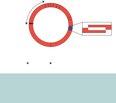
Figure 3.11. The circular and linear forms of the λ genome. λ DNA exists in a linear form in the bacteriophage and in a circular form upon entering the bacterium. The switch from the linear to the circular form occurs through complementation of the overhanging DNA ends at the cos sites. Many of the genes required for the integration of λ into the host chromosome, or for new phage replication and assembly, are grouped together on the λ chromosome. Some of these genes, or sets of genes, are shown. A region of the genome that is not required for lytic growth is indicated
frequently lysogenize. This behaviour makes sense, for in starved cells there will be less of the components necessary to make new phage particles.
The lytic pathway is characterized by a series of transcriptional events that produce different sets of proteins that are required for replication of the phage DNA and the production of new phage particles.
•Early transcription. Transcription of the N and cro genes occurs. This transcription is subject to repression by the product of the cI gene and in a lysogen this repression is the basis of immunity to superinfection.
•Delayed early transcription. The N protein product binds to the bacterial RNA polymerase and promotes transcription of the phage genes involved in DNA replication.
•Replication. Early replication proceeds from a single origin of replication
site. Later replication proceeds via a rolling circle mechanism to produce
132 |
VECTORS 3 |
|
|
long concatamers of the phage DNA that are connected to each other at the cos sites.
•Late transcription. The protein product of the cro gene builds up to a critical level and then stops early transcription. The product of the Q gene activates transcription, resulting in the production of the proteins required for the head and tail of the mature phage particle, and those required for bacterial cell lysis.
Finally, phage assembly occurs when a unit length of DNA is placed into the assembled head by cleavage of the concatameric DNA at the cos sites. The tail is added and the mature phage particle is completed. Upon cell lysis, approximately 100 newly synthesized phage particles are released from a single infected bacterial cell.
Wild-type λ DNA contains few unique restriction enzyme recognition sites into which foreign DNA fragments could be cloned, and is consequently not wholly suitable as a vector to carry such sequences. Additionally, the packaging of DNA into the λ phage is size limited. Efficient packaging will only occur with DNA fragments representing between 78 and 105 per cent of the wildtype genome size (37 – 51 kbp). These limits pose severe restrictions upon the amount of DNA that can be cloned into the phage genome. Two important developments, however, suggested that λ might be suitable as a cloning vector. Firstly it was determined that the gene products required for recombination could be removed from the λ genome and the lytic life cycle could still be completed and plaques would form. The remaining DNA, often referred to as the left-hand and right-hand arms of the λ genome, is capable of providing all necessary functions for the lytic pathway to occur. Secondly, naturally occurring restriction enzyme recognition sites could be eliminated without loss of gene function, which permitted the development of vectors with unique sites for the insertion of foreign DNA.
λ vectors could thus be constructed that lacked the genes required for recombination, and therefore could only enter the lytic cycle, but were capable of carrying much larger foreign DNA inserts. Two basic types of λ vector have been developed:
•insertional vector – DNA is inserted into a specific restriction enzyme recognition site;
•replacement vector – foreign DNA replaces a piece of DNA (stuffer fragment) of the vector.

3.3 λ VECTORS |
133 |
|
|
The advantage of replacement vectors is that they are capable of carrying larger DNA inserts. For example, λEMBL4 is a 42 kbp vector that contains 14 of kbp stuffer DNA between the left-hand and right-hand arms of λ. The ligation of just the λ arms would generate a 28 kbp λ genome. This is too small to be packaged into a λ particle. The insertion of foreign DNA between the two λ arms will, however, enable the genome to attain a suitable packaging size. The packaging size limit means that λEMBL4 is capable of holding foreign DNA fragments up to approximately 23 kbp in size (Figure 3.12).
l insertion vectors
cos |
Wild-type l DNA (48,502 bp) |
|
|
|
cos |
cos |
|
|
|
l vector (30–43 kbp) |
|
cos |
|||||||||||||||||||||||||||||||||
|
|
|
|
|
|
|
|
|
|
|
|
|
|
|
|
|
|
|
|
|
|
|
|
|
|
|
|
|
|
|
|
|
|
|
|
|
|
|
|
||||||
|
Tail |
Head |
|
Recombination |
CIII |
N |
cI |
cro |
cII |
DNA replication |
Q |
Lysis |
|
Cut and |
|
|
Tail |
|
|
|
Head |
N |
cI |
cro |
cII |
|
DNA replication |
Q |
Lysis |
|
|||||||||||||||
|
|
|
|
|
|
|
|
|
|
|
|
|
|
|
|
|
|
|
|
|
|
|
|
|
|
|
|
|
|
|
|
|
|
|
|
|
|
|
|
|
|||||
|
|
|
|
|
Non-essential |
|
|
|
|
|
|
|
|
|
|
|
|
|
|
|
|
|
|
|
|
|
|
|
|
|
|||||||||||||||
|
|
|
|
|
|
|
|
|
|
|
ligate |
|
|
|
|
|
|
|
|
|
|
|
|
|
|
|
|
|
|
|
|
||||||||||||||
|
|
|
|
|
for lytic growth |
|
|
|
|
|
|
|
|
|
|
|
|
|
|
|
|
|
|
|
|
|
|
|
|
|
|
|
|
|
|
|
|||||||||
|
|
cos |
|
|
lgt10 (43,430 bp) |
|
cos |
|
|
|
cos |
|
lZAPII (40,820 bp) |
|
cos |
|
|
|
|
||||||||||||||||||||||||||
|
|
|
|
|
|
|
|
|
|
|
|
|
|
|
|
|
|
|
|
|
|
|
|
|
|
|
|
|
|
|
|
|
|
|
|
|
|
|
|
|
|
|
|
|
|
|
|
|
|
|
|
|
|
cI |
|
|
|
|
|
|
|
|
|
|
|
|
|
|
|
|
lacZ' |
|
|
|
|
|
|
|
|
|
|
|
|
|
|
||||||
|
|
|
|
|
|
|
EcoRI |
|
|
|
|
|
|
|
|
|
|
|
|
|
MCS |
|
|
|
|
|
|
|
|||||||||||||||||
|
|
|
|
|
|
Accepts-8 kbp |
|
|
|
|
|
|
|
|
|
|
|
|
Accepts-10 kbp |
|
|
|
|
|
|
|
|||||||||||||||||||
|
|
|
|
|
|
|
|
|
|
|
|
|
|
l replacement vectors |
|
|
|
|
|
|
|
|
|
|
|
|
|
|
|
|
|
|
|||||||||||||
|
cos |
|
|
|
|
|
|
|
|
|
|
|
cos |
|
|
|
|
|
|
|
|
cos |
|
|
|
|
|
|
|
|
|
|
|
|
|
|
|
|
cos |
||||||
|
|
|
|
|
|
|
|
|
|
|
|
|
|
|
|
|
|
|
|
|
|
|
|
|
|
|
|
|
|
|
|
|
|
|
|
|
|
|
|
|
|
|
|
|
|
|
|
|
|
|
|
Stuffer DNA |
|
|
|
|
|
|
|
|
|
|
|
|
|
|
|
|
|
Insert DNA |
|
|
|
|
|
|
|
||||||||||||||
|
|
|
|
|
|
|
|
|
|
|
|
|
|
|
|
|
Cut and |
|
|
Selection on basis of size |
|
|
|
|
|||||||||||||||||||||
|
|
|
|
|
|
|
|
|
|
|
|
|
|
|
|
|
|
ligate |
|
|
|
|
|
||||||||||||||||||||||
|
|
|
|
|
|
|
|
|
|
|
|
|
|
|
|
|
|
|
|
|
|
|
|
|
|
|
|
|
|
|
|
|
|
|
|
|
|
||||||||
|
|
|
|
|
|
|
|
|
|
|
|
|
|
|
|
|
|
|
|
|
|
|
|
|
non-recombinant vector too small to |
||||||||||||||||||||
|
|
|
|
|
|
|
|
|
|
|
|
|
|
|
|
|
|
|
|
|
|
|
|
|
|
|
|
|
be packaged |
|
|
|
|
|
|
|
|||||||||
|
cos |
|
|
lWES.lB' |
cos |
|
|
|
|
|
|
|
|
|
|
cos |
|
|
|
|
|
|
|
|
|
|
|
|
|
|
|
cos |
|||||||||||||
|
|
|
|
|
|
|
|
|
|
|
|
|
|
|
|
|
|
|
|
|
|
|
|
|
|
|
|
|
|
|
|
|
|
|
|
|
|
|
|
|
|
|
|
|
|
|
|
|
|
|
|
|
|
|
|
|
|
|
|
|
|
|
|
|
|
|
|
|
|
|
|
|
|
|
|
|
|
|
|
Insert DNA |
|
|
|
|
|
||||||
|
|
|
|
|
|
|
|
|
|
|
|
|
|
|
|
Cut with EcoRI |
|
|
|
|
|
|
|
|
|
|
|
|
|
|
|
|
|
|
|
|
|||||||||
|
|
|
|
|
EcoRI |
|
|
|
|
|
|
|
|
|
|
|
|
|
|
|
|
|
|
|
|
|
|
|
|
|
|
|
|||||||||||||
|
|
|
|
|
|
|
|
|
|
EcoRI |
|
|
|
|
|
|
|
Accepts 4–17 kbp |
|
|
|
|
|||||||||||||||||||||||
|
|
|
|
|
|
|
|
|
|
|
|
|
|
|
|
|
and ligate with |
|
|
|
|
|
|
|
|
|
|||||||||||||||||||
|
|
|
|
|
|
|
|
|
|
|
|
|
|
|
|
|
|
|
|
|
|
|
|
|
|
|
|
|
|
|
|
|
|
|
|
|
|||||||||
insert DNA
cos |
lEMBL4 (42,360 bp) |
||||
|
|
|
|
|
|
|
|
|
|
14 kbp |
|
|
|
EcoRI BamHI SalI |
SalI BamHI EcoRI |
||
cos |
cos |
|
|
cos |
|||||
|
|
|
|
|
|
|
|
|
|
|
|
|
|
|
|
Insert DNA |
|
|
|
|
Cut with EcoRI, or BamHI |
Accepts-23 kbp |
|||||||
|
or SalI (or in combination) |
||||||||
and ligate with insert DNA
Figure 3.12. λ insertion and replacement vectors. All λ vectors have regions nonessential for lytic growth removed to increase the amount of DNA that will be packaged into the mature λ phage. Two λ insertion vectors are shown. λgt10 contains a unique EcoRI restriction enzyme recognition site in the cI gene. Recombinants will form clear rather than turbid plaques. λZAPII contains a multiple cloning site (MCS) in the lacZ gene and recombinants are identified using blue–white screening. Recombinants in λ replacement vectors are the only phages that will grow; if the two λ ends are ligated in the absence of insert DNA, the DNA is too small to be packaged
134 |
VECTORS 3 |
|
|
The size limitations of λ packaging thus provide a mechanism to ensure that foreign DNA has been inserted in between the λ arms to form a recombinant. Several other basic strategies have been devised to identify λ phage recombinants.
•Inactivation of the cI gene. Several λ phage vectors (e.g. λgt11) have unique restriction enzyme recognition sites contained within the cI gene. Phages in which the cI gene has been disrupted by foreign DNA insertion have an altered morphology, in which the plaques produced appear ‘clear’ as opposed to turbid. Screening of this type is technically difficult and requires a deal of skill on the part of the observer.
•Blue–white screening. λ phage vectors (e.g. λZAP) have been constructed to contain the lacZ gene expressing the α-fragment of β-galactosidase.
Screening for recombinant phages can then be preformed in E. coli cells
l lysogen BHB2688 |
l lysogen BHB2960 |
|
|
(mutant protein E) |
(mutant protein D) |
|
|
No protein E, |
|
No protein D, no |
|
no heads |
|
DNA packaging |
|
+ Protein D and other |
+ |
+ Protein E and other |
|
assembly proteins |
|
assembly proteins |
|
Tails |
Heads |
Tails |
|
cos |
cos |
cos |
cos |
Mix and add |
|
|
|
concatemerized l DNA |
|
|
|
|
37–51 kbp between cos sites |
|
|
Mature l phage
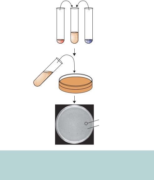
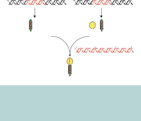
Figure 3.13. In vitro packaging of λ phage particles. Two different λ lysogens are used to produce the various components required for the packaging of λ particles. One of these lysogens (BHB2688) has a defective E gene, which results in no heads being produced. The other (BHB2960) has a defective D gene, resulting in a defect in DNA packaging. Mixing cell lysates of the two will result in an extract that is able to package concatamerized λ DNA. The multimerized DNA (37–51 kbp) will be cleaved at the cos sites and packaged into a mature λ phage particle
3.4 COSMID VECTORS |
135 |
|
|
expressing the ω-fragment of β-galactosidase in a similar way to screening for recombinants in pUC based plasmids.
Once a recombinant λ genome has been constructed, the problem arises of how to get the DNA into a viral particle so that it can be replicated in E. coli cells (Figure 3.13). Normal in vivo packaging of λ DNA involves first making pre-heads, structures composed of the major capsid protein encoded by gene E. A unit length of λ DNA is then inserted into the pre-head, with the unit length being prepared by cleavage of concatamerized λ genomes at neighbouring cos sites. A minor capsid protein D is then inserted in the pre-heads to complete head maturation, and the products of other genes serve as assembly proteins, ensuring joining of the completed tails to the completed heads. λ packaging of recombinant genomes can occur in vitro by utilizing two E. coli strains that bear λ lysogens containing different defects in the packaging pathway. A defect in producing protein E, resulting from a mutation introduced into gene E, prevents pre-heads being formed in strain BHB2688. A mutation in gene D prevents maturation of the pre-heads, with enclosed DNA, into complete heads in strain BHB2690. The components of the BHB2688/BHB2690 mixed lysate, however, complement each other’s deficiencies and provide all the products for correct packaging (Figure 3.13). Consequently, recombinant λ genomes can be constructed in vitro and packaged into mature λ phage particles before being propagated and replicated in E. coli cells.
3.4Cosmid Vectors
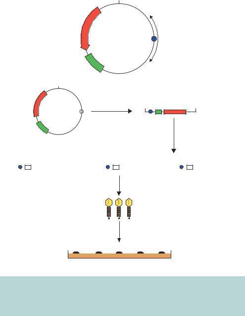
The only DNA requirements for in vitro packaging into λ phage are the presence of two cos sites that are separated by 37 –51 kbp of intervening sequence. Cosmids were developed in light of this observation, and are simply plasmids that contain a λ phage cos site (Collins and Bruning,¨ 1978). Figure 3.14 shows the overall architecture of a cosmid vector and a cloning scheme for the insertion of foreign DNA. As plasmids, cosmids contain an origin of replication and a selectable marker. Cosmids also possess a unique restriction enzyme recognition site into which DNA fragments can be ligated. After the packaging reaction has occurred, the newly formed λ particles are used to infect E. coli cells. The DNA is injected into the bacterium like normal λ DNA and circularizes through complementation of the cos ends. The lack of other λ sequences means, however, that λ infection will not proceed beyond this stage. The circularized DNA will, however, be maintained in the E. coli cell as a plasmid. Therefore selection of transformants is made on the basis of antibiotic resistance and bacterial colonies (rather than plaques) will form that contain
136 |
VECTORS 3 |
|
|
BamHI
AMPR
pJB8 |
cos |
l DNA |
|
5.4 kbp |
|||
|
|
ori
BamHI
AMPR |
Cut with BamHI BamHI |
BamHI |
cos 
BamHI BamHI ori Ligate 
Insert DNA
BamHI |
|
|
|
BamHI |
BamHI |
|
|
|
BamHI BamHI |
|
|
|
BamHI |
||||||
|
|
|
|
|
|
|
|
|
|
|
|
|
|
|
|
|
|
|
|
|
|
|
|
|
|
|
|
|
|
|
|
|
|
|
|
|
|
|
|
In vitro packaging
l particles
Infect E. coli
Colony containing circular
 recombinant cosmid
Petri dish containing agar with ampicillin
Figure 3.14. Cloning using a cosmid vector. The overall architecture of a cosmid vector, pJB8, is shown, together with a scheme for the insertion of foreign DNA into a cosmid. Since the cosmid lacks other λ genes, when the DNA is inserted into the E. coli cell it is maintained as a plasmid and selected for on the basis of antibiotic resistance
the recombinant cosmid. Since λ phage particles can accept between 37 and 51 kbp of DNA, and most cosmids are about 5 kbp in size, between 32 and 47 kbp of DNA can cloned into these vectors. This represents considerably more than could be cloned into a λ vector itself.
3.5 M13 VECTORS |
137 |
|
|
Cosmids, like plasmids, are very stable, but the insertion of large DNA fragments can mean that recombinant cosmids are difficult to maintain in a bacterial cell. Repeat DNA sequences are common in eukaryotic DNA, and DNA rearrangements can occur via recombination of the repeats present on the DNA inserted into the cosmid. The major difficulty in working with cosmids is, however, the production of linear, ligated DNA fragments in which the cosmid and insert are concatamerized together. Two basic problems exist.
•Ligation reactions of cosmid and insert DNA, like those shown in Figure 3.14, will generate circular DNA molecules that are unable to participate in the in vitro packaging reaction.
•More than one insert DNA molecule can be ligated between each cosmid DNA fragment. This could give a false impression of the DNA organization of the insert.
These difficulties can be overcome by cutting the cosmid with two different restriction enzymes to generate left-hand and right-hand ends that cannot religate to each other (Ish-Horowicz and Burke, 1981). Suitable phosphatase treatment of the insert DNA ensures that multiple inserts cannot be ligated to the cosmid DNA (see Chapter 2).
3.5M13 Vectors
M13, and its very close relatives f1 and fd, are filamentous E. coli bacteriophages. M13 is a male-specific lysogenic phage with a circular single-stranded DNA genome 6407 bp in length (Figure 3.15). M13 phage particles have dimensions of about 900 nm × 9 nm and contain a single-stranded circular DNA molecule (designated as the + strand). M13 infects bacteria that harbour the F pilus. The phage particle absorbs via one end to the F pilus, and the single-stranded phage DNA enters the bacterium (Figure 3.16). Very rapidly, the single-stranded DNA is converted into double-stranded (replicative form, RF) DNA by the synthesis of a complementary DNA strand (the – strand) using bacterial DNA polymerase. The RF form of the phage genome is rapidly multiplied until about 100 RF molecules are present within the bacterium. Transcription of the viral genes occurs to produce proteins required for the assembly of new viral particles. The production of a virally encoded singlestranded binding protein (the protein product of gene 2) eventually forces asymmetric replication of the RF DNA. This results in only one viral DNA strand being synthesized (the + strand). These single-stranded DNA molecules are assembled into new viral particles, and are released from the cell without