

John Wiley & Sons - 2004 - Analysis of Genes and Genomes
.pdf58 |
DNA: STRUCTURE AND FUNCTION 1 |
|
|
ribonucleoproteins (snRNPs, or snurps). The RNA molecules within these complexes are rich in uridine residues, and consequently the snRNPs have been designated by the names U1, U2, U4, U5 and U6. A model of the overall splicing process catalysed by the spliceosome is shown in Figure 1.29. The mechanism of splicing for exon coupling and intron extrusion depends on two transesterification reactions, which result in the formation of a lariat form of the intron and the fused exons. The branching associated with the A residue within the intron in the lariat form contains two phosphodiester linkages to the 2 and 3 of the A. The snRNPs play roles both in the catalytic process and in structurally maintaining the two exons in proximity with each other as the splicing reaction proceeds. Readers interested in the mechanism of splicing are directed to specific reviews on the topic (Sharp, 1994).
1.14.2Alternative Splicing
Since each exon in a eukaryotic gene encodes a portion of a protein, you can imagine that it is possible, by altering how the pre-mRNA is spliced, to produce different versions of the mRNA and ultimately, different proteins. Alternative splicing is a widely occurring phenomenon, with recent estimates suggesting that at least 30% of all human genes are subject to this type of processing (Sorek and Amitai, 2001). How do splice variants differ from the original sequence from which they are derived? The physiological activity of proteins produced from splice variants may be the same, opposite or completely different and unrelated. Alternative splicing therefore greatly increases the number of different protein activities that can be generated from a defined set of genes within the genome.
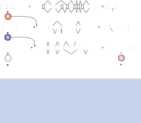
Perhaps one of the most dramatic examples of alternative splicing is found in the genes determining sex in the fruit fly Drosophila melanogaster. There are three genes that are involved in the sex determination process: sxl (sex lethal), tra (transformer) and dsx (double sex) (Chabot, 1996). Each of these genes produces a pre-mRNA that has two possible splicing patterns, depending upon whether the fly is male or female. Figure 1.30 shows these three genes and their splicing patterns. For males, the inclusion of two exons (exon3 in sxl and exon2 in tra) produces mRNA molecules that have termination (stop) codons and results in the formation of inactive proteins. The only active male product is the protein translated from the dsx gene. This protein inactivates all female-specific genes. The female, on the other hand, produces mRNAs without the stop codon containing exons. In the case of sxl and tra, the protein products have a positive effect on the splicing patterns observed, controlling the choice of introns removed in the spliceosome reaction.
|
|
|
|
|
|
|
|
|
|
|
|
|
|
|
|
|
|
|
|
|
|
|
|
|
|
|
|
|
|
|
|
|
|
|
|
|
|
1.15 |
|
TRANSLATION |
|
|
|
|
59 |
|
|||||||||||||||||||
|
|
|
|
|
|
|
|
|
|
|
|
|
|
|
|
|
|
|
|
|
|
|
|
|
|
|
|
|
|
|
|
|
|
|
|
|
|
|
|
|
|
|
|
|
|
|
|
|
|
|
|
|
|
|
|
|
|
|
|
|
|
|
|
|
|
|
|
|
|
|
|
|
|
|
|
|
|
|
|
|
|
|
|
|
|
|
|
|
|
|
|
|
|
|
Pre mRNA |
|
|
|
|
|
|
|
|
|
|
|
|
|
|
|
|
|
|
|
|
|
|
|
|
|
|
|
|
|
|
|
|||||
|
|
Female mRNA |
|
|
|
Female: |
|
|
|
|
|
|
|
sxl |
|
|
|
|
|
|
|
|
|
|
|
|
|
|
|
Male mRNA |
|
|
|
|
|
|
|
|
|||||||||||||||||||||||||||
|
|
|
|
|
|
|
|
|
|
|
|
|
|
|
|
|
|
|
|
|
|
|
|
|
|
|
|
|
|
|
|
|
|
|
|
|
|
|
|
|
|
|
|
|
|
|
|
|
|
|
|
|
|
|
|
|
|||||||||
|
|
|
|
|
|
|
|
|
|
AAAA |
|
|
|
|
|
|
|
|
|
|
|
|
|
|
|
|
|
|
|
|
|
|
|
|
|
|
|
|
|
|
|
|
|
|
|
|
|
|
|
|
|
|
|
|
|
AAAA |
|
|
|
|
|||||
|
1 |
2 |
4 |
5 |
6 |
7 |
8 |
|
|
|
|
|
|
1 |
|
|
|
2 |
|
3 |
|
4 |
|
5 |
|
6 |
|
7 |
|
8 |
|
|
|
|
|
1 |
2 |
|
3 |
4 |
5 |
6 |
7 |
8 |
|
|
|
|
|
||||||||||||||||
|
|
|
|
|
|
|
|
|
|
|
|
|
|
|
|
|
|
|
|
|
|
|
|
|
|||||||||||||||||||||||||||||||||||||||||
|
|
|
|
|
|
|
|
|
|
|
|
|
|
|
|
|
|
|
|
|
|
|
|
|
|
|
|
|
|
|
|
|
|
|
|
|
|
|
|
|
|
|
|
|
|
|
|
|
|
|
|
|
|
|
|
|
|
|
|
|
|
|
|
|
|
|
|
|
|
|
|
|
|
|
|
|
|
|
|
|
|
Male: |
|
|
|
|
|
|
|
|
|
|
|
|
|
|
|
|
|
|
|
|
|
|
|
|
|
|
|
Stop |
|
|
|
|
|
|
|
|
|
|
|
||||||||||
|
|
|
|
|
|
|
|
|
|
|
|
|
|
|
|
|
|
|
|
|
|
|
|
|
|
|
|
|
|
|
|
|
|
|
|
|
|
|
|
|
|
|
|
|
|
|
|
|
|
|
|
|
|||||||||||||
|
|
|
|
|
|
|
|
|
|
|
|
|
|
|
|
|
|
|
|
|
|
|
|
|
|
|
|
|
|
|
|
|
|
|
|
|
|
|
|
|
|
|
|
|
|
|
|
|
|
|
|
|
|
|
|
|
|
|
|
|
|
||||
|
|
|
|
sxl |
|
|
|
|
|
|
|
|
|
|
|
|
|
|
|
|
|
|
|
|
|
|
|
|
|
|
|
|
|
|
|
|
|
|
|
|
|
|
|
|
|
|
|
|
|
|
|
|
|
|
|
|
|
|
|
|
|
|
|||
|
|
|
|
|
|
|
|
|
|
|
|
|
|
|
|
|
|
Female: |
|
|
|
|
|
|
|
tra |
|
|
|
|
|
|
|
|
|
|
|
|
|
|
|
|
|
|
|
|
|
|
|
|
|
|
|
|
|
|
|
|
|
||||||
|
|
|
|
|
|
|
|
|
|
|
|
|
|
|
|
|
|
|
|
|
|
|
|
|
|
|
|
|
|
|
|
|
|
|
|
|
|
|
|
|
|
|
|
|
|
|
|
|
|
|
|
|
|
|
|
|
|
|
|
||||||
|
|
|
|
|
|
|
|
|
|
|
|
AAAA |
|
|
|
|
|
|
|
|
|
|
|
|
|
|
|
|
|
|
|
|
|
|
|
|
|
|
|
|
|
|
|
|
|
|
|
|
|
|
|
|
|
|
|
|
|
|
|
|
|
|
|||
|
1 |
|
3 |
|
|
|
4 |
|
|
|
|
|
|
|
1 |
|
|
|
2 |
|
3 |
|
|
|
|
4 |
|
|
|
|
|
|
|
|
|
|
1 |
2 |
|
|
|
3 |
|
|
4 |
|
AAAA |
||||||||||||||||||
|
|
|
|
|
|
|
|
|
|
|
|
|
|
|
|
|
|
|
|
|
|
|
|
|
|
|
|
|
|
|
|
|
|
|
|
||||||||||||||||||||||||||||||
|
|
|
|
|
|
|
|
|
|
|
|
|
|
|
|
|
|
|
|
|
|
|
|
|
|
|
|
|
|
|
|
|
|
|
|
|
|
|
|
|
|
|
|
|
|
|
|
|
|
|
|
|
|
|
|
|
|
|
|
|
|
|
|
|
|
|
|
|
|
|
|
|
|
|
|
|
|
|
|
|
|
|
|
|
|
Male: |
|
|
|
|
|
|
|
|
|
|
|
|
|
|
|
|
|
|
|
|
|
|
|
|
|
|
|
Stop |
|
|
|
|
|
|
|
|
|
|
|
||||||
|
|
|
|
|
|
|
|
|
|
|
|
|
|
|
|
|
|
|
|
|
|
|
|
|
|
|
|
|
|
|
|
|
|
|
|
|
|
|
|
|
|
|
|
|
|
|
|
|
|
|
|
|
|
|
|
|
|
||||||||
|
|
|
|
|
|
|
|
|
|
|
|
|
|
|
|
|
|
|
|
|
|
|
|
|
|
|
|
|
|
|
|
|
|
|
|
|
|
|
|
|
|
|
|
|
|
|
|
|
|
|
|
|
|
|
|
|
|
||||||||
|
|
|
|
|
|
|
|
|
|
|
|
|
|
|
|
|
|
|
|
|
|
|
|
|
|
|
|
|
|
|
|
|
|
|
|
|
|
|
|
|
|
|
|
|
|
|
|
|
|
|
|
|
|
|
|
|
|
|
|
|
|
||||
|
|
|
|
tra |
|
|
|
|
|
|
|
|
|
|
|
|
|
|
|
|
|
|
|
|
|
|
dsx |
|
|
|
|
|
|
|
|
|
|
|
|
|
|
|
|
|
|
|
|
|
|
|
|
|
|
|
|
|
|
|
|
|
|||||
|
|
|
|
|
|
|
|
|
|
|
|
|
|
|
|
Female: |
|
|
|
|
|
|
|
|
|
|
|
|
|
|
|
|
|
|
|
|
|
|
|
|
|
|
|
|
|
|
|
|
|
|
|
|
|
|
|
||||||||||
|
|
|
|
|
|
|
|
|
|
|
|
|
|
|
|
|
|
|
|
|
|
|
|
|
|
|
|
|
|
|
|
|
|
|
|
|
|
|
|
|
|
|
|
|
|
|
|
|
|
|
|
|
|
|
|
|
|
||||||||
|
|
|
|
|
|
|
|
|
|
|
|
|
AAAA |
|
|
|
|
|
|
|
|
|
|
|
|
|
|
|
|
|
|
|
|
|
|
|
|
|
|
|
|
|
|
|
|
|
|
|
|
|
|
|
|
|
|
|
|
|
|
|
|||||
|
1 |
|
2 |
|
3 |
|
4 |
|
|
|
|
|
|
1 |
|
|
2 |
|
|
|
3 |
|
|
4 |
|
|
5 |
|
|
|
6 |
|
|
|
|
|
|
1 |
|
2 |
|
3 |
|
5 |
|
|
6 |
|
|
AAAA |
|||||||||||||||
|
|
|
|
|
|
|
|
|
|
|
|
|
|
|
|
|
|
|
|
|
|
|
|
|
|
|
|
|
|
|
|
|
|
||||||||||||||||||||||||||||||||
|
|
|
|
|
|
|
|
|
|
|
|
|
|
|
|
|
Male: |
|
|
|
|
|
|
|
|
|
|
|
|
|
|
|
|
|
|
|
|
|
|
|
|
|
|
|
|
|
|
|
|
|
|
|
|
|
|
|
|
|
|
||||||
|
|
|
|
|
|
|
|
|
|
|
|
|
|
|
|
|
|
|
|
|
|
|
|
|
|
|
|
|
|
|
|
|
|
|
|
|
|
|
|
|
|
|
|
|
|
|
|
|
|
|
|
|
|
|
|
|
|
|
|||||||
|
|
|
|
dsx |
|
|
|
|
|
|
|
|
|
|
|
|
|
|
|
|
|
|
|
|
|
|
|
|
|
|
|
|
|
|
|
|
|
|
|
|
|
|
|
|
dsx |
|
|
|
|
|
|
|
|
||||||||||||
|
|
|
|
|
|
|
|
|
|
|
|
|
|
|
|
|
|
|
|
|
|
|
|
|
|
|
|
|
|
|
|
|
|
|
|
|
|
|
|
|
|
|
|
|
|
|
|
|
|
|
|
|
|
|
|
|
|
|
|||||||
|
|
|
|
|
|
|
|
|
|
|
|
|
|
|
|
|
|
|
|
|
|
|
|
|
|
|
|
|
|
|
|
|
|
|
|
|
|
|
|
|
|
|
|
|
|
|
|
|
|
|
|
|
|||||||||||||
|
|
|
|
|
|
|
|
|
|
|
|
|
|
|
|
|
|
|
|
|
|
|
|
|
|
|
|
|
|
|
|
|
|
|
|
|
|
|
|
|
|
|
|
|
|
|
|
|
|
|
|
|
|
|
|
|
|
|
|
|
|
|
|
|
|
|
Repression of |
|
|
|
|
|
|
|
|
|
|
|
|
|
|
|
|
|
|
|
|
|
|
|
|
|
|
|
|
|
|
|
|
|
|
|
|
|
|
|
|
|
Repression of |
|
|
|
|
|
|||||||||||||||||
male-specific genes |
|
|
|
|
|
|
|
|
|
|
|
|
|
|
|
|
|
|
|
|
|
|
|
|
|
|
|
|
|
|
|
|
|
|
|
|
female-specific genes |
||||||||||||||||||||||||||||
Figure 1.30. Alternative splicing during sex determination in Drosophila. In the centre of the diagram are the pre-mRNAs for three genes involved in the sex determination process (sxl, tra and dsx), with the splicing pattern for the female (top) and the male (bottom). The product mRNAs are shown to either side (female on the left, male on the right). In the female, the protein products of the sxl and tra mRNA control splicing site selection. These proteins are not produced in the male due to the inclusion of exons containing stop codons. Males, however, produce the dsx protein, which represses the transcription of other female-specific genes. The female version of the dsx protein represses the transcription of male-specific genes
1.15Translation
Translation is the process whereby the structural RNAs, and their associated proteins, decode the linear sequence of information contained within the mRNA to produce linear chains of amino acids, called polypeptides, that make up proteins. The nucleotide sequence of the mRNA is read as a series of triplets (a codon). Each codon specifies the insertion of a single amino acid into the growing peptide chain. Translation begins at an initiation codon, AUG, and ends at one of three different termination codons, UAA, UGA or UAG (see Appendix 1). Each triplet is read in turn from the initiation codon onwards (Figure 1.31). There are 20 different amino acids found within proteins. The triplets of the genetic code, however, provide up to 64 possible different codons (Appendix 1). Thus, there is degeneracy in the genetic code, with most amino acids being encoded by more than one codon. For example, methionine is

|
60 |
DNA: STRUCTURE AND FUNCTION 1 |
|
|
|
Sense strand |
Antisense strand |
|
DNA: 5′-AGCCTCCTGAAAGATGAAGCTACTGTCTTCTATCGAACAAGCATGCGATATTTGCTAATTTGAAGTCA...–3′ 3′-TCGGAGGACTTTCTACTTGCATGACAGAAGATAGCTTGTTCGTACGCTATAAACGATTAAACTTCAGT...–5′
mRNA: 5′-AGCCUCCUGAAAGAUGAAGCUACUGUCUUCUAUCGAACAAGCAUGCGAUAUUUGCUAAUUUGAAGUCA...–3′
Protein: MetLysLeuLeuSerSerIleGluGlnAlaCysAspIleCysStop
Point mutation:
DNA: 5′-AGCCTCCTGAAAGATGAAGCTACTGTCTGCTATCGAACAAGCATGCGATATTTGCTAATTTGAAGTCA...–3′ 3′-TCGGAGGACTTTCTACTTGCATGACAGACGATAGCTTGTTCGTACGCTATAAACGATTAAACTTCAGT...–5′
mRNA: 5′-AGCCUCCUGAAAGAUGAAGCUACUGUCUGCUAUCGAACAAGCAUGCGAUAUUUGCUAAUUUGUUCAGT...–3′
Protein: MetLysLeuLeuSerAlaIleGluGlnAlaCysAspIleCysStop
Insertion:
DNA: 5′-AGCCTCCTGAAAGATGAAGCTACTGTCTTGCTATCGAACAAGCATGCGATATTTGCTAATTTGAAGTCA...–3′ 3′-TCGGAGGACTTTCTACTTGCATGACAGAACGATAGCTTGTTCGTACGCTATAAACGATTAAACTTCAGT...–5′
mRNA: 5′-AGCCUCCUGAAAGAUGAAGCUACUGUCUUGCUAUCGAACAAGCAUGCGAUAUUUGCUAAUUUGAAGUCA...–3′
Protein: MetLysLeuLeuSerCysTyrArgThrSerMetArgTyrLeuLeuIleStop
Figure 1.31. The transcription and translation of a DNA sequence results in the formation of the encoded protein. Mutations in the DNA sequence can have dramatic effects on the protein produced. Shown are the effects of a point mutation on, and the insertion of a single nucleotide into, the coding region of a gene. The resulting changes in the sequence of the resulting protein are shown
encoded by a single codon (AUG), while leucine and arginine are encoded by six codons each. The genetic code is almost universal. The vast majority of genes from all organisms obey the same code; however, some mitochondrial encoded genes deviate from the code. For example, human and yeast mitochondria use the UGA termination codon to insert a tryptophan amino acid into an extending polypeptide chain.
Alterations in the DNA sequence can drastically affect the resulting protein sequence. In the example shown in Figure 1.31, mutation of the DNA sequence altering a sense strand T residue to a G results in the change of a serine codon (UCU) to an alanine codon (GCU). The insertion, or deletion, of nucleotides has an even more dramatic effect on the encoded protein. In the example shown, the insertion of a GC base pair alters the reading frame of the transcript so that the resulting protein is completely different from the original sequence after the point of insertion. Mutations like this are referred to as frame-shift mutations. Not all DNA mutations will result in changes to the protein sequence. For example, if the serine codon discussed above had been changed from UCU to UCG then this new codon would still encode serine. Mutations such as these are termed silent, as they do not alter the protein sequence.
The tRNAs act as intermediaries in the translation process. They transport a specific amino acid to the mRNA triplet it encodes. Enzymes
1.15 TRANSLATION |
61 |
|
|
called aminoacyl-tRNA synthetases couple the amino acid encoded by a particular codon to the tRNA that contains the appropriate anticodon. The codon –anticodon pairing therefore directs the addition of the correct amino acid to the growing polypeptide chain. The mRNA is translated upon ribosomes. These bipartite structures consist of a large and a small subunit. Each subunit contains structural RNAs (the rRNAs) complexed with the proteins that perform the various chemical reactions involved in translation. The mechanism of translation can, like DNA replication and transcription, be split into three steps – initiation, elongation and termination. In bacteria, initiation begins when the ribosome assembles on the purine-rich Shine–Dalgarno sequence (5 -AGGAGGU-3 ) 4 – 8 bases upstream of the initiator codon (Shine and Dalgarno, 1975). This sequence is complementary to the 3 -end of the 16S rRNA (5 -ACCUCCU-3 ) of the 30S ribosome subunit, and positions the ribosome to initiate translation. In eukaryotes, ribosome assembly occurs at the Kozak sequence (optimal consensus 5 -ACCAUGG-3 ) that surrounds the initiator codon (underlined) (Kozak, 1986). The ribosome then moves along the mRNA in a 5 to 3 direction (Figure 1.32). Translation starts at the AUG initiation codon and continues along the mRNA until the ribosome dissociates when it reaches the termination codon. No tRNAs exist for the terminator codons, and instead RF (release factor) protein binds to dissociate the ribosome. Under optimal conditions about 15 – 20 amino acids can be polymerized per second (Crowlesmith and Gamon, 1982). However, the actual rate of translation may be considerably higher than this since more than one ribosome may be bound to single mRNA at one time, forming a polysome. The number of ribosomes bound to a particular mRNA will influence the rate of its translation: the higher the number of ribosomes bound, then the higher will be the number of protein chains produced. The codon usage of an individual mRNA can also significantly influence the rate of elongation. The tRNA molecules for certain codons are more abundant than others, leading to a discrepancy in codon usage for highly expressed genes.
Translation has proven to be particularly important to the genetic engineer since many antibiotics target translation as their mechanism of inhibiting bacterial growth. The following antibiotics function by the inhibition of translation:
•chloramphenicol – inhibits peptidyl transferase on the 50S ribosomal subunit.
•erythromycin – inhibits translocation by 50S ribosomal subunit.
•fusidic acid – inhibits translocation by preventing the dissociation of an elongation factor from the ribosome.
62 |
DNA: STRUCTURE AND FUNCTION 1 |
|
|
•puromycin – an aminoacyl-tRNA analogue that causes premature chain termination.
•streptomycin – causes mRNA misreading and inhibits chain initiation
•tetracycline – inhibits binding of aminoacyl-tRNA to ribosomal A-site.
The protein chain contains information that directs post-translational processes and cellular compartmentalization in eukaryotes. All protein translation begins with a methionine residue at the amino-terminal end of the newly synthesized polypeptide (coded by the AUG codon). Many proteins, in both prokaryotes and eukaryotes, have this residue removed by an enzyme called methionine aminopeptidase such that the final protein sequence begins with the amino acid encoded by the second codon (Bradshaw, Briday and Walter, 1998). Soluble cytosolic proteins are simply released from the ribosome after polypeptide synthesis is complete, and are already in the correct location to undertake
(a)
TΨC loop
A
G C
|
3′ |
|
Alanine |
A |
|
|
|
|
|
C |
|
Acceptor stem |
C |
5′ |
A |
||
|
C |
G |
|
C |
G |
|
U |
G |
|
G |
C |
|
C |
G |
UU
U |
U |
|
C |
G |
|
|
U A |
|
|
C C G G |
A |
U |
|
|
G |
||
|
|
1 |
G C G C G |
|||||
|
|
|
|
|
m G |
|
DC D loop |
|
Ψ |
T |
G G C C |
U |
|
m2GC G C G A |
|
||
|
|
C |
|
|
G |
|||
|
|
D |
|
D G |
||||
|
|
G A G |
2 |
|
|
|||
|
|
|
C |
|
|
|
||
|
|
|
|
A |
U |
|
|
|
|
|
|
|
G |
C |
|
|
|
|
|
|
|
G |
C |
|
|
|
|
|
|
|
G |
C |
|
|
|
|
|
|
|
Ψ |
U |
|
|
|
|
|
|
|
m1I |
U |
|
|
|
|
|
|
|
C G |
I |
Anticodon |
|
|
|
mRNA 5′ |
|
G C C |
3′ |
|
|
||
Codon
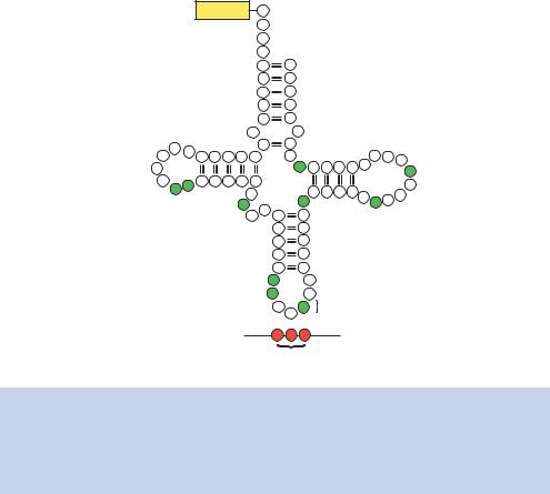
Figure 1.32. (a) Cloverleaf structure of yeast alanine tRNA showing the location of the unusual bases (shaded green). tRNA molecules can contain the following unusual bases: pseudouridine ( ), inosine (I), dihydrouridine (D), ribothymidine (T), methylguanosine (m1G), dimethylguanosine (m22G) and methylinosine (m1I). The tRNA anticodon base pairs with the appropriate codons in mRNA
|
|
|
|
1.15 TRANSLATION |
63 |
|
|
|
|
|
|
(b) |
Protein: |
Met Lys Leu Leu Ser Ser Ile Glu Gln Ala Cys Asp Ile Cys Stop |
|
||
mRNA: 5′-AGGAGGUCGAAAG AUG AAG CUA CUG UCU UCU AUC GAA CAA GCA UGC GAU AUU UGC UAA UUUGAAGUCA...–3′ |
|
||||
|
|
||||
|
|
|
|
Ribosome assembly |
|
|
|
|
|
|
|
|
|
50S |
|
|
|
|
|
|
|
||
mRNA: 5′-AGGAGGUCGAAAG AUG AAG CUA CUG UCU UCU AUC GAA CAA GCA UGC GAU AUU UGC UAA UUUGAAGUCA...–3′
30S
Addition of charged tRNA
MK
mRNA: 5′-AGGAGGUCGAAAG AUG AAG CUA CUG UCU UCU AUC GAA CAA GCA UGC GAU AUU UGC UAA UUUGAAGUCA...–3′
MK
Peptide bond formation
mRNA: 5′-AGGAGGUCGAAAG AUG AAG CUA CUG UCU UCU AUC GAA CAA GCA UGC GAU AUU UGC UAA UUUGAAGUCA...–3′
MK
Release of uncharged tRNA
mRNA: 5′-AGGAGGUCGAAAG AUG AAG CUA CUG UCU UCU AUC GAA CAA GCA UGC GAU AUU UGC UAA UUUGAAGUCA...–3′
MK 
Ribosome translocation by one codon
mRNA: 5′-AGGAGGUCGAAAG AUG AAG  CUA CUG UCU UCU AUC GAA CAA GCA UGC GAU AUU UGC UAA UUUGAAGUCA...–3′
M
K L
Addition of charged tRNA
mRNA: 5′-AGGAGGUCGAAAG AUG AAG  CUA CUG UCU UCU AUC GAA CAA GCA UGC GAU AUU UGC UAA UUUGAAGUCA...–3′
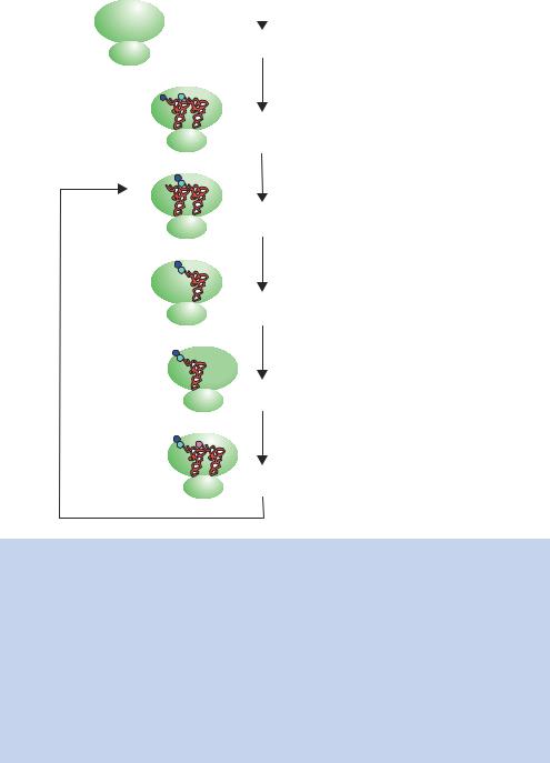
Figure 1.32. (b) Translation of an mRNA molecule on a ribosome in bacteria. The ribosome assembles on the mRNA just upstream of the initiation codon (AUG) at the Shine–Dalgarno sequence (shown in red). The 30S ribosomal subunit binds directly to this sequence and then recruits the 50S subunit. The assembled 70S ribosome is a large complex of proteins and rRNA that is of a physical size to cover approximately 35 bases of mRNA. A special initiator tRNA (fMet-tRNAf ) binds to the initiator codon (in the P site) through base pairing of the codon with the anticodon present in the tRNA. The next aminoacyl-tRNA (charged with its appropriate amino acid) binds to the next codon (in the A site) prior to peptide bond formation. The formation of a peptide bond involves the transfer of the amino acid(s) attached to the tRNA in the P site to the aminoacyl-tRNA in the A site. The uncharged tRNA then leaves the complex and the ribosome moves to the next codon. Subsequent amino acids are added in a similar fashion
64 |
DNA: STRUCTURE AND FUNCTION 1 |
|
|
their specific function. Many eukaryotic proteins are, however, destined for a particular compartment of the cell. The polypeptide itself encodes the information required for its final destination. Eukaryotic proteins that are destined to accumulate within the nucleus contain a nuclear localization signal (NLS), consisting of a short stretch of predominately basic amino acids (e.g. PKKKRLV), which direct the protein into the nucleus (Goldfarb et al., 1986). Proteins that are destined to be exported from the cell, or to be incorporated in cellular membranes, are transported into the endoplasmic reticulum (ER) during translation. Such proteins containing a stretch of predominantly hydrophobic amino acids at their amino terminus, called the signal sequence, which directs the polypeptide –ribosome complex to the ER, where the newly synthesized amino acid chain is inserted through the ER membrane. Inside the ER, the signal is cleaved off as the growing peptide chain is secreted into the ER. The protein is then transported by the Golgi apparatus to its final destination or, if it contains an ER retention signal (the amino acids KDEL) at its extreme carboxy-terminal end, it is retained within the ER itself (Munro and Pelham, 1987).

2Basic techniques in gene analysis
Key concepts
The coincident discovery of restriction enzymes, bacterial transformation, and agarose gel electrophoresis formed the basis of the explosion of molecular biology in the 1970s
Cutting DNA at defined sites and joining foreign DNA molecules together became possible
Cloning – the study of single genes in isolation – now became feasible
High-resolution analysis of DNA using gels
Blotting techniques to detect homologous nucleotide sequences
Rapid purification of DNA
One of the main problems in gene analysis is the relatively uniform nature of the DNA molecule itself. As we have already seen, the DNA that makes up the human genome is extremely long, with some 6.4 × 10⁹ base pairs of DNA in most cells, and is composed of only four different nucleotides. This size and relative lack of complexity makes isolating and studying single DNA fragments or genes appear a daunting task. We know that the base sequence of DNA is vital for encoding genes, but to investigate the function of a single gene it is essential that the gene be studied in isolation, freed from the rest of its native genome. Ideally, we would want to cut a single gene out of a genome. Until the 1970s, however, there were no methods available for cutting DNA at specific sequences. The ability to fragment DNA at specific sites became the cornerstone of molecular biology. The discovery of enzymes able to cleave DNA at specific
Analysis of Genes and Genomes |
Richard J. Reece |
2004 John Wiley & Sons, Ltd |
ISBNs: 0-470-84379-9 (HB); 0-470-84380-2 (PB) |
66 |
BASIC TECHNIQUES IN GENE ANALYSIS 2 |
|
|
sequences, restriction enzymes, led to the award of the 1978 Nobel Prize to Werner Arber, Daniel Nathans and Hamilton O. Smith.
When thinking about how we might like to isolate a particular piece of DNA, there are several important factors to take into account. We need to isolate the piece of DNA that we are interested in, and clone it so that it can be replicated and amplified in the absence of other human genes. How might this be achieved? The most favoured method of studying the function of a gene is to clone it into the molecular biologists’ favourite bacterium Escherichia coli. E. coli has several advantages that make it amenable for cloning – the cells grow quickly, the genetics are well characterized and strains have been engineered so that they are relatively harmless to ourselves. There are, however, many problems are associated with inserting foreign DNA sequences into bacteria. In general, DNA sequences will only be replicated if they contain a replicon. The genomes of bacteria and viruses usually contain a single replicon. So, simply putting a piece of foreign DNA into a bacteria cell will not result in the replication of that DNA. Indeed the most likely fate of foreign DNA in bacteria is degradation. As far back as the 1950s, it was noted that if a bacteriophage was prepared from a particular strain of E. coli cells (say strain C) then it would very efficiently infect cultures of the same E. coli strain. The bacteriophage would, however, infect other E. coli strains (strain K) at very low efficiency. Interestingly, the same bacteriophage prepared from an E. coli K strain would efficiently infect both E. coli K and C strains. The bacteriophage produced in E. coli C was in someway being restricted from entering E. coli K strains. Werner Arber noted that DNA from E. coli C prepared bacteriophages was rapidly degraded upon entering an E. coli K strain (Arber, 1965). Degradation occurs because the bacteria contain restriction –modification systems specifically designed to protect them from foreign DNA sequences.
2.1Restriction Enzymes
Bacterial restriction –modification systems have two components – a restriction endonuclease and a DNA methylase. The restriction enzyme (or restriction endonuclease) cleaves DNA at specific sequences. The term ‘endonuclease’ applies to sequence specific nucleases that break nucleic acid chains somewhere within the DNA, rather than at the ends of the molecule. The first restriction enzyme to be isolated was that from E. coli K laboratory strains in 1968 (Meselson and Yuan, 1968). This enzyme was able to cleave DNA, but the precise site of DNA cleavage remained unclear. The enzyme displayed a number of complex activities that made it difficult to study (Table 2.1).

2.1 RESTRICTION ENZYMES |
67 |
|
|
Table 2.1. Properties of restriction endonucleases. Adapted from Dryden, Murray and Rao (2001)
Property |
Type I |
Type II |
Type III |
|
|
|
|
Restriction and |
Single |
Separate nuclease |
Separate enzymes |
modification |
multifunctional |
and methylase |
sharing a |
|
enzyme |
|
common subunit |
Nuclease subunit |
Heterotrimer |
Homodimer |
Heterodimer |
structure |
|
|
|
Cofactors |
ATP, Mg2+, SAM |
Mg2+ |
Mg2+ (SAM) |
DNA cleavage |
Two recognition |
Single recognition |
Two recognition |
requirements |
sites in any |
site |
sites in a |
|
orientation |
|
head-to-head |
|
|
|
orientation |
Site of DNA |
Random, approx |
At or near |
24 – 26 bp to the |
cleavage |
1000 bp away |
recognition site |
3 -side of the |
|
from |
|
recognition site |
|
recognition site |
|
|
Enzymatic |
No |
Yes |
Yes |
turnover |
|
|
|
DNA translocation |
Yes |
No |
No |
Site of methylation |
At recognition site |
At recognition site |
At recognition site |
|
|
|
|
In 1970, Hamilton Smith and his co-workers isolated a restriction enzyme activity from the bacterium Haemophilus influenzae strain Rd and showed that it was able to cleave DNA at specific sites. The enzyme, called HindII, recognizes
a six-base-pair double-stranded DNA sequence of 5 -G – T – pyrimidine –purine – A – C-3 :
5′- GT(T/C) (G/A)AC-3′
3′-CA(A/G) (C/T)TG -5′
and cleaves DNA on both strands in the centre of the sequence (indicated by the dotted line). Smith found that his enzyme was unable to cleave Haemophilus influenzae genomic DNA, but it cleaved the bacteriophage T7 genome (39 937 bp in length) in over 40 places, to give a highly specific fragmentation pattern. The restriction enzyme HindII enzyme always recognizes the sequence above and always cuts directly in the centre of this sequence. This sequence is known